


Clinical value of cone-beam CT combined with panoramic radiography in impacted mandibular teeth extraction
-
摘要:
目的 分析锥形束CT(CBCT)联合全景片在下颌骨阻生牙拔除术前检查的临床价值。 方法 选取2019年1月~2020年1月于本院就诊的102例下颌骨阻生牙拔除患者(126颗下颌骨阻生牙)为研究对象。所有患者在术前均接受口腔影像学检查,记录阻生牙相关变量:牙根成角、牙根数、牙根远端和近中根形态、与下颌神经管的关系,在术中和术后评估以上变量以作为金标准。 结果 全景片图像无法清晰显示牙根与下牙槽神经管三维关系,CBCT图像中显示清晰;全景片、CBCT及CBCT联合全景片在评估成角、牙根数、远中根的弯曲情况、远中根向近中或远中弯曲、近中根弯曲、近中根近中或向远中弯曲中的差异无统计学意义(P>0.05);三者评估牙根与下颌管关系的差异有统计学意义(P < 0.05);患者拔牙时间为15.93±3.24 min,出血量为26.45± 3.15 mL,仅有1颗(0.79%)出现断根;术中仅有1例患者出现断根、根折,1例出现舌侧骨板骨折;术后无出血、感染患者,有2例患者出现感觉麻木、1例患者出现肿胀和受限。 结论 CBCT联合全景片在确定下颌第三磨牙与下颌管之间的关系方面有较大的优势,对改善患者术中情况和术中及术后并发症有积极意义。 Abstract:Objective To analyze the clinical value of cone- beam CT (CBCT) combined with panoramic radiography in the preoperative examination of impacted mandibular teeth extraction. Methods A total of 102 patients with mandibular impacted teeth (126 impacted teeth) treated in our hospital from January 2019 to January 2020 were selected. All patients underwent preoperative oral imaging, and the related variables of impacted teeth, including root angulation, root number, distal and mesial root morphology, and relationship with mandibular neural tube were recorded. and the above variables were evaluated as gold standard intraoperatively and postoperatively. Results The panoramic images could not clearly show the threedimensional relationship between the root and the inferior alveolar neural tube, which was clearly shown in the CBCT images. There were no significant differences in angle formation, root number, distal root bending, proximal root bending or distal root bending, proximal root bending or proximal root bending (P>0.05) in panoramic image, CBCT and CBCT combined panoramic image. There were statistically significant differences in the relationship between root and mandibular canal (P < 0.05). The extraction time was 15.93±3.24 min, the bleeding volume was 26.45±3.15 mL, only one (0.79%) had root breakage. During the operation, only one patient developed root breakage and root fracture, and one patient developed lingual bone plate fracture. there were no patients with bleeding or infection postoperatively, and two patients showed numbness and one showed swelling and limitation. Conclusion CBCT combined with panoramic radiography has great advantages in determining the relationship between the mandibular third molars and the mandibular canal, which has positive significance in improving intraoperative situation and intraoperative and postoperative complications of patients. -

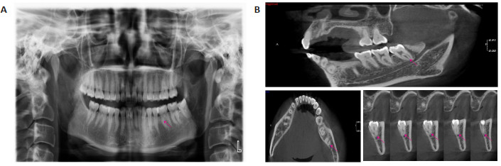
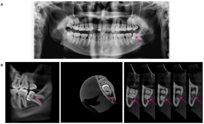
图 1 患者1全景片和CBCT
A:全景片显示左下第三磨牙牙根与下牙槽神经管相邻,具体位置不详;B:CBCT矢状位、水平位、冠状位显示下牙槽神经管位于第三磨牙根分叉下并于两牙根之间穿行.
Figure 1. Panoramic radiography and CBCT of patient 1.

图 2 患者2全景片和CBCT
A:全景片显示左下第三磨牙牙根与下牙槽神经管位置可能相连但未进入神经管,或者相邻但进入神经管;B:CBCT矢状位、水平位、冠状位显示下牙槽神经管位于第三磨牙根下方并且牙根根尖进入神经管上部.
Figure 2. Panoramic radiography and CBCT of patient 2.

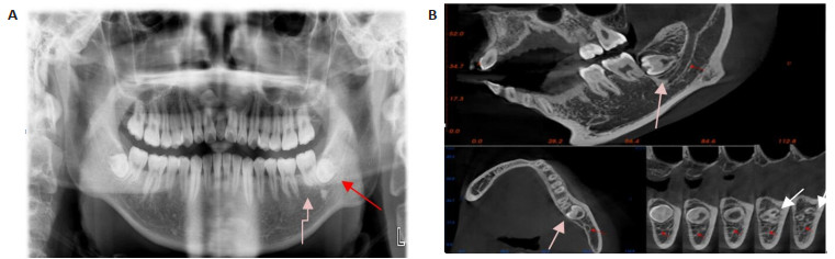
图 3 患者3全景片和CBCT
A:全景片显示左下第三磨牙水平低位骨埋伏阻生,但牙冠颊舌向位置不明,牙根情况不明确;B:CBCT矢状位、水平位、冠状位显示左下第三磨牙牙冠偏舌向阻生且根部偏颊侧,可见三个牙根并且牙根根尖未发育完善,下牙槽神经管位于第三磨牙根尖下并未与牙根相通(红色箭头:神经管;橙色箭头:牙冠;白色箭头:牙根).
Figure 3. Panoramic radiography and CBCT of patient 3.

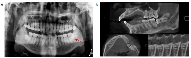
图 4 患者4全景片和CBCT
A:全景片显示左下第三磨牙近中牙根根尖与下牙槽神经管相邻,具体位置不详;B:CBCT矢状位、水平位、冠状位显示下牙槽神经管位于左下第三磨近中牙根下方并紧挨近中牙根根尖.
Figure 4. Panorama radiography and CBCT of patient 4.

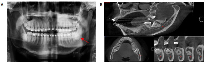
图 5 患者5全景片和CBCT
A:全景片显示左下第三磨牙颊舌向位置不明并且牙根与下牙槽神经管位置不清楚;B:CBCT矢状位、水平位、冠状位显示左下第三磨牙骨内位置偏颊侧,融合根,下牙槽神经管位于左下第三磨牙牙根舌侧,牙根根尖处与神经管相通.
Figure 5. Panorama radiography and CBCT of patient 5.
表 1 CBCT和全景片评估结果分布情况
Table 1. Distribution of evaluation results of CBCT and panoramic film[n(%)]
分布 全景片 CBCT CBCT联合全景片 χ2 P 成角 0.768 0.993 水平(n=27) 19(70.37) 23(85.19) 25(92.59) 远中角(n=18) 8(53.33) 10(55.56) 14(77.78) 近中角(n=43) 32(74.41) 37(86.05) 38(88.37) 垂直(n=38) 23(60.53) 26(68.42) 30(78.95) 牙根数 2.094 0.718 1(n=30) 9(30.00) 13(43.33) 16(53.33) 2(n=83) 62(74.70) 66(79.52) 73(87.95) ≥3(n=13) 3(23.08) 6(46.15) 8(61.54) 远端根弯曲 0.007 0.997 是(n=72) 38(52.78) 44(61.11) 49(68.06) 否(n=54) 35(64.81) 40(74.07) 44(81.48) 远端根近中或远端弯曲 0.174 0.917 是(n=54) 31(57.41) 32(59.26) 39(72.22) 否(n=72) 45(62.50) 53(73.61) 62(86.11) 近中根弯曲 0.607 0.738 是(n=85) 63(74.12) 71(83.53) 73(85.88) 否(n=41) 18(43.90) 20(48.78) 26(63.41) 近中根近中或远端弯曲 0.409 0.815 是(n=89) 66(74.16) 73(82.02) 76(85.39) 否(n=37) 21(56.76) 22(59.46) 28(75.68) 直接接触下颌管 6.205 0.045 是(n=21) 5(23.81) 15(71.43) 19(90.48) 否(n=105) 63(60.00) 60(57.14) 70(66.67) CBCT:锥形束CT.  下载: 导出CSV
-
[1] Kugelberg CF. Impacted lower third molars and periodontal health. An epidemiological, methodological, retrospective and prospective clinical, study[J]. Swed Dent J Suppl, 1990, 68: 1-52. [2] Hamasha AAH, Al Qudah MA, Bataineh AB, et al. Reasons for third molar teeth extraction in Jordanian adults[J]. J Contemp Dent Pract, 2006, 7(5): 88-95. doi: 10.5005/jcdp-7-5-88 [3] 陈全, 张晓, 张刚, 等.锥形束CT在下颌阻生第三磨牙拔除术前诊断应用的初步研究[J].中华口腔医学杂志, 2011, 46(7): 398-402. doi: 10.3760/cma.j.issn.1002-0098.2011.07.004 [4] 秦立业, 周忠伟.全景片对下颌智齿与下牙槽神经毗邻关系精确性判断的研究进展[J].口腔医学研究, 2019, 35(4): 319-21. https://www.cnki.com.cn/Article/CJFDTOTAL-KQYZ201904003.htm [5] 陈敏, 刘泉, 王凤琼, 等.全景片和锥形束CT对下颌骨阻生第三磨牙拔除术前诊断的准确性比较[J].临床口腔医学杂志, 2019, 35(4): 219-22. doi: 10.3969/j.issn.1003-1634.2019.04.009 [6] 刘晓华, 王恩博.锥形束CT在牙槽外科定量测量中精准性及应用的研究进展[J].口腔医学, 2020, 40(10): 955-9. https://www.cnki.com.cn/Article/CJFDTOTAL-KQYX202010018.htm [7] Mahmoud H, Amin H, Gaballah O, et al. Evaluation of the accuracy of cemental annulations versus con beam computed tomography for age estimation among adults[J]. Egypt Dent J, 2018, 64(1): 97P5. [8] Ishak MH, Zhun OC, Shaari R, et al. Panoramic radiography in evaluating the relationship of mandibular canal and impacted third molars in comparison with cone-beam computed tomography[J]. Mymensingh Med J, 2014, 23(4): 781-6. [9] Dawdy J, Halladay J, Carrasco-Labra A, et al. Efficacy of adjuvant laser therapy in reducing postsurgical complications after the removal of impacted mandibular third molars: a systematic review update and meta-analysis[J]. J Am Dent Assoc, 2017,148(12): 887-902.e4. doi: 10.1016/j.adaj.2017.06.043 [10] 周洁, 刘渊, 姜蕾, 等.锥形束CT在下颌阻生智齿拔除术前设计中的应用[J].第二军医大学学报, 2012, 33(4): 403-7. https://www.cnki.com.cn/Article/CJFDTOTAL-DEJD201204013.htm [11] Berghuis G, Cosyn J, de Bruyn H, et al. A controlled study on the diagnostic accuracy of panoramic and peri-apical radiography for detecting furcation involvement[J]. BMC Oral Health, 2021, 21(1): 115. doi: 10.1186/s12903-021-01460-z [12] 洪虓, 刘亮, 徐锦程.口腔全景片联合锥形束CT在上颌前部埋伏多生牙手术中的应用[J].中华解剖与临床杂志, 2018, 23(6): 551-3. doi: 10.3760/cma.j.issn.2095-7041.2018.06.020 [13] Alqerban A, Jacobs R, Fieuws S, et al. Comparison of two cone beam computed tomographic systems versus panoramic imaging for localization of impacted maxillary canines and detection of root resorption[J]. Eur J Orthod, 2011, 33(1): 93-102. doi: 10.1093/ejo/cjq034 [14] Schubert M, Proff P, Kirschneck C. Improved eruption path quantification and treatment time prognosis in alignment of impacted maxillary canines using CBCT imaging[J]. Eur J Orthod, 2018, 40(6): 597-607.[PubMed] [15] Moon HW, Nam W, Ahn HW, et al. Development of a maxillomandibular arch form based on the center of resistance of teeth using cone-beam computed tomography[J]. Am J Orthod Dentofacial Orthop, 2022,161(2): 208-19. doi: 10.1016/j.ajodo.2020.07.041 [16] Luo Q, Diao W, Luo L, et al. Comparisons of the computed tomographic scan and panoramic radiography before mandibular third molar extraction surgery[J]. Med Sci Monitor, 2018, 24: 3340-7. doi: 10.12659/MSM.907913 [17] Petersen LB, Vaeth M, Wenzel A. Neurosensoric disturbances after surgical removal of the mandibular third molar based on either panoramic imaging or cone beam CT scanning: a randomized controlled trial (RCT)[J]. Dentomaxillofac Radiol, 2016, 45(2): 20150224. doi: 10.1259/dmfr.20150224 [18] Arora A, Patil BA, Sodhi A. Validity of the vertical tube-shift method in determining the relationship between the mandibular third molar roots and the inferior alveolar nerve canal[J]. J Korean Assoc Oral Maxillofac Surg, 2015, 41(2): 66-73. doi: 10.5125/jkaoms.2015.41.2.66 [19] 韩倩, 张志宏.锥形束CT在下颌阻生第三磨牙微创拔牙中的临床应用[J].现代口腔医学杂志, 2016, 30(5): 283-6. https://www.cnki.com.cn/Article/CJFDTOTAL-XDKY201605008.htm [20] 丁水清, 周永强, 陈建荣, 等.锥形束CT在下颌阻生第三磨牙拔除术前定位和评估中的应用价值[J].广西医学, 2017, 39(11): 1687-9. https://www.cnki.com.cn/Article/CJFDTOTAL-GYYX201711022.htm [21] 陆璨, 尹乒.下颌阻生第三磨牙微创拔除法与传统拔除法临床并发症的对比研究[J].中国医师杂志, 2020, 22(9): 1410-3. [22] 朱志高, 谢春, 刘阳.锥形束CT介导下超声骨刀联合微创拔牙术拔除下颌阻生第三磨牙的效果[J].中国医药导报, 2015, 12(21): 93-6,101. https://www.cnki.com.cn/Article/CJFDTOTAL-YYCY201521026.htm -

 点击查看大图
计量
- 文章访问数: 213
- HTML全文浏览量: 129
- PDF下载量: 6
- 被引次数: 0