


Relationship between CT values of renal calculus and the outcome of holmium laser lithotripsy with percutaneous nephrolithotomy in patients
-
摘要:
目的 分析肾结石CT值与患者经皮肾镜钬激光碎石术治疗效果的关系。 方法 选取2016年10月~2021年8月于本院接受经皮肾镜钬激光碎石术治疗的90例肾结石患者的资料进行回顾性分析。所有患者术前均行螺旋CT扫描检查,明确结石大小并计算平均CT值,依据平均CT值大小分为两组:其中A组43例,CT值< 1000 Hu,B组47例,CT值≥1000 Hu。两组患者均经皮肾镜钬激光碎石术治疗,比较两组手术时间、单次结石清除率、术后住院时间以及并发症情况。采用Pearson相关分析CT值与手术时间的相关性,采用Spearman相关性分析结石CT值与单次结石清除率,采用Logistic多因素分析影响经皮肾镜钬激光碎石术术后结石残留的独立危险因素。 结果 A组手术时间短于B组,单次结石清除率高于B组,术后并发症低于B组,组间比较差异均有统计学意义(P < 0.05);两组术后住院时间比较差异无统计学意义(P>0.05)。相关性分析显示,CT值与手术时间呈正比,与患者结石清除率呈反比(P < 0.05);进一步Logistic回归分析显示,结石CT值、结石表面积为经皮肾镜钬激光碎石术术后结石残留的独立危险因素。 结论 肾结石CT值与患者经皮肾镜钬激光碎石术清石效率及结石清除率明显相关,且结石CT值、结石表面积是影响经皮肾镜钬激光碎石术后结石残留的独立危险因素。 -
关键词:
- 肾结石 /
- CT值 /
- 经皮肾镜钬激光碎石术 /
- 治疗效果 /
- 结石残留
Abstract:Objective To analyze the relationship between CT values of renal calculus and the treatment effect of percutaneous nephroscopic holmium laser lithotripsy in patients. Methods Ninety patients with renal calculus treated by percutaneous lithotripsy with holmium laser in our hospital from October 2016 to August 2021 were selected for retrospective analysis. All patients underwent preoperative spiral CT scan to determine the size of calculi and calculate the average CT value, and were divided into two groups according to the mean CT value: Group A (n=43) with CT value < 1000 Hu, group B (n=47) with CT value ≥1000 Hu. Patients in both groups were treated with percutaneous nephroscopic holmium laser lithotripsy. Operation time, single stone removal rate, postoperative hospital stay and complications were compared between the two groups. Pearson correlation was used to analyze the correlation between CT value and operation time, Spearman correlation was used to analyze the correlation between CT value and single stone removal rate, and Logistic multifactor analysis was used to analyze the independent risk factors affecting stone residual after percutaneous holmium laser lithotripsy. Results The operation time in group A was shorter than that in group B, the single stone removal rate was higher than that of group B, and the postoperative complications were lower than that in group B, with statistically significant differences (P < 0.05). There was no significant difference in postoperative hospital stay between the two groups (P>0.05). Correlation analysis showed that CT value was positively proportional to operation time and inversely proportional to stone removal rate (P < 0.05). Further Logistic regression analysis showed that CT value and stone surface area were independent risk factors for residual stones after percutaneous with holmium laser lithotripsy. Conclusion CT value of kidney stones is significantly correlated with stone removal efficiency and stone removal rate in patients undergoing percutaneous nephroscopic holmium laser lithotripsy, and CT value and stone surface area are independent risk factors affecting stone residual after percutaneous nephroscopic holmium laser lithotripsy. -

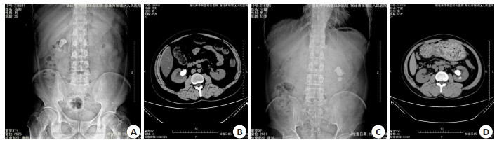
图 1 两患者术前腹部平片及术前肾脏CT影像
A: 患者1的术前腹部平片; B: 患者1的术前肾脏CT影像; C: 患者2的术前腹部平片; D: 患者2的术前肾脏CT影像.
Figure 1. Preoperative plain abdominal film and preoperative renal CT imaging of two patients.
表 1 两组患者一般资料比较
Table 1. Comparison of general data of the two groups
项目 A组(n=43) B组(n=47) t/χ2 P 性别(n,男/女) 25/18 27/20 0.004 0.947 年龄(岁, Mean±SD) 44.76±7.27 45.25±8.54 0.292 0.771 BMI(kg/m2, Mean±SD) 25.76±3.15 24.75±3.26 1.492 0.139 CT值(Hu, Mean±SD) 826.96±78.63 1103.45±78.23 16.707 < 0.01 结石部位(n) 0.143 0.706 右肾 23 27 左肾 20 20 A组: CT值< 1000 Hu; B组: CT值≥1000 Hu.  下载: 导出CSV
表 2 两组患者手术时间、术后住院时间、单次结石清除率比较
Table 2. Comparison of operation time, postoperative hospital stay and single stone clearance rate between the two groups
指标 A组(n=43) B组(n=47) χ2/t P 手术时间(min, Mean±SD) 60.23±7.45 83.45±8.63 13.604 < 0.01 术后住院时间(d, Mean±SD) 6.87±1.22 7.16±1.06 1.206 0.231 单次结石清除率[n(%)] 39(90.70) 34(72.34) 4.939 0.026
下载: 导出CSV
表 3 两组术后并发症发生情况比较
Table 3. Comparison of postoperative complications between the two groups [n(%)]
组别 发热 漏尿 出血 疼痛 总发生率 A组(n=43) 2(4.65) 0(0.00) 2(4.65) 1(2.32) 5(11.63) B组(n=47) 6(12.76) 3(6.38) 1(2.13) 4(8.51) 14(29.79) χ2 4.446 p 0.035
下载: 导出CSV
表 4 影响经皮肾镜钬激光碎石术术后结石残留的独立危险因素分析
Table 4. Analysis of independent risk factors affecting stone residual after percutaneous holmium laser lithotripsy
影响因素 β SE OR Wald χ2 P 95% CI 年龄 0.123 0.387 1.161 5.987 0.121 0.320~3.414 性别 0.056 0.347 1.927 8.674 1.039 0.598~1.824 结石表面积 0.522 0.716 3.259 5.420 0.024 1.168~3.624 结石位置 0.144 0.618 2.114 5.414 0.214 1.128~2.125 结石CT值 0.356 0.661 2.827 4.312 0.009 0.647~4.711 手术时间 0.126 0.452 1.842 7.589 0.184 0.489~1.597
下载: 导出CSV
-
[1] Kasote DM, Jagtap SD, Thapa D, et al. Herbal remedies for urinary stones used in India and China: a review[J]. J Ethnopharmacol, 2017, 203: 55-68. doi: 10.1016/j.jep.2017.03.038 [2] Cheungpasitporn W, Rossetti S, Friend K, et al. Treatment effect, adherence, and safety of high fluid intake for the prevention of incident and recurrent kidney stones: a systematic review and metaanalysis[J]. J Nephrol, 2016, 29(2): 211-9. doi: 10.1007/s40620-015-0210-4 [3] Nussberger F, Roth B, Metzger T, et al. A low or high BMI is a risk factor for renal hematoma after extracorporeal shock wave lithotripsy for kidney stones[J]. Urolithiasis, 2017, 45(3): 317-21. doi: 10.1007/s00240-016-0915-4 [4] 张江容, 罗旭, 李梦芝, 等. 肾结石患者经皮肾镜取石术后泌尿系感染的病原学特点及影响因素分析[J]. 中华医院感染学杂志, 2019, 29(2): 265-8. https://www.cnki.com.cn/Article/CJFDTOTAL-ZHYY201902026.htm [5] Hur I, Lee YK, Kalantar-Zadeh K, et al. Individualized hemodialysis treatment: a perspective on residual kidney function and precision medicine in nephrology[J]. Cardiorenal Med, 2019, 9 (2): 69-82. doi: 10.1159/000494808 [6] Chen ZJ, Yan YJ, Zhou JJ. Comparison of tubeless percutaneous nephrolithotomy and standard percutaneous nephrolithotomy for kidney stones: a meta-analysis of randomized trials[J]. Asian J Surg, 2020, 43(1): 60-8. doi: 10.1016/j.asjsur.2019.01.016 [7] Yilmazel FK, Cinislioglu AE, Karabulut I, et al. Ultra-mini flexible percutaneous nephrolithotomy in the treatment of moderate-size kidney stones: a new instrument, a preliminary prospective study[J]. Urolithiasis, 2021, 49(4): 345-50. doi: 10.1007/s00240-020-01225-3 [8] 杨林, 雷振涛, 史玉强, 等. 超声引导下微创单通道经皮肾镜钬激光碎石术治疗肾结石的临床分析[J]. 中国医学装备, 2019, 16 (5): 70-3. doi: 10.3969/J.ISSN.1672-8270.2019.05.019 [9] Hyams ES, Shah O. Percutaneous nephrostolithotomy versus flexible ureteroscopy/holmium laser lithotripsy: cost and outcome analysis[J]. J Urol, 2009, 182(3): 1012-7. doi: 10.1016/j.juro.2009.05.021 [10] 朱锦智, 宋宇, 姚志敏, 等. 上尿路结石CT值预测结石成分及对体外冲击波碎石术的指导意义[J]. 中国实用医药, 2018, 13(12): 54-6. https://www.cnki.com.cn/Article/CJFDTOTAL-ZSSA201812030.htm [11] El-Nahas AR, El-Assmy AM, Mansour O, et al. A prospective multivariate analysis of factors predicting stone disintegration by extracorporeal shock wave lithotripsy: the value of high-resolution noncontrast computed tomography[J]. Eur Urol, 2007, 51(6): 1688-94. doi: 10.1016/j.eururo.2006.11.048 [12] 《泌尿外科杂志(电子版)》编辑部. 泌尿系结石诊治指南解读(二): 肾结石治疗[J]. 泌尿外科杂志: 电子版, 2012, 4(1): 46-8. https://www.cnki.com.cn/Article/CJFDTOTAL-MNWZ201201016.htm [13] 韦巍, 黄剑华, 钟羽翔, 等. 2~3 cm肾结石的CT值对输尿管软镜碎石效果的预估价值[J]. 中华腔镜泌尿外科杂志: 电子版, 2019, 13(2): 95-8. doi: 10.3877/cma.j.issn.1674-3253.2019.02.006 [14] 黄思淮. 输尿管软镜联合经皮肾镜碎石术治疗复杂性肾结石的效果观察[J]. 临床合理用药杂志, 2019, 12(28): 142-3. https://www.cnki.com.cn/Article/CJFDTOTAL-PLHY201928085.htm [15] 倪大伟, 席俊华, 吴畏, 等. 经皮肾镜取石术和输尿管软镜碎石术治疗孤立肾结石有效性和安全性的Meta分析[J]. 中国医药导报, 2019, 16(3): 57-61. https://www.cnki.com.cn/Article/CJFDTOTAL-YYCY201903014.htm [16] 王进峰, 吴志坚, 刘鑫国, 等. 泌尿系结石CT值与其硬度值的关系[J]. 实用医学杂志, 2008, 24(3): 365-6. doi: 10.3969/j.issn.1006-5725.2008.03.013 [17] 罗林斌, 李美红, 朱斌, 等. CT预测泌尿系结石行体外冲击波及钬激光碎石时的易碎性研究[J]. 中国医药科学, 2020, 10(10): 212-4. doi: 10.3969/j.issn.2095-0616.2020.10.061 [18] 陈立杰, 吕学锋, 郭强, 等. 微通道经皮肾镜取石术与标准通道经皮肾镜取石术治疗上尿路结石的荟萃分析[J]. 现代泌尿外科杂志, 2019, 24(10): 837-42, 846. doi: 10.3969/j.issn.1009-8291.2019.10.012 [19] 曾鹏, 蒋重和, 李光明, 等. CT值预测经皮肾镜EMS碎石清石系统的取石效率[J]. 中国内镜杂志, 2016, 22(5): 17-20. https://www.cnki.com.cn/Article/CJFDTOTAL-ZGNJ201605005.htm [20] Olbert PJ, Hegele A, Schrader AJ, et al. Pre-and perioperative predictors of short-term clinical outcomes in patients undergoing percutaneous nephrolitholapaxy[J]. Urol Res, 2007, 35(5): 225-30. doi: 10.1007/s00240-007-0112-6 -

 点击查看大图
计量
- 文章访问数: 214
- HTML全文浏览量: 134
- PDF下载量: 4
- 被引次数: 0