


Model for predicting the benignity and malignancy of pulmonary sub-centimetre nodules based on enhanced dual-phase CT imaging
-
摘要:
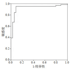
目的 分析基于增强双期CT成像的肺亚厘米结节良恶性预测模型。 方法 选择我院2019年1月~2021年3月收治的98例肺亚厘米结节患者作为研究对象,依照病理诊断结果分为良性病变组(n=64)和恶性病变组(n=34)。所有受试者行基于增强双期CT成像,采用Logistic回归模型分析增强双期CT成像预测结节良恶性预测模型,绘制ROC曲线分析增强双期CT成像的肺亚厘米结节良恶性预测模型的应用价值。 结果 良性病变组患者毛刺、结节边界清楚、上叶、分叶征、空泡征、胸膜凹陷征、血管集束征、磨玻璃密度发生率与恶性病变组的差异有统计学意义(P < 0.05);增强双期CT成像预测肺亚厘米结节良恶性预测模型为Log(P)=1.211×毛刺+2.843×分叶+1.981×磨玻璃+0.793×边界不清+1.326;增强双期CT成像预测肺亚厘米结节良恶性预测模型预测患者肺亚厘米结节良恶性的曲线下面积为0.930(P < 0.05)。 结论 基于增强双期CT成像预测肺亚厘米结节良恶性模型临床价值较高,具有较高的预测价值。 Abstract:Objective To analyze the prediction model of benign and malignant pulmonary sub-centimeter nodules based on enhanced dual-phase CT imaging. Methods Ninty-eight patients with pulmonary sub-centimeter nodules treated in our hospital from January 2019 to March 2021 were selected as the research objects, and divided into benign group (n=64) and malignant group (n=34) according to the pathological diagnosis. All the subjects underwent enhanced two-phase CT imaging, and a logistic regression model was used to analyze the predictive model of benign and malignant nodules by enhanced dual phase CT imaging, and ROC curve was drawn to analyze the application value of the predictive model of benign and malignant pulmonary sub-centimeter nodules by enhanced dual phase CT imaging. Results There were significant differences in the incidence of burr, clear nodule boundary, upper lobe, lobar sign, vacuole sign, pleural depression sign, vascular bundle sign and ground glass density between benign lesions and malignant lesions (P < 0.05). The prediction model for predicting the benignity and malignancy of pulmonary sub-centimeter nodules by enhanced duplex CT imaging was Log(P) =1.211×burr + 2.843×lobulation+1.981×groundglass+0.793×unclearboundary+1.326; The area under curve of enhanced dual phase CT imaging in predicting the benign and malignant of pulmonary sub-centimeter nodules was 0.930 (P < 0.05). Conclusion the model for predicting the benignity and malignancy of pulmonary sub-centimeter nodules based on enhanced dual phase CT imaging has high clinical value and high predictive value. -

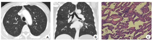
图 1 实性结节CT和病理检测
A: 肺部CT轴位影像; B: 肺部CT冠状位影像; C: 肺部活检病理图片(HE染色, ×400).
Figure 1. The CT imaging and pathological examination of solid nodules.

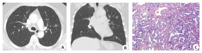
图 2 混合密度小结节CT和病理检测
A: 肺部CT轴位影像; B: 肺部CT冠状位影像; C: 肺部活检病理图片(HE染色, ×400).
Figure 2. The CT imaging and pathological examination of mixed density nodules
表 1 增强双期CT成像检测结果
Table 1. Results of enhanced dual phase CT imaging (n)
影响因素 良性病变组(n=64) 恶性病变组(n=34) χ2 P 毛刺 18.280 < 0.001 是 15 23 否 49 11 结节边界清楚 2.790 < 0.001 是 58 16 否 6 18 上叶 8.757 0.003 是 27 25 否 37 9 分叶征 55.742 < 0.001 是 6 29 否 58 5 空泡征 7.911 0.005 是 3 8 否 61 26 胸膜凹陷征 20.313 < 0.001 是 5 16 否 59 18 血管集束征 10.186 0.001 是 7 13 否 57 21 磨玻璃密度 7.159 0.008 是 12 15 否 52 19 钙化 0.008 0.928 是 6 3 否 58 31  下载: 导出CSV
表 2 增强双期CT成像预测肺亚厘米结节良恶性预测模型
Table 2. The prediction model of benign and malignant pulmonary subcentimeter nodules by enhanced dual phase CT imaging
指标 b SE χ2 P OR 95% CI 下限 上限 毛刺 1.211 0.231 27.483 < 0.001 3.357 2.135 5.279 分叶 2.843 0.392 52.599 < 0.001 17.167 7.962 37.015 磨玻璃 1.981 0.209 89.841 < 0.001 7.250 4.813 10.920 边界不清 0.793 0.189 17.604 < 0.001 2.210 1.526 3.201 常数项 1.326 0.199 44.400 < 0.001 3.766 2.550 5.56
下载: 导出CSV
-
[1] Abrishami A, Khalili N, Kooraki S, et al. Evaluation of cross-sectional imaging features that aid in the differentiation of benign and malignant splenic lesions[J]. Eur J Radiol, 2021, 136(5): 109549. http://www.sciencedirect.com/science/article/pii/S0720048X21000292 [2] Li Y, Yu JH, Du PJ, et al. High-score US-suspicious subcentimeter thyroid nodules: what factors affect adequate sampling of US-guided fine-needle aspiration biopsy?[J]. Int J Endocrinol, 2020, 3: 1-7. http://www.ncbi.nlm.nih.gov/pubmed/32377188 [3] Kim H, Lee M, Kim D, et al. Discrimination of benign and malignant microcalcifications using dual-energy imaging[J]. J Inst, 2020, 15(2): C02033. doi: 10.1088/1748-0221/15/02/C02033 [4] 杨艳艳, 马利军, 刘晓林. 孤立性肺结节的影像学研究进展与价值[J]. 世界最新医学信息文摘: 连续型电子期刊, 2020, 20(50): 69-71. [5] Boccatonda A, Susca V, Primomo GL, et al. Role of shear-wave and strain elastography to differentiate malignant vs benign subpleural lung lesions[J]. Medicine, 2021, 100(1): e24123. doi: 10.1097/MD.0000000000024123 [6] Homayounieh F, Singh R, Nitiwarangkul C, et al. Semiautomatic segmentation and radiomics for dual-energy CT: a pilot study to differentiate benign and malignant hepatic lesions[J]. AJR Am J Roentgenol, 2020, 215(2): 398-405. doi: 10.2214/AJR.19.22164 [7] Li MQ, Han RC, Song WJ, et al. Three dimensional volumetric analysis of solid pulmonary nodules on chest CT: cancer risk assessment[J]. Zhongguo Fei Ai Za Zhi, 2016, 19(5): 279-85. http://www.lungca.org/index.php?journal=01&page=article&op=download&path[]=10.3779/j.issn.1009-3419.2016.05.05&path[]=5333 [8] Hu X, Zhao JM, Qian HS, et al. Radiological and pathological analysis of LDCT screen detected and surgically resected sub-centimetre lung nodules in 44 asymptomatic patients[J]. Eur J Radiol Open, 2016, 3: 223-9. doi: 10.1016/j.ejro.2016.08.001 [9] Chen XM, Feng B, Chen YH, et al. A CT-based radiomics nomogram for prediction of lung adenocarcinomas and granulomatous lesions in patient with solitary sub-centimeter solid nodules[J]. Cancer Imaging, 2020, 20(1): 45. doi: 10.1186/s40644-020-00320-3 [10] 李辉贤, 左玉强, 段俊, 等. 亚厘米亚实性结节肺腺癌CT特征及经Hook-Wire定位切除的临床应用[J]. 临床肺科杂志, 2020, 25 (10): 1486-90. doi: 10.3969/j.issn.1009-6663.2020.10.007 [11] Peng MY, Xing GL, Zhang BY, et al. Genomic characterization of sub-centimeter pulmonary nodules[J]. J Clin Oncol, 2020, 38(15): e13530. http://www.researchgate.net/publication/341638302_Genomic_characterization_of_sub-centimeter_pulmonary_nodules [12] Ding JH, Jiang L, Fang JJ, et al. Predictors for malignancy risk in subcentimeter thyroid nodules categorized as atypia/follicular lesion of undetermined significance by fine needle aspiration[J]. Sci Rep, 2019, 9(1): 14973. doi: 10.1038/s41598-019-50597-z [13] Genere N, Hurtado MD, Cortes T, et al. Drivers of the decision to biopsy and follow-up of small suspicious thyroid nodules[J]. Endocr Pract, 2020, 26(8): 857-68. doi: 10.4158/EP-2019-0590 [14] 赫捷, 李霓, 陈万青, 等. 中国肺癌筛查与早诊早治指南(2021, 北京)[J]. 中华肿瘤杂志, 2021, 43(3): 243-68. https://www.cnki.com.cn/Article/CJFDTOTAL-ZHLU202102001.htm [15] 苏雷, 支修益, 张毅, 等. 亚厘米肺结节的外科诊疗分析[J]. 中国微创外科杂志, 2017, 17(1): 11-4. doi: 10.3969/j.issn.1009-6604.2017.01.004 [16] 严高武, 严高文, 孙清泉, 等. CT引导下经皮肺穿刺活检对肺部毛玻璃样病变良恶性的诊断价值[J]. 中国循证医学杂志, 2016, 16(4): 378-82. https://www.cnki.com.cn/Article/CJFDTOTAL-ZZXZ201604002.htm [17] 景瑞, 金鑫, 高洁, 等. 不同密度早期肺腺癌的影像特征与病理学分类[J]. 医学影像学杂志, 2019, 29(6): 937-40, 952. https://www.cnki.com.cn/Article/CJFDTOTAL-XYXZ201906014.htm [18] Reig B, Ha R. Editorial on "diagnosis of benign and malignant breast lesions on DCE-MRI by using radiomics and deep learning with consideration of peritumor tissue"[J]. J Magn Reson Imaging, 2020, 51(3): 810-1. doi: 10.1002/jmri.27025 [19] Vernuccio F, Porrello G, Cannella R, et al. Benign and malignant mimickers of infiltrative hepatocellular carcinoma: tips and tricks for differential diagnosis on CT and MRI[J]. Clin Imaging, 2021, 70: 33-45. doi: 10.1016/j.clinimag.2020.10.011 [20] 徐建平, 李会方, 叶伟, 等. 178例肺亚厘米结节(直径≤8 mm)影像学与临床病理分析[J]. 临床与实验病理学杂志, 2020, 36(3): 290-4. https://www.cnki.com.cn/Article/CJFDTOTAL-LSBL202003010.htm -

 点击查看大图
计量
- 文章访问数: 116
- HTML全文浏览量: 92
- PDF下载量: 9
- 被引次数: 0