


Imaging features of hepatic perivascular epithelioid cell tumors in different types
-
摘要:
目的 探讨不同类型肝脏血管周上皮样细胞分化的肿瘤(PEComa)的CT及MRI影像学特征。 方法 回顾性分析我院2014年7月~2018年7月经手术病理证实的12例肝脏PEComa患者的影像学资料,分析病灶在CT及MRI图像上的位置、密度及信号特点、强化形式。 结果 含脂肪型病灶肉眼可见的脂肪成分无明显强化,而软组织成分强化明显,动态增强扫描病灶动脉期显著强化并见到瘤内血管,门脉期及延时期出现中轻度强化,并可见环形强化的假包膜。无脂肪型呈实质性软组织肿块,肉眼不可见脂肪成分,动态增强扫描病灶动脉期呈明显均质强化,可见粗大扭曲的畸形血管和动脉瘤样结节状强化,门脉期及平衡期呈延时强化改变。由于病灶缺少正常肝细胞,肝胆特异期病灶对比剂摄取呈低信号改变。其中含脂肪型需与血管平滑肌脂肪瘤鉴别,但影像学缺乏特异性,无脂肪型几乎全部误诊为肝细胞癌。 结论 肝脏PEComa具有独特的免疫组化特征,如果依据病灶内是否含有脂肪成分来分型,影像学表现具有一定的特性,但是病变在表观扩散系数图及肝胆特异期没有特异性,不能作为单独的诊断依据。乏脂肪型PEComa临床术前误诊概率较大,因此放射科医生加强对此病的认识具有重要的意义。 -
关键词:
- 肝脏 /
- 血管周上皮样细胞肿瘤 /
- 计算机断层扫描 /
- 磁共振成像 /
- 分型
Abstract:Objective To investigate the CT and MRI features of hepatic perivascular epithelioid cell differentiation tumors (PEComa) in different types. Methods The imaging data of 12 patients with liver PEComa confirmed by surgical pathology from July 2014 to July 2018 in our hospital were retrospectively analyzed to analyze the location, density and signal characteristics, and form of enhancement of the lesions on CT and MRI images. Results The fat-containing lesions showed no significant enhancement of the fatty component with the naked eye, while the soft-tissue component was significantly enhanced. Dynamic enhancement scans showed significant enhancement in the arterial phase and intra-tumoural vessels, and moderate to mild enhancement in the portal and delayed phases, with a circularly enhanced pseudo-envelope. In fat-deficient type, which was mainly characterized by a substantial soft tissue mass, the fat components are invisible to the naked eye. Dynamic enhancement scan showed marked homogeneous enhancement in the arterial phase of the lesion. with coarse distorted malformed vessels and aneurysmal nodular enhancement visible in the portal and equilibrium phases, delayed enhancement in the portal and equilibrium phases. Due to lack of normal hepatocytes in the lesions, the hepatobiliary specific phase showed low signal without contrast uptake. The fat-containing type tumors need to be differentiated from angiomyolipoma, but the lack of specificity in imaging means that the non- fatty type is almost always misdiagnosed as hepatocellular carcinoma. Conclusion There is unique immunohistochemical features in hepatic PEComa. If it is staged according to the presence or absence of a fat component, the imageology manifestations have certain characteristics. However, the lesions have no specificity in ADC or hepatobiliary specific phase, and cannot be used as a separate diagnostic basis. The probability of misdiagnosis before clinical procedures is relatively high in fat-deficient type, so it is important for radiologists to be more aware of this condition. -
Key words:
- liver /
- perivascular epithelial cell tumors /
- computed tomography /
- magnetic resonance imaging /
- typing
-

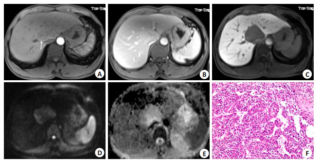
图 1 男性,47岁,含脂肪型PEComa
A: CT平扫,病灶内可见脂肪成分; B~C: 分别为正反相位, 反相位见病灶信号明显减低, 含脂丰富; D~E:分别为MRI动脉期、门脉期, 明显强化的包膜和病灶内血管样强化显示清晰(箭头); F: 病理图(HE, ×200), 混合型PEComa, 脂肪细胞及肌样PEComa细胞混合组成.
Figure 1. A47-year-old male patient, fat-containing type PEComa.
-
[1] 毕颖文, 袁一飞, 张锐, 等. 眼内血管周上皮样细胞肿瘤二例[J]. 中华眼科杂志, 2021, 57(11): 857-60. doi: 10.3760/cma.j.cn112142-20201123-00767 [2] 赵宁, 杨劼, 王飞, 等. 肺血管周上皮细胞分化肿瘤自发破裂出血1例[J]. 中华胸心血管外科杂志, 2021, 37(9): 562-3. doi: 10.3760/cma.j.cn112434-20200221-00059 [3] 李新敏, 古雅丽, 张瑶, 等. 子宫血管周上皮样细胞肿瘤1例[J]. 临床与实验病理学杂志, 2021, 37(6): 756-7. https://www.cnki.com.cn/Article/CJFDTOTAL-LSBL202106035.htm [4] Zamboni G, Pea M, Martignoni G, et al. Clear cell"sugar"tumor of the pancreas. A novel member of the family of lesions characterized by the presence of perivascular epithelioid cells[J]. Am J Surg Pathol, 1996, 20(6): 722-30. doi: 10.1097/00000478-199606000-00010 [5] Kirnap M, Ozgun G, Moray G, et al. Perivascular epithelioid cell tumor outgrowth from the liver[J]. Int J Surg Case Rep, 2018, 53: 295-8. doi: 10.1016/j.ijscr.2018.10.046 [6] Nie P, Wu J, Wang HX, et al. Primary hepatic perivascular epithelioid cell tumors: imaging findings with histopathological correlation[J]. Cancer Imaging, 2019, 19(1): 32. doi: 10.1186/s40644-019-0212-x [7] Low SCS, Peh WCG, Muttarak M, et al. Imaging features of hepatic angiomyolipomas[J]. J Med Imaging Radiat Oncol, 2008, 52(2): 118-23. doi: 10.1111/j.1440-1673.2008.01927.x [8] Martignoni G, Pea M, Reghellin D, et al. PEComas: the past, the present and the future[J]. Virchows Arch, 2008, 452(2): 119-32. doi: 10.1007/s00428-007-0509-1 [9] 宣兰兰, 魏建国, 刘红刚. 血管周上皮样细胞肿瘤的病理诊断及新进展[J]. 中华病理学杂志, 2021, 50(3): 282-7. doi: 10.3760/cma.j.cn112151-20200721-00579 [10] Hao BB, Rao JH, Fan Y, et al. Hepatic perivascular epithelioid cell tumor in three patients[J]. Hepatobiliary Pancreat Dis Int, 2016, 15 (6): 660-4. doi: 10.1016/S1499-3872(16)60077-2 [11] 何璐, 覃玲艳, 周梦琦, 等. 肝脏血管周上皮样细胞肿瘤的多模态影像学表现与病理对照分析[J]. 临床放射学杂志, 2021, 40(5): 913-7. https://www.cnki.com.cn/Article/CJFDTOTAL-LCFS202105018.htm [12] 贺亚琼, 姚景江, 刘建滨, 等. 肝脏血管周上皮样细胞肿瘤的病理与MSCT表现[J]. 临床放射学杂志, 2019, 38(1): 105-9. https://www.cnki.com.cn/Article/CJFDTOTAL-LCFS201901023.htm [13] Selvaggi F, Risio D, Claudi R, et al. Malignant PEComa: a case report with emphasis on clinical and morphological criteria[J]. BMC Surg, 2011, 11: 3. doi: 10.1186/1471-2482-11-3 [14] 马英腾, 沈丹华. 肝脏血管周上皮样细胞肿瘤临床与病理学特征分析[J]. 中华普通外科杂志, 2017, 32(12): 1007-9. doi: 10.3760/cma.j.issn.1007-631X.2017.12.008 [15] 王国禹, 赵闯, 侯登峰, 等. 肝脏血管周上皮样细胞瘤11例临床诊治分析[J]. 中华医学杂志, 2018, 98(34): 2715-7. doi: 10.3760/cma.j.issn.0376-2491.2018.34.007 [16] Son HJ, Kang DW, Kim JH, et al. Hepatic perivascular epithelioid cell tumor (PEComa): a case report with a review of literatures[J]. Clin Mol Hepatol, 2017, 23(1): 80-6. doi: 10.3350/cmh.2016.0034 [17] Tan Y, Zhang H, Xiao EH. Perivascular epithelioid cell tumour: dynamic CT, MRI and clinicopathological characteristics: analysis of 32 cases and review of the literature[J]. Clin Radiol, 2013, 68(6): 555-61. doi: 10.1016/j.crad.2012.10.021 [18] 陈翼, 董丽华, 李江城, 等. 肝血管周上皮细胞瘤的多种影像表现[J]. 医学影像学杂志, 2017, 27(7): 1405-9. https://www.cnki.com.cn/Article/CJFDTOTAL-XYXZ201707060.htm [19] Wang SY, Kuai XP, Meng XX, et al. Comparison of MRI features for the differentiation of hepatic angiomyolipoma from fat-containing hepatocellular carcinoma[J]. Abdom Imaging, 2014, 39(2): 323-33. doi: 10.1007/s00261-013-0070-0 [20] Jafari A, Fischer HP, von Websky M, et al. Primary perivascular epitheloid cell tumour (PEComa) of the liver: case report and review of the literature[J]. Z Gastroenterol, 2013, 51(9): 1096-100. doi: 10.1055/s-0033-1350123 [21] Zhu QQ, Niu ZF, Yu FD, et al. Epithelioid angiomyolipoma of the pancreas: a case report and review of the literature[J]. World J Clin Cases, 2021, 9(8): 1931-9. doi: 10.12998/wjcc.v9.i8.1931 [22] Liu J, Zhang CW, Hong DF, et al. Primary hepatic epithelioid angiomyolipoma: a malignant potential tumor which should be recognized[J]. World J Gastroenterol, 2016, 22(20): 4908-17. doi: 10.3748/wjg.v22.i20.4908 -

 下载:
下载:
 点击查看大图
图(2)
计量
- 文章访问数: 170
- HTML全文浏览量: 162
- PDF下载量: 10
- 被引次数: 0