


Differential diagnosis value of SPECT/CT bone quantitative SUVmax analysis in the bone metastases of elderly prostate cancer patients
-
摘要:
目的 探讨SPECT/CT骨定量分析获得最大标准摄取值(SUVmax)对老年前列腺癌骨转移的鉴别诊断价值。 方法 回顾性分析2019年1~12月我院158例确诊前列腺癌患者的99mTc-MDP SPECT/CT骨显像资料。对异常放射性浓聚灶及182个正常椎体行SPECT/CT检查及骨定量分析,比较良恶性病灶与正常椎体之间SUVmax的差异性。选取2019年1~4月46例患者的144个异常浓聚灶,通过绘制ROC曲线得到SUVmax的最佳临界值;选取2019年5~12月293个异常浓聚灶,使用SUVmax的最佳临界值比较SPECT/CT骨定量分析与常规SPECT/CT的诊断效能。 结果 以病理或随访作为判断标准,所有老年前列腺癌患者共437个异常浓聚灶(恶性254个,良性183个),恶性病灶的SUVmax明显高于良性病灶(43.93±19.09 vs 15.26±6.81,P < 0.01)和正常椎体(6.54±1.19,P < 0.01)。通过ROC曲线得到SUVmax≥19.2为诊断恶性病灶的最佳临界值,常规SPECT/CT与SPECT/CT骨定量诊断的准确度、敏感度、特异性、阳性预测率、阴性预测率分别为81.6%和94.5%、93.0%和96.5%、65.2%和91.7%、79.2%和94.3%、86.8%和94.9%,SPECT/CT骨定量诊断的准确度、特异性、阳性预测率和阴性预测率均明显高于常规SPECT/CT。 结论 SPECT/CT骨定量分析在老年前列腺癌骨转移的鉴别诊断中具有重要临床应用价值,具有临床应用的潜力。 -
关键词:
- 前列腺癌 /
- 骨转移 /
- SPECT/CT定量分析 /
- 鉴别诊断 /
- 标准摄取值
Abstract:Objective To investigate the differential diagnosis value of SPECT/CT bone quantitative analysis in bone metastases of elderly prostate cancer patients. Methods From January to December in 2019, 158 patients with elderly prostate cancer who underwent whole body bone scan were enrolled. SPECT/CT bone quantitative analysis were preformed on abnormal radioactive concentration lesions and 182 normal vertebral. The differences of SUVmax among the benign, malignant, and normal vertebral were compared. A total of 144 lesions of 46 patients from January to April in 2019 are plotted ROC curve to obtain cut-off values of SUVmax. A total of 293 lesions from May to December 2019 were selected to compare the diagnostic efficacy between SPECT/CT bone quantitative analysis and conventional SPECT/CT. Results A total of 437 lesions (254 malignants/183 benigns) were determined with pathology or follow-up results. The SUVmax of malignant lesions was significantly higher than benign lesions (43.93±19.09 vs 15.26±6.81, P < 0.01) and normal vertebral (6.54±1.19, P < 0.01). Using the best cut-off value of SUVmax≥19.2 as the diagnostic criterion, the accuracy, sensitivity, specificity, positive prediction rate, and negative prediction rate of conventional SPECT/CT and SPECT/CT bone quantitative analysis were 81.6% and 94.5%, 93.0% and 96.5%, 65.2% and 91.7%, 79.2% and 94.3%, 86.8% and 94.9%, respectively. The accuracy, specificity, positive prediction rate and negative prediction rate of SPECT/CT bone quantitative diagnosis were significantly higher than conventional SPECT/CT. Conclusion SPECT/CT bone quantitative analysis has important differential diagnosis value in bone metastases of elderly prostate cancer, and it has the potential for clinical application. -

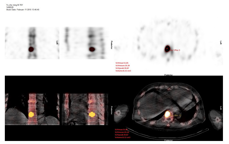
图 1 利用自动VOI技术测量病灶SUV及体积信息
Figure 1. Using automatic VOI software to measure the standard uptake value and volume.

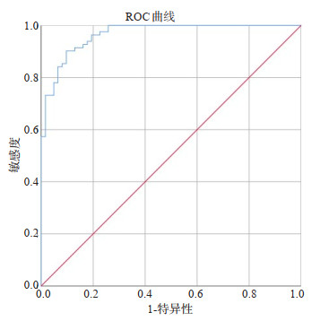
图 2 骨转移良恶性病灶SUVmax ROC曲线
Figure 2. The ROC curve of the SUVmax between malignant and benign lesions.
表 1 前列腺癌患者骨转移病灶分布
Table 1. Distribution of bone metastases in prostate cancer patients (n)
病灶分布 恶性病灶 良性病灶 合计 脊柱 94 75 169 肋骨 42 34 76 骨盆 106 72 178 胸骨 8 0 8 四肢 4 2 6  下载: 导出CSV
表 2 常规SPECT/CT、SPECT/CT骨定量诊断结果与金标准对比
Table 2. Comparison of the results of conventional SPECT/CT, SPECT/CT quantitative analysis and the gold standard (n)
诊断结果 常规SPECT/CT 合计 SPECT/CT骨定量 合计 恶性 良性 恶性 良性 病理或随访 恶性 160 12 172 166 6 172 良性 42 79 121 10 111 293 合计 202 91 293 176 117 293
下载: 导出CSV
表 3 常规SPECT/CT与SPECT/CT骨定量诊断效能对比
Table 3. Comparison of the diagnostic efficiency of conventional SPECT/CT and SPECT/CT quantitative analysis (%)
诊断方式 准确度 敏感度 特异性 阳性预测率 阴性预测率 常规SPECT/CT 81.6 93 65.2 79.2 86.8 SPECT/CT骨定量 94.5 96.5 91.7 94.3 94.9
下载: 导出CSV
-
[1] 周毅, 姚远, 杨剑文, 等. 老年前列腺癌并存代谢综合征患者的临床相关性研究[J]. 中华老年医学杂志, 2014, 33(2): 172-4. doi: 10.3760/cma.j.issn.0254-9026.2014.02.016 [2] 孙海燕, 方呈祥, 刘艳娥, 等. 盆腔磁共振T2加权像在老年前列腺癌中的诊断价值[J]. 中国老年学杂志, 2015, 35(7): 1798-800. doi: 10.3969/j.issn.1005-9202.2015.07.034 [3] Boellaard R. Standards for PET image acquisition and quantitative data analysis[J]. J Nucl Med, 2009, 50(Suppl 1): 11S-20S. doi: 10.2967/jnumed.108.057182 [4] Bailey DL, Willowson KP. An evidence-based review of quantitative SPECT imaging and potential clinical applications[J]. J Nucl Med, 2013, 54(1): 83-9. doi: 10.2967/jnumed.112.111476 [5] Bailey DL, Willowson KP. Quantitative SPECT/CT: SPECT joins PET as a quantitative imaging modality[J]. Eur J Nucl Med Mol Imaging, 2014, 41(Suppl 1): S17-S25. doi: 10.1007/s00259-013-2542-4 [6] Armstrong IS, Hoffmann SA. Activity concentration measurements using a conjugate gradient (Siemens xSPECT) reconstruction algorithm in SPECT/CT[J]. Nucl Med Commun, 2016, 37(11): 1212-7. doi: 10.1097/MNM.0000000000000586 [7] Yamane T, Fukushima K, Shirotake S, et al. Test-retest repeatability of quantitative bone SPECT/CT[J]. Ann Nucl Med, 2021, 35(3): 338-46. doi: 10.1007/s12149-020-01568-2 [8] Bae S, Kang Y, Song YS, et al. Maximum standardized uptake value of foot SPECT/CT using Tc-99m HDP in patients with accessory navicular bone as a predictor of surgical treatment[J]. Medicine: Baltimore, 2019, 98(2): e14022. doi: 10.1097/MD.0000000000014022 [9] Suh MS, Lee WW, Kim YK, et al. Maximum standardized uptake value of 99mTc hydroxymethylene diphosphonate SPECT/CT for the evaluation of temporomandibular joint disorder[J]. Radiology, 2016, 280(3): 890-6. doi: 10.1148/radiol.2016152294 [10] Kim J, Lee HH, Kang Y, et al. Maximum standardised uptake value of quantitative bone SPECT/CT in patients with medial compartment osteoarthritis of the knee[J]. Clin Radiol, 2017, 72(7): 580-9. doi: 10.1016/j.crad.2017.03.009 [11] Ryoo HG, Lee WW, Kim JY, et al. Minimum standardized uptake value from quantitative bone single-photon emission computed tomography/computed tomography for evaluation of femoral head viability in patients with femoral neck fracture[J]. Nucl Med Mol Imaging, 2019, 53(4): 287-95. doi: 10.1007/s13139-019-00600-2 [12] Cachovan M, Vija AH, Hornegger J, et al. Quantification of 99mTcDPD concentration in the lumbar spine with SPECT/CT[J]. EJNMMI Res, 2013, 3(1): 45. doi: 10.1186/2191-219X-3-45 [13] Kaneta T, Ogawa M, Daisaki H, et al. SUV measurement of normal vertebrae using SPECT/CT with Tc- 99m methylene diphosphonate [J]. Am J Nucl Med Mol Imaging, 2016, 6(5): 262-8. http://www.ajnmmi.us/files/ajnmmi0027797.pdf [14] Kuji I, Yamane T, Seto A, et al. Skeletal standardized uptake values obtained by quantitative SPECT/CT as an osteoblastic biomarker for the discrimination of active bone metastasis in prostate cancer[J]. Eur J Hybrid Imaging, 2017, 1(1): 2. doi: 10.1186/s41824-017-0006-y [15] Mohd Rohani MF, Mat Nawi N, Shamim SE, et al. Maximum standardized uptake value from quantitative bone single-photon emission computed tomography/computed tomography in differentiating metastatic and degenerative joint disease of the spine in prostate cancer patients[J]. Ann Nucl Med, 2020, 34(1): 39-48. doi: 10.1007/s12149-019-01410-4 [16] Zhang YQ, Li BL, Yu HJ, et al. The value of skeletal standardized uptake values obtained by quantitative single-photon emission computed tomography-computed tomography in differential diagnosis of bone metastases[J]. Nucl Med Commun, 2021, 42(1): 63-7. doi: 10.1097/MNM.0000000000001311 [17] 杜芬, 罗小毛, 刘明, 等. 定量SPECT/CT(SUVmax)在骨良恶性病灶中的鉴别诊断及增益价值[J]. 分子影像学杂志, 2021, 44(3): 435- 40. doi: 10.12122/j.issn.1674-4500.2021.03.04 [18] Seret A, Nguyen D, Bernard C. Quantitative capabilities of four stateof-the-art SPECT-CT cameras[J]. EJNMMI Res, 2012, 2(1): 45. doi: 10.1186/2191-219X-2-45 [19] Tabotta F, Jreige M, Schaefer N, et al. Quantitative bone SPECT/CT: high specificity for identification of prostate cancer bone metastases [J]. BMC Musculoskelet Disord, 2019, 20(1): 619. doi: 10.1186/s12891-019-3001-6 -

 点击查看大图
计量
- 文章访问数: 460
- HTML全文浏览量: 209
- PDF下载量: 19
- 被引次数: 0