


Correlation between carotid arteriosclerosis by ultrafast pulse wave velocity technology and retinopathy in patients with essential hypertension
-
摘要:
目的 探讨超极速脉搏波(UFPWV)技术检测颈动脉硬化程度与原发性高血压视网膜病变程度的相关性分析。 方法 利用UFPWV技术测量120例原发性高血压患者颈动脉内-中膜厚度、收缩开始时脉搏波传导速度(PWV)、收缩结束时PWV,采集患者双眼眼底图像并进行临床分级。收集患者的年龄、性别、高血压病程、血压值、吸烟史、BMI、高密度脂蛋白胆固醇、低密度脂蛋白胆固醇、总胆固醇、甘油三酯、血尿酸、血肌酐和尿素指标。分析以上各因素与眼底改变的相关性及比较UFPWV、颈动脉内-中膜厚度对眼底分级的预测效果。 结果 高血压患者收缩结束时PWV、收缩开始时PWV、颈动脉内-中膜厚度与眼底分级呈正相关(P < 0.001)。多因素Logistic回归分析显示,收缩结束时PWV、收缩开始时PWV和颈动脉内-中膜厚度是高血压视网膜病变的独立预测因子。收缩结束时PWV的预测效果优于收缩开始时PWV、颈动脉内-中膜厚度,差异有统计学意义(P < 0.05)。当收缩结束时PWV大于截断值8.97时,收缩结束时PWV提示高血压视网膜病变严重(P < 0.05)。 结论 原发性高血压患者颈动脉硬化程度与视网膜病变程度具有相关性。UFPWV技术将可能作为临床辅助预测高血压视网膜病变的简便安全、高效准确的诊断指标。 Abstract:Objective To investigate the correlation between the degree of carotid arteriosclerosis by the ultrafast pulse wave velocity (UFPWV) technology and the degree of essential hypertensive retinopathy. Methods The UFPWV technology was used to measure carotid artery intima-media thickness (IMT), the carotid PWV at the beginning of systole (PWV-BS), and the carotid PWV at the end of systole (PWV-ES) in 120 patients with essential hypertension and to collect the images of the patient' s binocular fundus for clinical grading. We collected data on patients' age, gender, course of hypertension, blood pressure, smoking history, BMI, HDL-C, LDL-C, TC, TG, UA, Cr and urea. The correlation between the above factors and fundus changes and compared the predictive effects of IMT and UFPWV values on the severity of fundus grading were analyzed. Results PWV-ES, PWV-BS, and IMT in hypertensive patients were found to be positively correlated with the degree of retinopathy (P < 0.001). In multivariate logistic regression analysis, PWV-ES, PWV-BS, and IMT were identified as important independent predictors of hypertensive retinopathy. The prediction effect of PWV-ES was better than that of PWV-BS and IMT, and the difference was significant (all P < 0.05). When the value of PWV-ES was greater than the cut-off value of 8.97, PWV-ES indicated that hypertensive retinopathy lesions were serious (P < 0.05). Conclusion The degree of carotid arteriosclerosis in patients with essential hypertension is correlated with the degree of retinopathy. The UFPWV technology may be used as a simple, safe, efficient, and accurate diagnostic index for clinical auxiliary prediction of hypertensive retinopathy. -

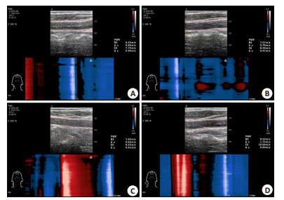
图 1 颈总动脉UFPWV采集图
A: HR Ⅰ级患者左侧颈动脉PWV-BS、PWV-ES分别为6.55 m/s、7.74 m/s; B: HR Ⅱ级患者左侧颈动脉PWV-BS、PWV-ES分别为7.57 m/s、8.59 m/s; C: HR Ⅲ级患者左侧颈动脉PWV-BS、PWV-ES分别为7.40 m/s、9.35 m/s; D: HR Ⅳ级患者左侧颈动脉PWV-BS、PWV-ES分别为9.55 m/s、10.99 m/s.
Figure 1. Images of UFPWV collection in common carotid artery.

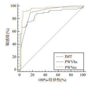
图 2 IMT、PWV-BS、PWV-ES预测严重眼底分级ROC曲线
Figure 2. ROC curve of IMT, PWV- BS and PWV- ES predicting severe HR classification.
表 1 HR分级与各变量的相关分析
Table 1. Correlation analysis between HR classification and various variables
变量 rs P PWV-BS 0.823 < 0.001 PWV-ES 0.894 < 0.001 IMTs 0.727 < 0.001 年龄 0.168 0.067 高血压分级 0.820 < 0.001 高血压病程 0.607 < 0.001 总胆固醇 0.186 0.042 甘油三酯 0.219 0.016 HDL-C 0.071 0.443 LDL-C -0.014 0.878 血肌酐 0.224 0.014 尿素 0.266 0.003 血尿酸 0.325 < 0.001 BMI 0.731 < 0.001 PWV-BS: 收缩期开始时PWV; PWV-ES: 收缩期结束时PWV; IMT: 内-中膜厚度; HDL-C: 高密度脂蛋白胆固醇; LDL-C: 低密度脂蛋白胆固醇.  下载: 导出CSV
表 2 性别、吸烟史、饮酒史对HR分级的影响
Table 2. The influence of gender, smoking history, and drinking history on HR classification (n)
变量 正常组(n=18) Ⅰ级(n=19) Ⅱ级(n=23) Ⅲ级(n=29) Ⅳ级(n=31) Z P 性别 1.687 0.092 男 10 11 8 17 23 女 8 8 15 12 8 吸烟史 4.211 < 0.001 有 0 3 2 8 15 无 18 16 21 21 16
下载: 导出CSV
表 3 不同眼底分级患者UFPWV比较
Table 3. Comparison of UFPWV in patients with different fundus classifications (n=60)
变量 眼底分级严重 眼底分级非严重+正常 检验值 P PWV-BS (m/s) 8.7±1.1 6.6±0.7 7.873 < 0.001 PWV-ES (m/s) 10.7±1.6 7.6±1.1 10.225 < 0.001 IMT (cm) 0.08 (0.08, 0.09) 0.06 (0.06, 0.07) 7.718 < 0.001a 年龄(岁) 55.6±10.3 51.3±9.1 2.432 0.017 高血压病史(年) 9.5 (6.5, 10.5) 5.0 (3.5, 7.5) 5.598 < 0.001a 总胆固醇(mmol/L) 4.5±1.4 4.2±1.1 1.441 0.152 甘油三酯(mmol/L) 1.70 (1.23, 2.19) 1.45 (1.23, 2.07) 1.231 0.218a HDL-C(mmol/L) 1.01 (0.86, 1.37) 1.11 (0.78, 1.25) 0.407 0.684a LDL-C(mmol/L) 2.30 (1.83, 3.33) 2.54 (1.89, 3.22) 0.344 0.731a 血肌酐(μmol/L) 66.6 (60.5, 92.2) 63.9 (53.0, 71.0) 2.433 0.015a 尿素(mmol/L) 6.23 (5.09, 7.16) 5.22 (4.44, 6.52) 2.955 0.003a 血尿酸(μmol/L) 363.8±92.4 317.4±89.5 2.795 0.006 BM(I kg/m2) 23.5±1.1 21.4±1.3 9.266 < 0.001 性别(男/女) 40/20 29/31 4.126 0.042b 吸烟史(年) 23 (38.3) 5 (8.3) 15.093 < 0.001b 注: a两独立样本秩和检验(Mann-Whitney U检验); b卡方检验.
下载: 导出CSV
表 4 HR的多因素logistic回归分析
Table 4. Multivariate logistic regression analysis of HR
变量 B S.E Wald P OR 95% CI PWV-BS 1.040 0.340 9.362 0.002 2.829 0.374, 1.707 PWV-ES 1.629 0.286 32.476 < 0.001 5.099 1.069, 2.189 IMT 44.921 21.119 4.524 0.033 3.228 3.529, 86.314 高血压病史 0.110 0.097 1.911 0.167 1.116 -0.046, 0.265 总胆固醇 0.097 0.197 0.244 0.621 1.102 -0.289, 0.483 甘油三酯 0.095 0.267 0.127 00.722 1.100 -0.428, 0.617 血肌酐 -0.003 0.010 0.107 0.743 0.997 -0.024, 0.017 尿素 0.014 0.129 0.012 0.914 1.014 -0.239, 0.267 血尿酸 -0.001 0.003 0.165 0.685 0.999 -0.008, 0.005 BMI 0.953 0.224 18.157 < 0.001 2.593 0.515, 1.391 吸烟史 -0.097 0.039 6.080 0.014 0.909 -0.173, -0.020 饮酒史 0.128 0.040 10.161 0.001 1.136 0.049, 0.206
下载: 导出CSV
表 5 不同HR分级的PWV对应值
Table 5. Corresponding values of PWV for different HR classifications (m/s, Mean±SD)
指标 正常组(n=18) Ⅰ级(n=19) Ⅱ级(n=23) Ⅲ级(n=29) Ⅳ级(n=31) F P PWV-BS 6.11±0.50 6.81±0.70 6.89±0.55 8.01±1.03a 9.30±0.83b 64.157 < 0.001 PWV-ES 6.50±0.62 7.65±0.60 8.53±0.74 9.80±1.32a 11.51±1.30b 83.723 < 0.001 aP < 0.05 vs正常组、Ⅰ级、Ⅱ级; bP < 0.05 vs正常组、Ⅰ级、Ⅱ级、Ⅲ级.
下载: 导出CSV
表 6 不同指标对HR分级(严重)预测效果
Table 6. The effect of different indicators on severe HR classification prediction
指标 截断点 AUC 敏感度(%) 特异性(%) 约登指数 IMT (cm) > 0.07 0.903 (0.835, 0.949) 90.0 (79.5, 96.2) 83.3 (71.5, 91.7) 0.733 PWV-BS (m/s) > 8.00 0.869 (0.795, 0.923) 75.0 (62.1, 85.3) 88.3 (77.4, 95.2) 0.633 PWV-ES (m/s) > 8.97 0.955 (0.901, 0.984) 91.7 (81.6, 97.2) 88.3 (77.4, 95.2) 0.867
下载: 导出CSV
-
[1] Bruno RM, Cartoni G, Stea F, et al. Carotid and aortic stiffness in essential hypertension and their relation with target organ damage: the CATOD study[J]. J Hypertens, 2017, 35(2): 310-8. doi: 10.1097/HJH.0000000000001167 [2] 张真, 王彤, 徐强, 等. 高血压视网膜病变的研究进展[J]. 中国慢性病预防与控制, 2016, 24(7): 541-3. https://www.cnki.com.cn/Article/CJFDTOTAL-ZMXB201607018.htm [3] Chen X, Meng Y, Li J, et al. Serum uric acid concentration is associated with hypertensive retinopathy in hypertensive Chinese adults[J]. BMC Ophthalmol, 2017, 17(1): 83. doi: 10.1186/s12886-017-0470-y [4] Mancia G, De Backer G, Dominiczak A, et al. 2007 guidelines for the management of arterial hypertension: the task force for the management of arterial hypertension of the European society of hypertension (esh) and of the European society of cardiology (esc) [J]. Eur Heart J, 2007, 28(12): 1462-536. http://med.wanfangdata.com.cn/Paper/Detail/PeriodicalPaper_PM24107724 [5] Kario K, Kanegae H, Oikawa T, et al. Hypertension is predicted by both large and small artery disease[J]. Hypertension, 2019, 73(1): 75-83. doi: 10.1161/HYPERTENSIONAHA.118.11800 [6] Dąbrowska E, Harazny JM, Miszkowska-Nagórna E, et al. Aortic stiffness is not only associated with structural but also functional parameters of retinal microcirculation[J]. Microvasc Res, 2020, 129: 103974. doi: 10.1016/j.mvr.2020.103974 [7] 臧鹏程, 赵磊, 王云慧, 等. 视网膜动脉硬化Ⅱ级以上高血压患者眼底病变与颈动脉血流参数、斑块情况的相关性[J]. 岭南心血管病杂志, 2020, 26(5): 570-3, 579. doi: 10.3969/j.issn.1007-9688.2020.05.17 [8] Zhang W, Li J, Zhao L, et al. Positive relationship of hypertensive retinopathy with carotid in Tima: media thickness in hypertensive patients[J]. J Hypertens, 2020, 38(10): 2028-35. doi: 10.1097/HJH.0000000000002509 [9] Zhu ZQ, Chen LS, Wang H, et al. Carotid stiffness and atherosclerotic risk: non-invasive quantification with ultrafast ultrasound pulse wave velocity[J]. Eur Radiol, 2019, 29(3): 1507-17. doi: 10.1007/s00330-018-5705-7 [10] van Bortel LM, Laurent S, Boutouyrie P, et al. Expert consensus document on the measurement of aortic stiffness in daily practice using carotid-femoral pulse wave velocity[J]. J Hypertens, 2012, 30 (3): 445-8. doi: 10.1097/HJH.0b013e32834fa8b0 [11] 李宏波, 王晗, 殷立平, 等. 极速成像技术测定高血压患者的脉搏波传导速度及相关影响因素[J]. 中华高血压杂志, 2017, 25(5): 477-81. https://www.cnki.com.cn/Article/CJFDTOTAL-ZGGZ201705019.htm [12] Pan FS, Yu L, Luo J, et al. Carotid artery stiffness assessment by ultrafast ultrasound imaging: feasibility and potential influencing factors[J]. J Ultrasound Med, 2018, 37(12): 2759-67. doi: 10.1002/jum.14630 [13] 中国高血压防治指南修订委员会, 高血压联盟(中国), 中华医学会心血管病学分会, 等. 中国高血压防治指南(2018年修订版) [J]. 中国心血管杂志, 2019, 24 (1): 24-56. doi: 10.3969/j.issn.1007-5410.2019.01.002 [14] Keith NM, Wagener HP, Barker NW. Some different types of essential hypertension[J]. Am J Med Sci, 1939, 197(3): 332-43. doi: 10.1097/00000441-193903000-00006 [15] Kayange PC, Schwering MS, Manda CS, et al. Prevalence and clinical spectrum of hypertensive retinopathy among hypertension clinic patients at queen elizabeth central hospital in malawi[J]. Malawi Med J, 2018, 30(3): 180-3. doi: 10.4314/mmj.v30i3.9 [16] 边波, 万征, 李永乐, 等. 高血压患者视网膜病变调查及其临床价值评价[J]. 中国慢性病预防与控制, 2011, 19(2): 170-1. https://www.cnki.com.cn/Article/CJFDTOTAL-ZMXB201102023.htm [17] Katsi V, Vlachopoulos C, Souretis G, et al. Association between retinal microcirculation and aortic stiffness in hypertensive patients [J]. Int J Cardiol, 2012, 157(3): 370-3. doi: 10.1016/j.ijcard.2010.12.074 [18] 关玉波. 颈动脉超声检测对原发性高血压患者颈动脉粥样硬化的临床诊断价值研究[J]. 中国医药指南, 2018, 16(36): 63. https://www.cnki.com.cn/Article/CJFDTOTAL-YYXK201836046.htm [19] Sobey CG, Faraci FM, Piegors DJ, et al. Effect of short-term regression of atherosclerosis on reactivity of carotid and retinal arteries[J]. Stroke, 1996, 27(5): 927-33. doi: 10.1161/01.STR.27.5.927 [20] Wong TY, Klein R, Klein BE, et al. Retinal microvascular abnormalities and their relationship with hypertension, cardiovascular disease, and mortality[J]. Surv Ophthalmol, 2001, 46 (1): 59-80. doi: 10.1016/S0039-6257(01)00234-X [21] Laurent S, Briet M, Boutouyrie P. Large and small artery cross-talk and recent morbidity-mortality trials in hypertension[J]. Hypertension, 2009, 54(2): 388-92. doi: 10.1161/HYPERTENSIONAHA.109.133116 [22] Yin LX, Ma CY, Wang S, et al. Reference values of carotid ultrafast pulse-wave velocity: a prospective, multicenter, population-based study[J]. J Am Soc Echocardiogr, 2021, 34(6): 629-41. doi: 10.1016/j.echo.2021.01.003 [23] 黄辉, 朱正球, 栾云, 等. 超极速超声成像脉搏波技术在早期颈动脉粥样硬化风险动态评估中的应用价值[J]. 东南大学学报: 医学版, 2017, 36(1): 9-13. doi: 10.3969/j.issn.1671-6264.2017.01.003 [24] Williams B, Mancia G, Spiering W, et al. 2018 ESC/ESH Guidelines for the management of arterial hypertension[J]. Eur Heart J, 2018, 39 (33): 3021-104. doi: 10.1093/eurheartj/ehy339 [25] Pan FS, Xu M, Yu L, et al. Relationship between carotid intimamedia thickness and carotid artery stiffness assessed by ultrafast ultrasound imaging in patients with type 2 diabetes[J]. Eur J Radiol, 2019, 111: 34-40. doi: 10.1016/j.ejrad.2018.12.016 [26] Li XP, Jiang J, Zhang H, et al. Measurement of carotid pulse wave velocity using ultrafast ultrasound imaging in hypertensive patients [J]. J Med Ultrason, 2017, 44(2): 183-90. doi: 10.1007/s10396-016-0755-4 [27] 回亚男, 刘梦堃, 王焕程. 颈动脉彩色多普勒超声检查老年女性高血压患者颈动脉病变与血压变异性的相关性研究[J]. 中国超声医学杂志, 2018, 34(12): 1061-5. doi: 10.3969/j.issn.1002-0101.2018.12.003 [28] Naqvi TZ, Lee MS. Carotid intima-media thickness and plaque in cardiovascular risk assessment[J]. JACC Cardiovasc Imaging, 2014, 7(10): 1025-38. doi: 10.1016/j.jcmg.2013.11.014 [29] Lorenz MW, Markus HS, Bots ML, et al. Prediction of clinical cardiovascular events with carotid intima-media thickness: a systematic review and meta-analysis[J]. Circulation, 2007, 115(4): 459-67. doi: 10.1161/CIRCULATIONAHA.106.628875 [30] Andrus B, Lacaille D. 2013 ACC/AHA guideline on the assessment of cardiovascular risk[J]. J Am Coll Cardiol, 2014, 63(25): 2886. http://www.sciencedirect.com/science/article/pii/s0735109713060312 [31] Hodis S. Pulse wave velocity as a diagnostic index: The effect of wall thickness[J]. Phys Rev E, 2018, 97(6): 062401. doi: 10.1103/PhysRevE.97.062401 [32] Gotschy A, Bauer E, Schrodt C, et al. Local arterial stiffening assessed by MRI precedes atherosclerotic plaque formation[J]. Circ Cardiovasc Imaging, 2013, 6(6): 916-23. doi: 10.1161/CIRCIMAGING.113.000611 [33] Yin LX, Ma CY, Wang S, et al. Reference values of carotid ultrafast pulse-wave velocity: a prospective, multicenter, population- based study[J]. J Am Soc Echocardiogr, 2021, 34(6): 629-41. doi: 10.1016/j.echo.2021.01.003 [34] Yang W, Wang Y, Yu Y, et al. Establishing normal reference value of carotid ultrafast pulse wave velocity and evaluating changes on coronary slow flow[J]. Int J Cardiovasc Imaging, 2020, 36(10): 1931-9. doi: 10.1007/s10554-020-01908-3 -

 点击查看大图
计量
- 文章访问数: 221
- HTML全文浏览量: 147
- PDF下载量: 3
- 被引次数: 0