


MRI findings of hepatobiliary phase doughnut-like nodules in Gd-EOB-DTPA and their relationship with cholestasis
-
摘要:
目的 分析肝硬化患者普美显肝胆期(HBP)甜甜圈结节的MRI影像表现及HBP结节信号与胆汁淤积关系。 方法 回顾性分析2019年1月1日~2021年7月31日在本院行MR普美显检查的551例肝硬化患者的影像和临床资料。对存在HBP甜甜圈结节的60例患者进行分析,分析HBP甜甜圈结节在不同原因肝硬化出现频率,结节在MR平扫、扩散加权成像(DWI)及增强各序列信号特点及随访情况。分析HBP结节信号与胆汁淤积等的关系,60例HBP甜甜圈结节患者中31例肝背景伴发肝癌、部分行手术治疗,为避免该因素对结果干扰故被排除,另外3例因资料不全被排除;最终纳入112例患者,其中HBP甜甜圈结节患者26例,高信号结节患者27例,等信号结节患者59例;分析HBP甜甜圈结节、高信号结节与等信号结节患者组胆汁淤积标志物(总胆红素、直接胆红素、间接胆红素、碱性磷酸酶、谷氨酰转移酶、总胆汁酸)差异。 结果 551例肝硬化患者中HBP甜甜圈结节患者占10.9%(60/551),不同原因肝硬化甜甜圈结节出现频率的差异有统计学意义(P=0.032)。60例患者共计688个甜甜圈结节,单发占18.3%,多发占81.7%,病灶径线5~32 mm;Fs-T2WI序列上77%结节呈等信号,22.5%结节为稍高信号,0.4%结节呈低信号。同反相位T1WI上89.1%结节为等信号,7%呈稍高信号,3.9%呈低信号。DWI序列上4.5%结节DWI呈稍高信号,但无弥散受限;余95.5%结节DWI呈等信号。所有结节动脉期均无强化,48.5%结节自门脉期开始强化,42.8%自延迟期开始强化,8.7%结节多期无强化。25例患者随访无恶性转化。HBP甜甜圈结节、单纯高信号结节及等信号结节组年龄、性别、直接胆红素、间接胆红素、碱性磷酸酶及肝功能分级组间差异无统计学意义(P > 0.05)。总胆红素、谷氨酰转移酶及总胆汁酸组间差异存在统计学意义(P < 0.05),多重比较显示甜甜圈组与等信号组总胆红素、甜甜圈结节与高信号结节组谷氨酰转移酶差异有统计学意义(P < 0.05)。 结论 HBP甜甜圈结节并不少见,具有无动脉期强化、门脉供血且无弥散受限的特征性影像表现,随访结节没有恶变;HBP甜甜圈结节、高信号结节形成与胆汁淤积无关。 Abstract:Objective To analyze the MRI findings of hepatobiliary phase (HBP) doughnut-like nodules in patients with liver cirrhosis and the relationship between HBP nodule signal with cholestasis. Methods The imaging and clinical data of 551 patients with liver cirrhosis who underwent MRI with Gd-EOB-DTPA from January 1st, 2019 to July 31st, 2021 were analyzed retrospectively. Sixty patients with HBP doughnut nodules were analyzed. The frequency of HBP doughnut-like nodules in different causes of liver cirrhosis, the signal characteristics on MR plain scan, diffusion weighted imaging (DWI) and enhanced sequence and follow-up of nodules were analyzed. The relationship between HBP nodule signal and cholestasis was analyzed. Of the 60 patients with HBP doughnut nodules, 31 cases had liver cancer in liver background and some underwent surgical treatment. In order to avoid the interference of this factor, they were excluded, and the other 3 cases were excluded due to incomplete data. A total of 112 patients were finally included, 26 patients with HBP doughnut-like nodules, 27 patients with high signal nodules and 59 patients with isosignal nodules were included. Whether there were differences in cholestasis markers such as total bilirubin, direct bilirubin, indirect bilirubin, alkaline phosphatase, glutamyltransferase, total bile acid between HBP doughnut-like nodules, high signal nodules and iso-signal nodules were analyzed. Results Among 551 patients with liver cirrhosis, HBP doughnut-like nodules accounted for 10.9% (60/551). There were significant differences in the frequency of doughnut-like nodules in different causes of liver cirrhosis (P=0.032). There were 688 doughnut-like nodules in 60 patients, single in 18.3% and multiple in 81.7%. On fs-T2WI sequence, 77% of the nodules showed isointensity, 22.5% showed hyperintensity and 0.4% showed hypointensity. On in/out phase T1WI, 89.1% of the nodules showed isointensity, 7% showed hyperintensity and3.9% showed hypointensity. On DWI, 4.5% nodules showed hyperintensity, but there was no limited diffusion. The remaining 95.5% of nodules showed isointensity. All nodules had no enhancement in arterial phase, 48.5% of nodules began to enhancement in portal phase, 42.8% began to enhancement in delayed phase, and 8.7% nodules had no enhancement in multiple phases. 25 patients were followed up without malignant transformation. There was no significant difference (P > 0.05) in age, sex, direct bilirubin, indirect bilirubin, alkaline phosphatase and liver function between HBP doughnut- like nodules, high signal nodules and iso-signal nodules group. The differences in total bilirubin, glutamyltransferase and total bile acid between the groups were significant (P < 0.05). Multiple comparisons showed that there were significant differences in total bilirubin between HBP doughnut-like nodules group and iso-signal group, in glutamyltransferase between HBP doughnut-like nodules and high signal nodules group (P < 0.05). Conclusion HBP doughnut-like nodules in are not uncommon. They have characteristic imaging manifestations of no arterial phase enhancement, portal blood supply and no limited diffusion, and have no malignant tendency during follow-up. The formation of HBP doughnut-like nodules and high signal nodules are not related to cholestasis. -

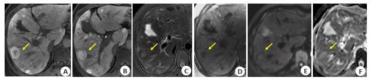
图 1 女,41岁,乙肝肝硬化失代偿期(Child-PughA级)
A、B: 普美显肝胆期图像显示肝实质内多发甜甜圈样结节, 较大结节(黄箭)中心呈瘢痕状, 下一个层面病灶内条状低信号为门脉分支结构; C: T2WI压脂序列示病灶中心呈稍高信号, 边缘呈低信号; D: 同相位T1WI图显示病灶中心为低信号; E、F: DWI及ADC图示结节呈稍高信号, 无弥散受限.
Figure 1. A41-year-old female, decompensated cirrhosis of hepatitis B (Child-PughA).
表 1 各变量统计分析结果
Table 1. Statistical analysis results of each variable (n)
分组 性别 年龄(岁, Mean±SD) 总胆红素 直接胆红素 间接胆红素 ALP R-GT 总胆汁酸 肝功能分级 男 女 + - + - + - + - + - + - A B C 甜甜圈 18 8 54.15±10.997 7 19 21 5 6 20 5 21 6 20 8 18 15 10 1 高信号 18 9 51.15±10.553 13 14 20 7 8 19 12 15 18 9 17 10 17 9 1 等信号 42 17 51.56±10.336 35 24 49 10 20 39 15 44 26 33 34 25 3 30 6 统计值 0.182 0.68 7.593 0.949 1.009 4.734 10.2 6.732 5.286 P 0.913 0.509 0.022 0.622 0.604 0.094 0.006 0.035 0.238 ALP: 碱性磷酸酶(ALP); R-GT: 谷酰转肽酶.  下载: 导出CSV
-
[1] Hanna RF, Aguirre DA, Kased N, et al. Cirrhosis-associated hepatocellular nodules: correlation of histopathologic and MR imaging features[J]. Radiographics, 2008, 28(3): 747-69. doi: 10.1148/rg.283055108 [2] 赵玉娇, 沈文. 肝胆特异性MRI对比剂钆塞酸二钠临床应用专家共识解读与病例分析[J]. 实用器官移植电子杂志, 2020, 8(5): 337-41. doi: 10.3969/j.issn.2095-5332.2020.05.003 [3] Joo I, Kim SY, Kang TW, et al. Radiologic-pathologic correlation of hepatobiliary phase hypointense nodules without arterial phase hyperenhancement at gadoxetic acid-enhanced MRI: a multicenter study[J]. Radiology, 2020, 296(2): 335-45. doi: 10.1148/radiol.2020192275 [4] Yoneda N, Matsui O, Kitao A, et al. Benign hepatocellular nodules: hepatobiliary phase of gadoxetic acid-enhanced MR imaging based on molecular background[J]. RadioGraphics, 2016, 36(7): 2010-27. doi: 10.1148/rg.2016160037 [5] 周小娇, 龙莉玲. 肝脏结节在Gd-EOB-DTPA增强MRI肝胆期不同表现的分子病理基础[J]. 临床放射学杂志, 2020, 39(12): 2549-52. https://www.cnki.com.cn/Article/CJFDTOTAL-LCFS202012043.htm [6] 李晓明, 蔡萍, 朱琳. Gd-EOB-DTPA MRI肝胆期高信号病变的研究进展[J]. 临床放射学杂志, 2021, 40(1): 181-4. https://www.cnki.com.cn/Article/CJFDTOTAL-LCFS202101042.htm [7] Kozaka K, Kobayashi S, Yoneda N, et al. Doughnut-like hyperi-ntense nodules on hepatobiliary phase without arterial-phase hyperenhancement in cirrhotic liver: imaging and clinico- pathological features[J]. Eur Radiol, 2019, 29(12): 6489-98. doi: 10.1007/s00330-019-06329-y [8] 中华医学会肝病学分会, 中华医学会消化病学分会, 中华医学会感染病学分会. 胆汁淤积性肝病诊断和治疗共识(2015)[J]. 肝脏, 2015, 20 (12): 950-9. https://www.cnki.com.cn/Article/CJFDTOTAL-LCGD201512007.htm [9] 陈勇, 张颖. 肝胆特异性磁共振对比剂Gd-EOB-DTPA在肝脏局灶性结节增生的诊断价值[J]. 中国医学计算机成像杂志, 2017, 23(5): 422-6. doi: 10.3969/j.issn.1006-5741.2017.05.008 [10] Shimamatsu K, Kage M, Nakashima O, et al. Pathomorphological study of HCV antibody-positive liver cirrhosis[J]. J Gastroenterol Hepatol, 1994, 9(6): 624-30. doi: 10.1111/j.1440-1746.1994.tb01572.x [11] Ferrell LD, Kakar S, Terracciano LM, et al. Tumours and tumour-like lesions of the liver[M]//Macsween's Pathology of the Liver. Amsterdam: Elsevier, 2018: 780-879. [12] Reshamwala PA, Kleiner DE, Heller T. Nodular regenerative hyperplasia: not all nodules are created equal[J]. Hepatology, 2006, 44(1): 7-14. doi: 10.1002/hep.21258 [13] Yoneda N, Matsui O, Kitao A, et al. Hepatocyte transporter expression in FNH and FNH-like nodule: correlation with signal intensity on gadoxetic acid enhanced magnetic resonance images[J]. Jpn J Radiol, 2012, 30(6): 499-508. doi: 10.1007/s11604-012-0085-4 [14] Yoneda N, Matsui O, Kitao A, et al. Beta-catenin-activated hepatocellular adenoma showing hyperintensity on hepatobiliary-phase gadoxetic-enhanced magnetic resonance imaging and overexpression of OATP8[J]. Jpn J Radiol, 2012, 30(9): 777-82. doi: 10.1007/s11604-012-0115-2 [15] Reizine E, Amaddeo G, Pigneur F, et al. Quantitative correlation between uptake of Gd-BOPTA on hepatobiliary phase and tumor molecular features in patients with benign hepatocellular lesions[J]. Eur Radiol, 2018, 28(10): 4243-53. doi: 10.1007/s00330-018-5438-7 [16] Inchingolo R, Faletti R, Grazioli L, et al. MR with Gd-EOB-DTPA in assessment of liver nodules in cirrhotic patients[J]. World J Hepatol, 2018, 10(7): 462-73. doi: 10.4254/wjh.v10.i7.462 [17] Sciarra A, Schmidt S, Pellegrinelli A, et al. OATPB1/B3 and MRP3 expression in hepatocellular adenoma predicts Gd-EOB-DTPA uptake and correlates with risk of malignancy[J]. Liver Int, 2019, 39 (1): 158-67. doi: 10.1111/liv.13964 -

 点击查看大图
图(1) / 表(1)
计量
- 文章访问数: 401
- HTML全文浏览量: 230
- PDF下载量: 6
- 被引次数: 0