


Correlation of coronal features of automatic breast volume and Ki67 and C-erbB-2 in breast cancer
-
摘要:
目的 分析乳腺癌自动乳腺容积(ABVS)冠状面图像特征与患者血清Ki67抗原及C-erbB-2的关系。 方法 收集我院2016年1月~2020年12月的78例乳腺癌患者,获取81个病灶的ABVS冠状面特征(病灶大小、边界、虫蚀征、汇聚征、微钙化、高回声环),并采用免疫组织化学方法进行检测,比较Ki67抗原与C-erbB-2与乳腺癌ABVS冠状面超声特征以及患者年龄之间的差异,对差异有统计学意义的指标及临床考虑可能有意义的指标进行多因素Logisitc回归分析。 结果 病灶大小(≥2 cm,OR= 4.400)是Ki67高表达的危险因素;微钙化(OR=2.741)是C-erbB-2阳性的危险因素;边界、虫蚀征、汇聚征、高回声环、年龄与Ki67、C-erbB-2均无相关性(P > 0.05)。 结论 乳腺癌ABVS冠状面的特征征象间接反映肿瘤细胞的分子生物学行为,以此来评估乳腺癌的治疗及预后。 Abstract:Objective To analyze the relationship between breast cancer automatic breast volume (ABVS) coronal image features, serum Ki67 antigen and C-erbB-2. Methods A total of 78 breast cancer patients in our hospital from January 2016 to December 2020, 81 lesions were collected. ABVS coronal features, including size, boundary, worm erosion sign, convergent sign, microcalcification, hyperechoic ring and immunization were obtained. The immunohistochemical method was used for detection. The difference between Ki67 antigen and C- erbB- 2 in breast cancer ABVS coronal ultrasound features and age of patients was compared. Multivariate Logisitc regression analysis was conducted for indicators with statistically significant differences and for clinically considered indicators that might be significant. Results Size (≥2 cm, OR=4.400) was a risk factor for the high expression of Ki67. Microcalcification (OR=2.741) was a risk factor for C-erbB-2 positive. Boundary, worm-eaten sign, convergence sign, hyperechoic ring and age had no correlation with Ki67 and C-erbB-2 (P > 0.05). Conclusion The characteristic signs of the ABVS coronal surface of breast cancer indirectly reflect the molecular biological behavior of tumor cells, which can be used to evaluate the treatment and prognosis of breast cancer. -
Key words:
- breast cancer /
- automatic breast volume /
- Ki67 /
- C-erbB-2
-

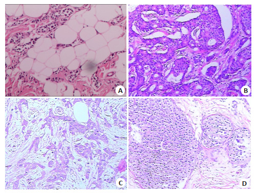
图 1 乳腺癌免疫组化染色图
A: 浸润性导管癌Ⅱ级, Ki67低表达, C-erbB-2(-)(HE染色, ×100); B: 浸润性导管癌II级, Ki67低表达, C-erbB-2(-)(HE染色, ×400); C: 乳腺浸润性导管癌Ⅱ级, Ki67高表达, C-erbB-2(+)(HE染色, ×40); D: 乳腺浸润性导管癌III级, KI67高表达, C-erbB-2(+)(HE染色, ×100).
Figure 1. Immunohistochemical staining of breast cancer.
表 1 乳腺癌ABVS表现与Ki67表达的关系
Table 1. Relationship between ABVS expression and Ki67 expression in breast cancer [n(%)]
因素 Ki67 χ2/t P 低 高 年龄(岁, Mean±SD) 51.83±10.53 51.52±12.17 -0.107 0.915 大小(cm) 8.022 0.005 < 2(n=21) 11 10 ≥2(n=60) 12 48 边界 0.281 0.596 不清(n=42) 13 29 清(n=39) 10 29 虫蚀征 0.107 0.744 有(n=47) 14 33 无(n=34) 9 25 微钙化 0.060 0.807 有(n=44) 12 32 无(n=37) 11 26 汇聚征 1.312 0.252 有(n=21) 8 13 无(n=60) 15 45 高回声环 0.404 0.525 有(n=14) 3 11 无(n=67) 20 47  下载: 导出CSV
表 2 乳腺癌ABVS表现与ER、PR表达的关系
Table 2. Relationship between ABVS expression and expression of ER and PR in breast cancer
因素 C-erbB-2 χ2/t P 阴性 阳性 年龄(岁, Mean±SD) 50.66±11.50 52.73±11.91 -0.794 0.429 大小(cm) 0.657 0.418 < 2(n=21) 13 8 ≥2(n=60) 31 29 边界 0.656 0.418 不清(n=42) 21 21 清(n=39) 23 16 虫蚀征 0.479 0.489 有(n=47) 24 23 无(n=34) 20 14 微钙化 4.817 0.028 有(n=44) 19 25 无(n=37) 25 12 汇聚征 1.741 0.187 有(n=21) 14 7 无(n=60) 30 30 高回声环 1.966 0.158 有(n=14) 10 4 无(n=67) 34 33
下载: 导出CSV
表 3 Ki67多因素Logistic回归结果
Table 3. Ki67 multivariate logistic regression results
项目 B S.E Wald P OR 95% CI 大小 1.482 0.543 7.439 0.006 4.400 1.517~12.759 常量 -1.386 0.323 18.449 < 0.001 0.250
下载: 导出CSV
表 4 C-erbB-2多因素Logistic回归结果
Table 4. C-erbB-2 multivariate logistic regression results
项目 B S.E Wald P OR 95% CI 微钙化 1.008 0.465 4.709 0.030 2.741 1.102~6.816 常量 -0.734 0.351 4.368 0.037 0.480
下载: 导出CSV
-
[1] Torre LA, Islami F, Siegel RL, et al. Global cancer in women: burden and trends[J]. Cancer Epidemiol Biomarkers Prev, 2017, 26(4): 444-57. doi: 10.1158/1055-9965.EPI-16-0858 [2] Zuo TT, Zheng RS, Zeng HM, et al. Female breast cancer incidence and mortality in China, 2013[J]. Thorac Cancer, 2017, 8(3): 214-8. doi: 10.1111/1759-7714.12426 [3] Elwy F, Helwa R, El Leithy AA, et al. PIK3CA mutations in HER2-positive breast cancer patients; frequency and clinicopathological perspective in Egyptian patients[J]. Asian Pac J Cancer Prev, 2017, 18(1): 57-64. http://journal.waocp.org/article_43156_ba488390ca088dd905a94afd0905f03b.pdf [4] van Egdom LSE, Lagendijk M, Heijkoop EHM, et al. Three-dimensional ultrasonography of the breast; An adequate replacement for MRI in neoadjuvant chemotherapy tumour response evaluation?-RESPONDER trial[J]. Eur J Radiol, 2018, 104: 94-100. doi: 10.1016/j.ejrad.2018.05.005 [5] Ferlay J, Soerjomataram I, Dikshit R, et al. Cancer incidence and mortality worldwide: Sources, methods and major patterns in GLOBOCAN 2012[J]. Int J Cancer, 2015, 136(5): E359-86. DOI: 10.1002/ijc.29210. [6] 李贺, 郑荣寿, 张思维, 等. 2014年中国女性乳腺癌发病与死亡分析[J]. 中华肿瘤杂志, 2018, 40(3): 166-71. doi: 10.3760/cma.j.issn.0253-3766.2018.03.002 [7] Brentnall AR, Veen EM, Harkness EF, et al. A case-control evaluation of 143 single nucleotide polymorphisms for breast cancer risk stratification with classical factors and mammographic density[J]. Int J Cancer, 2020, 146(8): 2122-9. doi: 10.1002/ijc.32541 [8] Collaborative group on hormonal factors in breast cancer. Type and timing of menopausal hormone therapy and breast cancer risk: individual participant meta-analysis of the worldwide epidemiological evidence[J]. Lancet, 2019, 394(10204): 1159-68. doi: 10.1016/S0140-6736(19)31709-X [9] 赵倩, 李莹, 刘光, 等. 乳腺浸润性导管癌Ki-67表达与ER、PR、HER-2及MRI表现的相关性[J]. 中国介入影像与治疗学, 2016, 13 (8): 481-5. https://www.cnki.com.cn/Article/CJFDTOTAL-JRYX201608007.htm [10] Olayioye MA, Beuvink I, Horsch K, et al. ErbB receptor-induced activation of stat transcription factors is mediated by src tyrosine kinases[J]. J Biol Chem, 1999, 274(24): 17209-18. doi: 10.1074/jbc.274.24.17209 [11] Tsutsui S, Ohno S, Murakami S, et al. Prognostic value of c- erbB2 expression in breast cancer[J]. J Surg Oncol, 2002, 79(4): 216-23. doi: 10.1002/jso.10079 [12] Bullwinkel J, Baron-Lühr B, Lüdemann A, et al. Ki-67 protein is associated with ribosomal RNA transcription in quiescent and proliferating cells[J]. J Cell Physiol, 2006, 206(3): 624-35. doi: 10.1002/jcp.20494 [13] Haroon S, Hashmi AA, Khurshid A, et al. Ki67 index in breast cancer: correlation with other prognostic markers and potential in Pakistani patients[J]. Asian Pac J Cancer Prev, 2013, 14(7): 4353-8. doi: 10.7314/APJCP.2013.14.7.4353 [14] Stavros AT, Thickman D, Rapp CL, et al. Solid breast nodules: use of sonography to distinguish between benign and malignant lesions[J]. Radiology, 1995, 196(1): 123-34. doi: 10.1148/radiology.196.1.7784555 [15] 包凌云, 朱罗茜, 孔凡雷, 等. 自动乳腺全容积成像和常规超声对乳腺微钙化诊断的对比研究[J]. 中华超声影像学杂志, 2012(3): 220-3. doi: 10.3760/cma.j.issn.1004-4477.2012.03.013 [16] 徐易, 刘莹, 杨先, 等. 乳腺癌超声显像簇状钙化点与c-erbB-2表达的相关性研究[J]. 中国超声医学杂志, 2011, 27(12): 1057-9. doi: 10.3969/j.issn.1002-0101.2011.12.001 [17] 韦瑶, 姜玉新, 张璟, 等. 自动乳腺全容积成像对乳腺病变的诊断价值分析[J]. 中华健康管理学杂志, 2017, 11(6): 514-8. doi: 10.3760/cma.j.issn.1674-0815.2017.06.007 [18] Xiao YM, Zhou QC, Chen ZH. Automated breast volume scanning versus conventional ultrasound in breast cancer screening[J]. Acad Radiol, 2015, 22(3): 387-99. doi: 10.1016/j.acra.2014.08.013 [19] Chen L, Chen Y, Diao XH, et al. Comparative study of automated breast 3-D ultrasound and handheld B-mode ultrasound for differentiation of benign and malignant breast masses[J]. Ultrasound Med Biol, 2013, 39(10): 1735-42. doi: 10.1016/j.ultrasmedbio.2013.04.003 [20] Ichikawa S, Motosugi U, Hernando D, et al. Histological grading of hepatocellular carcinomas with intravoxel incoherent motion diffusion-weighted imaging: inconsistent results depending on the fitting method[J]. Magn Reson Med Sci, 2018, 17(2): 168-73. doi: 10.2463/mrms.mp.2017-0047 [21] 王怡, 费小春, 周庆华, 等. 乳腺癌超声BI-RADS与ER、PR、CerbB-2的相关性分析[J]. 中国超声医学杂志, 2013, 29(2): 123-6. https://www.cnki.com.cn/Article/CJFDTOTAL-ZGCY201302012.htm -

 点击查看大图
计量
- 文章访问数: 304
- HTML全文浏览量: 341
- PDF下载量: 9
- 被引次数: 0