


Comparation of the value of digital periapical film and cone-beam CT in measuring tooth length
-
摘要:
目的 比较数字化根尖片与口腔锥形束CT(CBCT)测量牙齿长度的价值。 方法 回顾性分析本院2017年6月~2020年6月收治103例拔牙正畸患者(共200颗前磨牙)的临床资料,均行数字化根尖片与口腔CBCT检查,以游标卡尺实际测量为金标准,采用InVivoDental软件及平行长焦距平行投照技术拍摄测量不同检查方法牙齿长度,比较不同牙位上牙齿口腔CBCT检查轴位、矢状位和冠状位时及数字化根尖检测的牙齿影像长度,计算其与实际长度的差值。 结果 数字化根尖测量不同牙位牙齿长度与实际牙齿长度差异无统计学意义(P > 0.05);口腔CBCT轴位测量下颌磨牙区、下颌尖牙区、下颌前牙区牙位牙齿长度小于实际牙齿长度(P < 0.05),上颌磨牙区、上颌前磨牙区、上颌尖牙区、上颌前牙区、下颌前磨牙区牙位牙齿长度与实际牙齿长度差异无统计学意义(P > 0.05);口腔CBCT矢状位测量上颌磨牙区、下颌磨牙区、下颌前磨牙区、下颌尖牙区、下颌前牙区与实际牙齿长度比较有明显差异(P < 0.001),上颌前磨牙区、上颌尖牙区、上颌前牙区与实际牙齿长度差异无统计学意义(P > 0.05);口腔CBCT冠状位测量上颌磨牙区、上颌尖牙区、上颌前牙区、下颌前磨牙区、下颌尖牙区、下颌前牙区小于实际牙齿长度(P < 0.001),上颌前磨牙区、下颌磨牙区与实际牙齿长度差异无统计学意义(P > 0.05)。 结论 平行长焦距平行投照下数字化根尖片较口腔CBCT更能准确测量牙齿长度。 Abstract:Objective To compare the value of digital periapical film and cone- beam CT (CBCT) in measuring tooth length. Methods Clinical data of 103 cases of tooth extraction orthodontic patients (a total of 200 premolars) admitted to our hospital from June 2017 to June 2020 were retrospectively analyzed. All patients underwent digital periapical film and CBCT examination. The actual measurement with a vernier caliper was taken as the gold standard. InVivoDental software and parallel long-focus parallel projection technology were used to measure the tooth lengths of different examination methods. The image lengths of teeth in axially, sagittally and coronally examined by CBCT and those by digital periapical detection were compared in different dental positions, and the difference between the actual length and image lengths was calculated. Results There was no significant difference between the tooth length measured by digital periapical method and the actual tooth length in different positions (P > 0.05). The differences between the tooth length of mandibular molar area, canine area, anterior area axially measured by the CBCT and the actual tooth length were significant (P < 0.05). There was no significant difference in the tooth length between the maxillary molar area, maxillary premolar area, maxillary canine area, maxillary anterior area and mandibular premolar area and the actual tooth length (P > 0.05). The differences between the tooth length, sagittally measured by CBCT, of the maxillary molar area, the mandibular molar area, the mandibular premolar area, the mandibular canine area and the mandibular anterior area with the actual tooth length were significant (P < 0.05). There was no significant difference between the tooth length of the maxillary premolar area, the maxillary canine area and the maxillary anterior area with the actual tooth length (P > 0.05). The differences between the tooth length, coronally measured by CBCT, of the maxillary molar area, the maxillary canine area, the maxillary anterior area, the mandibular premolar area, the mandibular canine area and the mandibular anterior area, and the actual tooth length were significant (P < 0.05). There was no significant difference between the tooth length of the maxillary premolar area and the mandibular molar area and the actual tooth length (P > 0.05). Conclusion Compared with CBCT, digital periapical film under the parallel long- focus parallel projection technology can measure the tooth length more accurately. -
Key words:
- digital periapical film /
- cone-beam CT /
- tooth length /
- orthodontics /
- tooth extraction
-

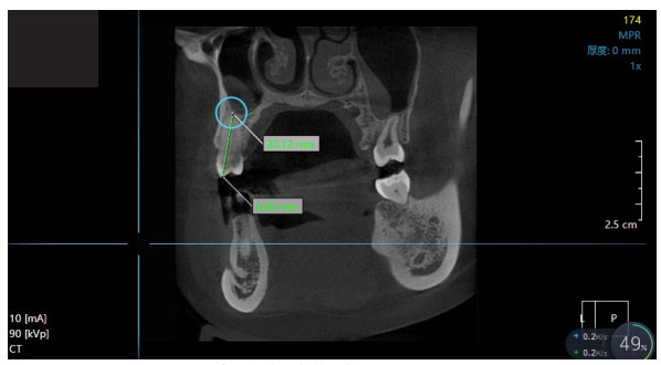
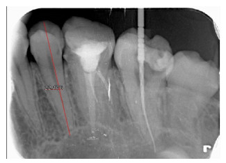
图 1 数字化根尖测量牙齿长度的影像学图像
Figure 1. Imaging image of digital apical measurement of tooth length.
表 1 数字化根尖测量牙齿长度比较
Table 1. Comparison of tooth length measured by digital apical (Mean±SD, mm, n=25)
牙位 实际牙齿长度 数字化根尖 t P 上颌磨牙区 21.72±1.34 21.46±1.07 0.060 0.953 上颌前磨牙区 21.89±1.25 22.02±1.41 0.345 0.732 上颌尖牙区 21.10±1.45 21.13±1.34 0.076 0.940 上颌前牙区 21.52±1.40 21.50±1.46 0.049 0.961 下颌磨牙区 21.30±1.05 21.36±1.27 0.182 0.856 下颌前磨牙区 21.67±1.38 21.74±1.45 0.175 0.862 下颌尖牙区 21.45±1.28 21.51±1.30 0.164 0.870 下颌前牙区 21.43±1.37 21.52±1.31 0.237 0.813  下载: 导出CSV
表 2 口腔CBCT轴位测量牙齿长度比较
Table 2. Comparison of dental length measured by axial CBCT (Mean±SD, mm, n=25)
牙位 实际牙齿长度 口腔CBCT轴位 t P 上颌磨牙区 21.72±1.34 21.25±1.42 1.204 0.235 上颌前磨牙区 21.89±1.25 21.74±1.45 0.450 0.654 上颌尖牙区 21.10±1.45 20.56±1.28 1.396 0.169 上颌前牙区 21.52±1.40 21.23±1.37 0.740 0.463 下颌磨牙区 21.30±1.05 20.56±1.25 2.235 0.030 下颌前磨牙区 21.67±1.38 20.99±1.41 1.723 0.091 下颌尖牙区 21.45±1.28 20.56±1.37 2.373 0.022 下颌前牙区 21.43±1.37 20.23±1.41 3.052 0.004
下载: 导出CSV
表 3 口腔CBCT矢状位测量牙齿长度比较
Table 3. Comparison of dental length measured by CBCT in sagittal position (Mean±SD, mm, n=25)
牙位 实际牙齿长度 口腔CBCT矢状位 t P 上颌磨牙区 21.72±1.34 20.21±1.23 4.151 < 0.001 上颌前磨牙区 21.89±1.25 21.58±1.42 0.819 0.417 上颌尖牙区 21.10±1.45 20.89±1.27 0.545 0.588 上颌前牙区 21.52±1.40 21.25±1.41 0.679 0.500 下颌磨牙区 21.30±1.05 18.96±1.23 7.235 < 0.001 下颌前磨牙区 21.67±1.38 18.79±1.41 7.299 < 0.001 下颌尖牙区 21.45±1.28 20.01±1.25 4.024 < 0.001 下颌前牙区 21.43±1.37 20.00±1.18 3.954 < 0.001
下载: 导出CSV
表 4 口腔CBCT冠状位测量牙齿长度比较
Table 4. Comparison of dental length measured by coronal CBCT (Mean±SD, mm, n=25)
牙位 实际牙齿长度 口腔CBCT冠状位 t P 上颌磨牙区 21.72±1.34 20.10±1.10 4.672 < 0.001 上颌前磨牙区 21.89±1.25 21.68±1.45 0.548 0.586 上颌尖牙区 21.10±1.45 20.12±1.23 2.577 0.013 上颌前牙区 21.52±1.40 19.41±1.28 5.562 < 0.001 下颌磨牙区 21.30±1.05 21.14±1.45 0.447 0.657 下颌前磨牙区 21.67±1.38 20.10±1.24 4.231 < 0.001 下颌尖牙区 21.45±1.28 19.25±1.12 6.467 < 0.001 下颌前牙区 21.43±1.37 19.34±1.25 5.635 < 0.001
下载: 导出CSV
-
[1] Najeeb S, Siddiqui F, Khurshid Z, et al. Effect of bisphosphonates on root resorption after tooth replantation - a systematic review[J]. Dent Traumatol, 2017, 33(2): 77-83. doi: 10.1111/edt.12316 [2] An JK, Matsumoto Y, Ono T. The relationships between the arrangement of teeth, root resorption, and dental maturity in bovine mandibular incisors[J]. Korean J Orthod, 2017, 47(6): 365-74. doi: 10.4041/kjod.2017.47.6.365 [3] Singh RK, Pandey RP, Purohit S, et al. Morphological and digital radiographical dental anatomy of adult buffaloes[J]. Buffal Bulletin, 2017, 36(2): 407-14. [4] 刘学军, 谢玲. 正畸治疗青少年患者的心理状况、认知、需求调查与配合度的相关性[J]. 中国健康心理学杂志, 2020, 28(6): 844-8. [5] Hikita Y, Yamaguchi T, Tomita D, et al. Growth hormone receptor gene is related to root length and tooth length in human teeth[J]. Angle Orthod, 2018, 88(5): 575-81. doi: 10.2319/092917-659.1 [6] Shahid F, Nowrin SA, Rahman NA, et al. Validity and reliability of external apical root resorption (EARR) measurements: A 3D cone beam computed tomography (CBCT) study[J]. Sains Malaysiana, 2020, 49(2): 343-8. doi: 10.17576/jsm-2020-4902-12 [7] 李惠玲. CBCT检测口腔正畸中牙槽骨、牙根吸收状况的效果[J]. 解放军预防医学杂志, 2018, 36(11): 1451-3. [8] 陆一平. 自锁托槽和传统托槽矫治后切牙牙根吸收差异的影像学研究[J]. 口腔医学, 2017, 37(7): 635-7. [9] Ruetters M, Hagenfeld D, ElSayed N, et al. Ex vivo comparison of CBCT and digital periapical radiographs for the quantitative assessment of periodontal defects[J]. Clin Oral Investig, 2020, 24 (1): 377-84. doi: 10.1007/s00784-019-02933-w [10] Ramos Brito AC, Verner FS, Junqueira RB, et al. Detection of fractured endodontic instruments in root canals: comparison between different digital radiography systems and cone-beam computed tomography[J]. J Endod, 2017, 43(4): 544-9. doi: 10.1016/j.joen.2016.11.017 [11] 崔旋旋, 申静, 高静, 等. 锥形束CT与根尖片识别根尖周膜宽度的对比性研究[J]. 口腔医学研究, 2018, 34(4): 393-6. [12] 孙媛元, 邬春兰, 李超伦. 不同测量方法对牙槽骨角形吸收评价的比较[J]. 口腔医学, 2019, 39(2): 117-20. [13] 陈建萍, 王林红, 杨帆. 数字化技术评估上颌前牙区唇侧牙龈厚度与骨板厚度的相关性研究[J]. 口腔材料器械杂志, 2020, 29(2): 32-7. [14] Kobayashi-Velasco S, Salineiro FC, Gialain IO, et al. Diagnosis of alveolar and root fractures: an in vitro study comparing CBCT imaging with periapical radiographs[J]. J Appl Oral Sci, 2017, 25 (2): 227-33. doi: 10.1590/1678-77572016-0332 [15] 王智, 邹立东. 微创技术在上颌窦内提升植骨中应用效果的影像学评价[J]. 口腔医学研究, 2020, 36(7): 688-92. [16] 赵娜, 陈鑫, 周广超, 等. 锥形束CT和数字化牙片测量牙齿长度的准确性研究[J]. 口腔医学, 2019, 39(11): 1038-41. [17] 杨艺强, 刘琪, 庄东鹏, 等. 数字化根尖片、曲面断层片、CBCT测量牙齿长度准确性的比较研究[J]. 临床放射学杂志, 2014, 33(9): 1434-7. [18] 邹晨, 邹道星, 艾毅龙. 口内三维扫描结合CBCT建立数字化模型的研究[J]. 口腔医学研究, 2019, 35(9): 87-90. [19] 童丽, 顾卫平, 陈岗, 等. 基于CBCT的下颌第一磨牙区即刻种植相关的影像学研究[J]. 口腔医学, 2020, 40(3): 227-31. -

 点击查看大图
计量
- 文章访问数: 289
- HTML全文浏览量: 230
- PDF下载量: 9
- 被引次数: 0