


Plaque distribution, composition and related characteristics in patients with basilar artery stenosis by high-resolution MRI imaging
-
摘要:
目的采用高分辨MRI成像分析有症状和无症状基底动脉狭窄患者斑块分布、成分及相关特征。 方法回顾性分析2018年3月~2021年3月我院收治的106例行高分辨MRI检查的基底动脉狭窄患者,根据是否存在后循环卒中分为症状性组(n=45)和无症状组(n=61),收集所有纳入对象影像学资料,分析两组斑块分布、成分、重构等相关特征。 结果症状性组斑块主要分布在背侧壁,无症状组斑块主要分布在腹侧壁,两组斑块分布差异存在统计学意义(P < 0.05);症状性组合并斑块内出血、纤维帽断裂概率高于无症状组(P < 0.05);症状性组患者最狭窄层的血管面积、管壁面积、管腔面积、最大管壁厚度、狭窄率、血管重构指数(RI)均高于无症状组(P < 0.05),两组斑块负荷差异无统计学意义(P > 0.05);两组斑块强化等级分布存在明显差异,其中症状性组斑块2级强化率高于无症状组(P < 0.05)。 结论有症状和无症状基底动脉狭窄患者斑块分布、成分、重构等特征存在一定差异,而高分辨MRI成像能为识别斑块特征和及早干预提供可靠影像学参考。 Abstract:ObjectiveTo analyze the distribution, composition and related characteristics of plaque in patients with symptomatic and asymptomatic basilar artery stenosis using high-resolution MRI imaging. MethodsA retrospective analysis of 106 patients with basilar artery stenosis who underwent high-resolution MRI examinations in our hospital was conducted from March 2018 to March 2021. They were divided into symptomatic group (n=45) and asymptomatic group (n=61) according to the presence or absence of posterior circulation stroke with the imaging data collected. The distribution, composition, reconstruction and other related characteristics of the two groups of plaques were analyzed. ResultsThe plaques of the symptomatic group mainly localized on the dorsal wall, the plaques of the asymptomatic group mainly localized on the ventral wall, the distribution and composition of the plaques in the two groups were significantly different (P < 0.05). The probability of bleeding and fiber cap rupture in symptomatic group was higher than that in asymptomatic group (P < 0.05). The vascular area, wall area, lumen area, maximum wall thickness, stenosis rate, and vascular remodeling index of the most stenosis layer in the symptomatic group were higher than those in the asymptomatic group (P < 0.05). There was no significant difference in plaque load between the two groups (P > 0.05). There was a significant difference in the distribution of plaque enhancement grades between the two groups, and the level 2 plaque enhancement rate of the symptomatic group was higher than that of the asymptomatic group (P < 0.05). ConclusionThere are massive differences between symptomatic and asymptomatic patients with basilar artery stenosis in plaque distribution, composition, and remodeling. High-resolution MRI imaging can give a reliable imaging reference for identifying plaque characteristics and early intervention. -
Key words:
- high-resolution MRI /
- basilar artery stenosis /
- plaque distribution /
- plaque composition
-
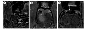
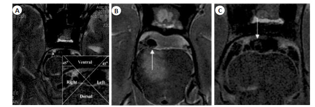
图 1 基于HR-MRI图像的基底动脉斑块分布
A: 基底动脉在图像上被分为4个象限,上下左右分别为背侧、腹侧、左侧和右侧; B: 典型症状性患者,女,47岁,HR-MRI显示斑块位于基底动脉背侧壁; C: 典型无症状性患者,男,52岁,HR-MRI显示斑块位于基底动脉腹侧壁.
Figure 1. Basilar artery plaque distribution based on HR-MRI image.
表 1 两组一般资料比较
Table 1. Comparison of general information between the two groups[n(%)]
组别 性别(男/女) 年龄(岁, Mean±SD) 合并高血压 合并高血脂 合并糖尿病 症状性组(n=45) 36/9 63.15±9.24 24(53.33) 26(57.78) 20(44.44) 无症状组(n=61) 44/17 60.23±9.78 31(50.82) 30(49.18) 27(44.26) χ2/t 0.866 0.159 0.066 0.768 0.001 P 0.352 0.824 0.798 0.381 0.985  下载: 导出CSV
表 2 两组斑块分布比较
Table 2. Comparison of plaque distribution between the two groups[n(%)]
组别 腹侧壁 背侧壁 左侧壁 右侧壁 症状性组
(n=238)36(15.13) 102(42.86) 48(20.17) 52(21.85) 无症状组
(n=272)108(39.71) 92(33.82) 27(9.93) 45(16.54) χ2 40.815 P < 0.001
下载: 导出CSV
表 3 两组斑块成分特点比较
Table 3. Comparison of the characteristics of plaque composition between the two groups[n(%)]
组别 斑块内出血 纤维帽断裂 症状性组(n=238) 64(26.89) 33(13.87) 无症状组(n=272) 45(16.54) 25(9.19) χ2 4.514 2.802 P 0.034 0.045
下载: 导出CSV
表 4 两组基底动脉血管面积和厚度比较
Table 4. Comparison of the area and thickness of the basilar artery between the two groups (Mean±SD)
组别 血管面积(mm2) 管壁面积(mm2) 管腔面积(mm2) 最大管壁厚度(mm2) 狭窄率(%) 斑块负荷 RI 症状性组(n=45) 24.15±3.62 21.42±3.95 4.61±1.82 2.34±0.31 62.72±14.58 -0.24±0.05 1.20±0.43 无症状组(n=61) 22.49±4.42 18.32±3.16 2.86±1.07 1.92±0.36 71.86±8.52 0.06±0.01 0.34±0.09 t 2.060 4.487 6.202 6.2909 4.051 1.209 18.302 P 0.042 < 0.001 < 0.001 < 0.001 < 0.001 0.237 < 0.001
下载: 导出CSV
表 5 两组斑块强化情况比较
Table 5. Comparison of plaque enhancement between the two groups[n(%)]
组别 0级 1级 2级 症状性组(n=45) 5(11.11) 22(48.89) 18(40.00) 无症状组(n=61) 33(54.10) 16(26.23) 12(19.67) Z 20.839 P < 0.001
下载: 导出CSV
-
[1] 周莹雪, 崔英哲, 南东, 等. 后循环脑缺血的基底动脉高分辨磁共振成像研究[J]. 磁共振成像, 2021, 12(2): 15-8, 23. [2] 张帆, 汪明佳, 王婉. 急性脑梗死患者颈动脉硬化斑块稳定性的影响因素[J]. 西部医学, 2019, 31(6): 917-21. doi: 10.3969/j.issn.1672-3511.2019.06.020 [3] Kang HG, Lee CH, Shin BS, et al. Characteristics of symptomatic basilar artery Stenosis using high-resolution magnetic resonance imaging in ischemic stroke patients[J]. J Atheroscler Thromb, 2020, 15(11): 164-7. http://www.researchgate.net/publication/346978952_Characteristics_of_Symptomatic_Basilar_Artery_Stenosis_Using_High-Resolution_Magnetic_Resonance_Imaging_in_Ischemic_Stroke_Patients [4] 王勇胜, 张文杰, 杨明瑞, 等. 高分辨MRI测量大脑中动脉狭窄率的准确性评价[J]. 中华全科医师杂志, 2019, 18(6): 553-7. doi: 10.3760/cma.j.issn.1671-7368.2019.06.009 [5] 中国后循环缺血专家共识组. 中国后循环缺血的专家共识[J]. 中华内科杂志, 2006(9): 786-7. doi: 10.3760/j.issn:0578-1426.2006.09.034 [6] 症状性颅内动脉粥样硬化性狭窄血管内治疗专家共识组. 症状性颅内动脉粥样硬化性狭窄血管内治疗中国专家共识[J]. 中华内科杂志, 2013, 52(3): 271-5. doi: 10.3760/cma.j.issn.0578-1426.2013.03.028 [7] 杨雪, 胡勇, 向波, 等. CTA探讨颈动脉几何形态与粥样硬化斑块的关系[J]. 西部医学, 2019, 31(8): 1273-7, 1282. doi: 10.3969/j.issn.1672-3511.2019.08.028 [8] 张志勇, 张海波, 焦劲松, 等. 基底平行解剖扫描磁共振成像在颅内椎-基底动脉病变中的应用价值[J]. 中国脑血管病杂志, 2020, 17(9): 531-7. doi: 10.3969/j.issn.1672-5921.2020.09.005 [9] Zhu C, Tian X, Degnan AJ, et al. Clinical significance of intraplaque hemorrhage in low- and high-grade basilar artery Stenosis on high-resolution MRI[J]. AJNR Am J Neuroradiol, 2018, 39(7): 1286-92. doi: 10.3174/ajnr.A5676 [10] 沙丽塔娜提·达列力, 贾文霄, 韩秉艳, 等. 基底动脉狭窄高分辨率磁共振管壁成像研究[J]. 中国医学装备, 2020, 17(9): 60-4. doi: 10.3969/J.ISSN.1672-8270.2020.09.014 [11] 陈旭高, 邹建勋, 叶国伟, 等. 高分辨率磁共振血管壁成像在基底动脉狭窄的临床应用[J]. 医学影像学杂志, 2019, 29(9): 1463-6. https://www.cnki.com.cn/Article/CJFDTOTAL-XYXZ201909008.htm [12] Wang W, Yang Q, Li D, et al. Incremental value of plaque enhancement in patients with moderate or severe basilar artery Stenosis: 3.0 T high-resolution magnetic resonance study[J]. Biomed Res Int, 2017, 2017: 428-39. http://pubmedcentralcanada.ca/pmcc/articles/PMC5623789/ [13] 张梁, 周志斌. 高分辨率磁共振血管壁成像在颅内动脉粥样硬化性疾病中的应用[J]. 分子影像学杂志, 2020, 43(1): 45-8. doi: 10.12122/j.issn.1674-4500.2020.01.10 [14] 王旭超, 徐敏, 孙兆男, 等. 基于HR-MRI对轻度狭窄的基底动脉斑块特征与临床症状关系的研究[J]. 磁共振成像, 2019, 10(8): 561-5. https://www.cnki.com.cn/Article/CJFDTOTAL-CGZC201908002.htm [15] Zhang M, Ye GF, Liu YD, et al. Clinical application of high-resolution MRI in combination with digital subtraction angiography in the diagnosis of vertebrobasilar artery dissecting aneurysm: an observational study (STROBE compliant)[J]. Medicine, 2019, 98 (14): e14857. doi: 10.1097/MD.0000000000014857 [16] 符念霞, 宋建勋, 陈思静, 等. 高分辨磁共振血管壁成像诊断椎基底动脉延长扩张症并基底动脉末端不稳定斑块一例[J]. 磁共振成像, 2020, 11(11): 1034-5. doi: 10.12015/issn.1674-8034.2020.11.016 [17] 张明宇, 陈红燕, 马丁. 高分辨率磁共振血管壁成像评估椎-基底动脉硬化血管壁对急性脑梗死患者预后的评估价值[J]. 卒中与神经疾病, 2020, 27(3): 323-6. doi: 10.3969/j.issn.1007-0478.2020.03.009 [18] Xu ZQ, Li MY, Hou ZK, et al. Association between basilar artery configuration and Vessel Wall features: a prospective high-resolution magnetic resonance imaging study[J]. BMC Med Imaging, 2019, 19(1): 1-7. http://www.ncbi.nlm.nih.gov/pubmed/31878890 [19] 王宽. HR-MRI检测在大脑中动脉与基底动脉脑梗死患者中的应用[J]. 心脑血管病防治, 2019, 19(5): 444-6. doi: 10.3969/j.issn.1009-816x.2019.05.019 [20] 林惠花, 杨本强, 段阳. 基底动脉弯曲角度与其斑块形成相关性研究[J]. 磁共振成像, 2019, 10(6): 410-4. https://www.cnki.com.cn/Article/CJFDTOTAL-CGZC201906006.htm -

 点击查看大图
计量
- 文章访问数: 301
- HTML全文浏览量: 158
- PDF下载量: 7
- 被引次数: 0