


Limb necrosis of rabbit model injured by high voltage electric injury based on magnetic resonance imaging
-
摘要:
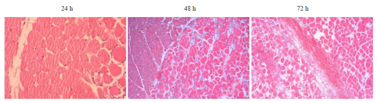
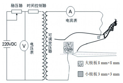
目的探究磁共振成像常规序列及多b值扩散加权成像(DWI)序列对电击伤模型电击损伤的评价效果。 方法选取30只普通家兔,根据电击时间将家兔随机分为3组:A组(电击0.2 s)、B组(电击0.5 s)、C组(电击1 s),10只/组;电击伤后根据标准对电击家兔进行肢体损伤分级评价;H&E染色观察家兔电击伤后24、48和72 h下肢肌肉组织病理学变化;采用磁共振T2WI平扫和T1WI-FS增强扫描观察家兔电击伤后24、48和72 h下肢损伤情况;采用DWI扫描观察不同b值下家兔电击伤不同损伤程度的表观扩散系数值。 结果3种不同电击时间条件下,共计造成“轻”度损伤模型家兔为7只,“中”度损伤模型家兔为7只,“重”度损伤模型家兔为6只,“特重”度损伤模型家兔为10只(χ2=21.486,P=0.002)。组织学结果显示,电击后24 h后可见家兔下肢肌肉间质存在出血、肌细胞凝固性坏死、肌溶解及炎性浸润情况;48 h后则可观察到肌肉间质血管肿胀且有血栓形成;72 h后肌肉坏死组织进一步扩大加重;T2WI平扫及T1WI-FS增强扫描信号显示电击家兔呈渐进性坏死情况,与组织学结果一致;不同b值DWI扫描结果显示,b值在等于600 s/mm2和800 s/mm2条件下,“轻”、“中”、“重”和“特重”度损伤表观扩散系数值差异均有统计学意义(P < 0.01)。 结论T2WI平扫及T1WI-FS增强MRI扫描联合多b值DWI序列扫描对于临床电击损伤的早期诊断具有重要参考意义。 Abstract:ObjectiveTo explore the evaluation effect of conventional magnetic resonance imaging sequence and multi-B-value diffusion weighted imaging (DWI) sequence on electric shock injury model. MethodsAccording to the electric shock time, 30 rabbits were randomly divided into three groups: Group A (0.2 s), Group B (0.5 s) and Group C (1 s), with 10 rabbits in each group. After electric injury, the limb injury of rabbits was graded according to the standard. H&E staining was used to observe the histopathological changes of lower limb muscles in rabbits at 24 h, 48 h and 72 h after electric injury. The lower limb injuries of rabbits after electric injury at 24 h, 48 h and 72 h were observed by using MRI T2WI plain scan and T1WI-FS enhanced scan. Using DWI scanning, the ADC values of rabbits with different degrees of electrical injury under different B values were observed. ResultsUnder three different electric shock time conditions, 7 rabbits suffered from mild injury, 7 rabbits suffered from moderate injury, 6 rabbits suffered from severe injury and 10 rabbits suffered from severe injury (χ2= 21.486, P=0.002). Histological results showed that there were bleeding, coagulation necrosis, myolysis and inflammatory infiltration in muscle interstitium of lower limbs of rabbits 24 h after electric shock. After 48 h, swelling of muscle interstitial blood vessels and thrombosis were observed. After 72 h, the necrotic tissue of muscle was further enlarged and aggravated. The signals of T2WI plain scan and T1WI-FS enhanced scan showed progressive necrosis of rabbits after electric shock, which was consistent with the histological results. DWI scanning results with different B values showed that ADC values of "light", "medium", "heavy" and "extra-heavy" injuries were significantly different when B values were equal to 600 s/mm2 and 800 s/mm2 (P < 0.01). ConclusionT2WI plain scan and T1WI-FS enhanced MRI combined with multi-B-value DWI sequence scan have important reference significance for early diagnosis of clinical electric shock injury. -
Key words:
- magnetic resonance /
- electric injury /
- multi-b diffusion weighted imaging /
- rabbit
-
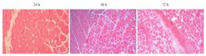
图 2 家兔电击后不同时间下肢H&E染色
Figure 2. H&E staining of lower extremities at different times after electric shock in rabbits (×400).

图 3 家兔电击后不同时间下MRI图像扫描
Figure 3. MRI image scanning at different time after electric shock in rabbits.
表 1 电击伤后肢体损伤程度分级标准
Table 1. Grading standard of limb injury after electrical injury
损伤程度 创口大小 创面深度 轻 限于腿腹侧,R值< 3 皮肤或部分浅层肌群坏死 中 明显向近端扩大,R值:3~5 浅层肌群或部分深层肌群坏死 重 小腿环形创面,R值:5~8 深层肌群坏死或伴少许骨质外露 特重 小腿全部坏死,R值> 8 骨质外露坏死 肢体损伤由两名医师分别评估,结果不一致时取平均值;R值为电击后小极板下创面面积与小极板面积之比.  下载: 导出CSV
表 2 电击伤后各组家兔损伤程度评价
Table 2. Evaluation of injury degree of rabbits in each group after electric shock(n)
损伤程度 A组 B组 C组 总计 轻 6 1 0 7 中 2 5 0 7 重 1 2 3 6 特重 1 2 7 10 χ2 21.486 P 0.002
下载: 导出CSV
表 3 各b值条件下家兔电击伤不同损伤程度的ADC值
Table 3. ADC values of different degrees of electrical injury in rabbits under the condition of b values (×10-3 mm2/s, Mean±SD)
组别 b=400 s/mm2 b=600 s/mm2 b=800 s/mm2 b=1000 s/mm2 b=1200 s/mm2 轻(n=7) 2.32±0.10 2.15±0.10 2.07±0.13 2.03±0.21 1.90±0.11 中(n=7) 1.82±0.15# 1.55±0.11# 1.48±0.20# 1.48±0.11# 1.47±0.12# 重(n=6) 1.74±0.12# 1.31±0.16#* 1.24±0.10#* 1.11±0.31#* 1.10±0.22#* 特重(n=10) 1.03±0.13#*&& 0.99±0.14#*& 0.98±0.15#*& 0.94±0.22#* 0.94±0.15#* F 149.62 113.69 74.48 37.21 60.98 P < 0.01 < 0.01 < 0.01 < 0.01 < 0.01 #P < 0.001 vs“轻”度;*P < 0.01 vs“中”度; & P < 0.01 vs“重”度, && P < 0.001 vs“重”度.
下载: 导出CSV
-
[1] Gentges J, Schieche C. Electrical injuries in the emergency department: an evidence-based review[J]. Emerg Med Pract, 2018, 20(11): 1-20. [2] 胡皖玲, 姜娇慧. 电击伤的急救与防范[J]. 保健医苑, 2019(10): 34-5. https://www.cnki.com.cn/Article/CJFDTOTAL-YYBJ201910016.htm [3] Shih JG, Shahrokhi S, Jeschke MG. Review of adult electrical burn injury outcomes worldwide: an analysis of low-voltage vs high-voltage electrical injury[J]. J Burn Care Res, 2017, 38(1): e293-8. doi: 10.1097/BCR.0000000000000373 [4] 李利根, 柴家科. 肢体高压电烧伤软组织及血管损伤的影像学判断及临床意义[J]. 中华烧伤杂志, 2020, 36(11): 1009-12. doi: 10.3760/cma.j.cn501120-20190904-00371 [5] Babu SH, Chandrasekhar D, Noone M, et al. Magnetic resonance imaging findings in brain resulting from high-voltage electrical shock injury of the scalp[J]. Indian J Radiol Imaging, 2018, 28(3): 312. doi: 10.4103/ijri.IJRI_368_17 [6] Li SJ, Wang ZL, Zhu WP, et al. Clinical research of features of magnetic resonance imaging of high-voltage electrical burns in limbs at early stage[J]. Zhonghua Shao Shang Za Zhi, 2017, 33 (12): 750-6. http://www.ncbi.nlm.nih.gov/pubmed/29275616 [7] 文一臻, 张丕红, 任利成, 等. 136例上肢电烧伤患者的临床特征及修复效果[J]. 中华烧伤杂志, 2019, 35(11): 784-9. doi: 10.3760/cma.j.issn.1009-2587.2019.11.004 [8] 欧志强, 陈松, 梁佩虹, 等. Diffusion-weighted MRI在家兔电烧伤后应用初步研究[J]. 中国医疗前沿, 2013, 8(20): 1-2. doi: 10.3969/j.issn.1673-5552.2013.20.0001 [9] Chai JK, Li LG, Gao QW, et al. Establishment of soft-tissue-injury model of high-voltage electrical burn and observation of its pathological changes[J]. Burns, 2009, 35(8): 1158-64. doi: 10.1016/j.burns.2009.02.010 [10] 张伟, 谢卫国, 赵超莉, 等. 大鼠单侧肢体高压电击伤模型的建立[J]. 中华损伤与修复杂志: 电子版, 2008, 3(4): 426-32. doi: 10.3969/j.issn.1673-9450.2008.04.005 [11] Zhao JC, Shi K, Hong L, et al. Retrospective review of free anterolateral thigh flaps for limb salvage in severely injured high-voltage electrical burn patients[J]. Ann Plast Surg, 2018, 80 (3): 232-7. doi: 10.1097/SAP.0000000000001283 [12] Xing PP, Guo HN, Di HP, et al. Clinical effect of free anterolateral thigh flap combined with arterial vascular reconstruction on repairing high-voltage electrical burn wound on the wrist[J]. Zhonghua Shao Shang Za Zhi, 2020, 36(6): 419-25. [13] Daskal Y, Beicker A, Dudkiewicz M, et al. High voltage electric injury: mechanism of injury, clinical features and initial evaluation. [J]. Harefuah, 2019, 158(1): 65-9. http://www.ncbi.nlm.nih.gov/pubmed/30663297 [14] 舒锦尔, 仇旭光, 李惠民, 等. 前臂肌群急性高压电击伤的MRI研究[J]. 中华放射学杂志, 2001, 35(1): 63-7 doi: 10.3760/j.issn:1005-1201.2001.01.019 [15] 李楠, 李俊海, 黄梅, 等. 下肢创伤中动脉损伤平面对肢体远端血供的影响[J]. 中华骨科杂志. 2019, 39(7): 429-435. [16] Zhou HM, Xu SJ, Wang L, et al. Influences of high-voltage electrical burns on the pulmonary microcirculation in rabbits[J]. Clin Hemorheol Microcirc, 2016, 62(3): 193-203. doi: 10.3233/CH-141921 [17] 单连强, 瞿色华, 石士奎, 等. 磁共振表观扩散系数对前列腺癌的筛查价值[J]. 分子影像学杂志, 2019, 42(2): 151-4. doi: 10.12122/j.issn.1674-4500.2019.02.02 [18] Lin X, Lee M, Buck O, et al. Diagnostic accuracy of T1-weighted dynamic contrast-enhanced-MRI and DWI-ADC for differentiation of glioblastoma and primary CNS lymphoma[J]. AJNR Am J Neuroradiol, 2017, 38(3): 485-91. http://pubmedcentralcanada.ca/pmcc/articles/PMC5352508/ [19] 赵印生, 衣服新, 梁峰, 等. DWI和ADC值在脑脓肿包膜期与脑胶质瘤鉴别诊断中的应用[J]. 锦州医科大学学报, 2017, 38(2): 34-6, 113. https://www.cnki.com.cn/Article/CJFDTOTAL-JZYX201702012.htm [20] 马莉, 裴亚亚, 孙鹏飞. ADC联合DWI鉴别诊断中枢神经细胞瘤与室管膜瘤的应用价值[J]. 磁共振成像, 2017, 8(4): 283-8. https://www.cnki.com.cn/Article/CJFDTOTAL-CGZC201704014.htm [21] 鲁辛健, 班允清, 王晓燕, 等. IVIM-DWI和DCE-MRI评估高强度聚焦超声刀治疗子宫肌瘤的疗效[J]. 分子影像学杂志, 2020, 43(4): 557-62. doi: 10.12122/j.issn.1674-4500.2020.04.01 -


 点击查看大图
计量
- 文章访问数: 302
- HTML全文浏览量: 102
- PDF下载量: 2
- 被引次数: 0