


Application value of high-frequency ultrasound and its image features in the diagnosis of superficial lymph node lesions
-
摘要:
目的探究高频超声及其图像特征在浅表淋巴结病变诊断中的应用价值。 方法回顾2019年6月~2021年2月于我院收治的106例浅表淋巴结病变患者的临床资料,患者均进行高频超声检查,以病理检查结果作为金标准。将患者根据病理诊断良恶性结果进行分组,比较良性组(n=45)、恶性组(n=61)患者高频超声图像特征差异,包括:淋巴结长径(L)/短径(S)、血管化评分(VS)、淋巴结最大血流速度(Vmax)、最小流速(Vmin)、阻力指数(RI)。比较高频超声与病理检查结果差异,使用ROC曲线分析高频超声评估浅表淋巴结病变的价值。 结果良性组、恶性组患者淋巴结L/S < 2、VS、淋巴结Vmax、RI差异均有统计学意义(P < 0.05)。高频超声诊断浅表淋巴结病变的准确率为85.85%,敏感度为90.16%,特异性为80.00%,阳性预测值为85.94%,阴性预测值为85.71%。ROC曲线结果显示,淋巴结Vmax、RI诊断价值均较高(P < 0.05)。 结论高频超声评估浅表淋巴结病变的价值较高,可区分良恶性浅表淋巴结病变。 Abstract:ObjectiveTo explore the application value of high-frequency ultrasound and its image features in the diagnosis of superficial lymph node lesions. MethodsThe clinical data of 106 patients with superficial lymph node lesions admitted to the hospital between June 2019 and February 2021 were reviewed. All patients underwent high-frequency ultrasound examination, and pathological examination Results were used as the gold standard. The patients were grouped based on the pathological diagnosis of benign and malignant results. The features of high-frequency ultrasound images [lymph node long diameter (L)/short diameter (S), vascularization score (VS), lymph node maximum blood flow velocity (Vmax), minimum flow velocity (Vmin), resistance index (RI)] were compared between benign group (n=45) and malignant group (n=61). The differences in Results of high-frequency ultrasound and pathological examination were compared. ROC curve was used to analyze the value of high-frequency ultrasound in evaluating superficial lymph node lesions. ResultsThe difference in lymph node L/S < 2, VS, lymph node Vmax, and RI between benign group and malignant group were significant(P < 0.05). The accuracy rate, sensitivity, specificity, positive predictive value and negative predictive value of high-frequency ultrasound in the diagnosis of superficial lymph node lesions were 85.85%, 90.16%, 80.00%, 85.94% and 85.71%. ROC curve Results showed that the diagnostic value of lymph node Vmax and RI were both high (P < 0.05). ConclusionHigh-frequency ultrasound is of higher value in evaluating superficial lymph node lesions, and can distinguish benign and malignant superficial lymph node lesions. -
Key words:
- high-frequency ultrasound /
- lymph nodes /
- superficial lymph nodes /
- lesions /
- diagnosis
-
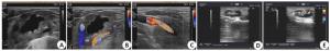
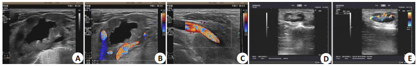
图 1 淋巴结高频超声表现
A~C:左侧颈部探及多个淋巴结回声,较大者大小约42 mm×23 mm,皮髓质分界不清,内可见片状无回声区,其内可见点状血流信号,较大者包绕部分左侧颈动脉及锁骨下动脉;D~E:左肘部及左腋窝探及多个淋巴结回声,较大者大小约25 mm×10 mm,皮髓质分界清晰,血流信号丰富。病理结果:(左肘部淋巴结)猫抓病.
Figure 1. Image of high frequency ultrasonography of lymph nodes.
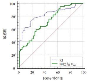
图 2 淋巴结Vmax、RI在浅表淋巴结病变中的诊断效能
Figure 2. Diagnostic efficacy of lymph node Vmax and RI in superficial lymph node lesions.
表 1 两组患者高频超声图像特征比较
Table 1. Comparison of high-frequency ultrasound images features between the two groups of patients (Mean±SD)
指标 恶性组(n=61) 良性组(n=45) Z/t/χ2 P VS[n(%)] 22.04 < 0.01 Ⅰ 3(4.92) 28(62.22) Ⅱ 32(52.46) 7(15.56) Ⅲ 16(26.23) 5(11.11) Ⅳ 10(16.39) 5(11.11) L/S < 2 [n(%)] 46(75.41) 4(8.89) 45.98 < 0.01 淋巴结Vmax(cm/s) 26.85±5.71 21.06±5.42 5.27 < 0.01 淋巴结Vmin(cm/s) 7.23±1.52 7.69±1.56 1.52 0.13 RI 0.75±0.18 0.56±0.13 6.01 < 0.01  下载: 导出CSV
表 2 高频超声与病理检查结果
Table 2. Results of high-frequency ultrasound and pathological examination (n)
高频超声诊断 病理诊断 合计 恶性 良性 恶性 55 9 64 良性 6 36 42 合计 61 45 106
下载: 导出CSV
表 3 淋巴结Vmax、RI在浅表淋巴结病变中的诊断效能
Table 3. Diagnostic efficacy of lymph node Vmax and RI in superficial lymph node lesions
指标 最佳预测临界点 AUC 95%CI 敏感性(%) 特异度(%) Z P 淋巴结Vmax 24.37 0.723 0.628~0.805 59.02 73.33 4.541 < 0.05 RI 0.64 0.823 0.737~0.890 77.05 77.78 8.016 < 0.05
下载: 导出CSV
-
[1] 王涛, 高转转, 张良西. 高频彩色多普勒超声对甲状腺微小癌的诊断价值[J]. 分子影像学杂志, 2017, 40(1): 16-9. doi: 10.3969/j.issn.1674-4500.2017.01.05 [2] Ben ZF, Gao SS, Wu WJ, et al. Clinical value of the VTIQ technology in the differential diagnosis of superficially enlarged lymph nodes[J]. Acta Radiol, 2018, 59(7): 836-44. doi: 10.1177/0284185117732601 [3] Dobson CM, Stringfellow EA, Banks ME. Lymph-node palpation— no laughing matter[J]. N Engl J Med, 2016, 374(10): 996-7. doi: 10.1056/NEJMc1514211 [4] Wang GZ, Li XG, Li L, et al. Clinical value of ultrasonic imaging in diagnosis of hypopharyngeal cancer with cervical lymph node metastasis[J]. Oncol Lett, 2019, 18(6): 5917-22. http://www.ncbi.nlm.nih.gov/pubmed/31788065 [5] Yin SS, Cui QL, Fan ZH, et al. Diagnostic value of arrival time parametric imaging using contrast- enhanced ultrasonography in superficial enlarged lymph nodes[J]. J Ultrasound Med, 2019, 38 (5): 1287-98. doi: 10.1002/jum.14809 [6] 高立霓, 刘秉彦, 莫泽来. 彩色多普勒超声诊断颈部淋巴结结核的临床价值[J]. 海南医学, 2017, 28(1): 102-4. doi: 10.3969/j.issn.1003-6350.2017.01.032 [7] 白云, 杨凤, 刘俊平, 等. 高频彩色多普勒超声对浅表淋巴结病变的相关因素定量分析[J]. 河北医药, 2008, 30(8): 1132-4. doi: 10.3969/j.issn.1002-7386.2008.08.019 [8] Chammas MC, Macedo TAA, Lo VW, et al. Predicting malignant neck lymphadenopathy using color duplex sonography based on multivariate analysis[J]. J Clin Ultrasound, 2016, 44(9): 587-94. doi: 10.1002/jcu.22380 [9] de Stefano G, Scognamiglio U, di Martino F, et al. The role of CEUS in characterization of superficial lymph nodes: a single center prospective study[J]. Oncotarget, 2016, 7(32): 52416-22. doi: 10.18632/oncotarget.9385 [10] Mei M, Ye LG, Quan J, et al. Contrast-enhanced ultrasound for the differential diagnosis between benign and metastatic superficial lymph nodes: a meta- analysis[J]. Cancer Manag Res, 2018, 10: 4987-97. doi: 10.2147/CMAR.S174751 [11] Turgut E, Celenk C, Tanrivermis Sayit A, et al. Efficiency of Bmode ultrasound and strain elastography in differentiating between benign and malignant cervical lymph nodes[J]. Ultrasound Q, 2017, 33(3): 201-7. doi: 10.1097/RUQ.0000000000000302 [12] 高侠, 周展, 罗庆春. 高频超声联合彩色多普勒对颈部淋巴结病变的诊断价值研究[J]. 解放军预防医学杂志, 2018, 36(1): 107-9. https://www.cnki.com.cn/Article/CJFDTOTAL-JYYX201801032.htm [13] 王娟, 魏春红, 鲁一兵, 等. 超声弹性成像结合颈部淋巴结超声检查在甲状腺结节良恶性鉴别诊断中的价值[J]. 临床超声医学杂志, 2016, 18(10): 670-2. https://www.cnki.com.cn/Article/CJFDTOTAL-LCCY201610008.htm [14] Wu QX, Zheng DD, Shi LG, et al. Differentiating metastatic from nonmetastatic lymph nodes in cervical cancer patients using monoexponential, biexponential, and stretched exponential diffusion-weighted MR imaging[J]. Eur Radiol, 2017, 27(12): 5272- 9. doi: 10.1007/s00330-017-4873-1 [15] 满育平, 马隆佰, 周平婷, 等. 多模态MRI对颈部良恶性淋巴结鉴别诊断的临床应用价值[J]. 临床放射学杂志, 2019, 38(8): 1385-90. https://www.cnki.com.cn/Article/CJFDTOTAL-LCFS201908009.htm [16] 褚巍, 杨沪, 宋燕, 等. 高频彩色多普勒超声早期诊断乳腺癌腋窝淋巴结转移的临床应用价值[J]. 癌症进展, 2018, 16(3): 309-11, 338. https://www.cnki.com.cn/Article/CJFDTOTAL-AZJZ201803013.htm [17] 李漪, 陈志刚, 李煜华, 等. 高频彩色多普勒超声对猫抓病性淋巴结炎的诊断价值[J]. 西部医学, 2017, 29(8): 1158-60, 1164. doi: 10.3969/j.issn.1672-3511.2017.08.030 [18] 柯晓丽, 沈浩霖, 吕国荣, 等. 颈部淋巴结超声良恶性风险预测模型的构建及价值[J]. 中国超声医学杂志, 2020, 36(4): 314-7. doi: 10.3969/j.issn.1002-0101.2020.04.008 [19] 王志民, 刘剑, 杜月明, 等. 高频二维超声及彩色多普勒超声声像图对甲状腺癌颈部淋巴结转移的诊断价值[J]. 癌症进展, 2018, 16(13): 1612-5. https://www.cnki.com.cn/Article/CJFDTOTAL-AZJZ201813012.htm [20] 王海璇, 陈廷财, 陈东玲. 高频彩色多普勒超声联合MRI在乳腺癌腋下转移性淋巴结鉴别诊断中的应用[J]. 检验医学与临床, 2018, 15 (13): 1952-5, 1959. doi: 10.3969/j.issn.1672-9455.2018.13.026 [21] 杨红玲, 杨清. 高频超声联合磁共振可提高对乳腺癌早期诊断的价值[J]. 分子影像学杂志, 2020, 43(3): 520-4. doi: 10.12122/j.issn.1674-4500.2020.03.32 -

 点击查看大图
计量
- 文章访问数: 421
- HTML全文浏览量: 244
- PDF下载量: 3
- 被引次数: 0