


The Correlation analysis between MRI enterography and Crohn's disease activity index on Crohn's disease activity
-
摘要:
目的探讨MRI小肠造影(MRE)对克罗恩病活动度分级的评估及其与临床克罗恩病活动指数(CDAI)的相关性。 方法对22例临床或病理确诊并经临床活动度分级的活动性克罗恩病患者行MRE检查。扫描后的图像由2名副主任及以上诊断医师对克罗恩病进行主观评分,并按其得分进行影像学活动度分级,比较评估结果与该患者临床分级结果相比较,观察其一致性。 结果22例活动性克罗恩病患者均完成MRE检查,其中轻度患者7例,得分6.5±1.7分,中位数6.0分;中度患者9例,得分11.9±1.6分,中位数12.0分;重度患者6例,得分24.0±5.4分,中位得分23.5分。Spearman相关分析显示,MRE克罗恩病轻度、中度、重度与临床CDAI相关系数分别为r=0.873、0.826、0.899(P < 0.05)。 结论MRE与临床CDAI具有相关性,可以对活动性克罗恩病的活动度分级进行准确评估,为临床治疗提供参考。 Abstract:ObjectiveTo explore the correlation between magnetic resonance imaging enterography (MRE)on the classification of Crohn's disease activity and clinical Crohn's disease activity index (CDAI). MethodsTwenty-two patients with active Crohn's disease diagnosed clinically or pathologically and graded by clinical activity were examined by MRE. The scanned images were scored subjectively by 2 deputy director or above diagnostic physicians, and the classification of image activity was conducted according to their scores. Then the Results were compared with the clinical classification Results of the patient, and the consistency was observed. ResultsAll 22 patients with active Crohn's disease completed MRE, of which 7 were mild patients with a score of 6.5±1.7 and a median of 6.0. Nine were moderate patients with a score of 11.9±1.6. The median was 12.0 points; 6 severe patients had a score of 24.0±5.4 points and the median was 23.5 points. Spearman correlation analysis was used to compare Crohn's disease activity grading criteria with clinical CDAI calculation methods. The correlations were r=0.873、0.826、0.899, respectively(P < 0.05). ConclusionMRE has a significant correlation with CDAI. It can assess the activity grade of active Crohn's disease accurately and provide reference for clinical treatment. -
Key words:
- MRI enterography /
- crohn's disease /
- activity grading /
- clinical activity index
-
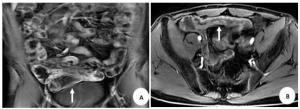
图 1 男,28岁,临床诊断为回肠末端克罗恩病,其影像学表现
A~B:增强扫描,病变区肠壁明显分层强化,呈“靶征”改变(箭).
Figure 1. Male, 28 years old, the clinical diagnosis was Crohn's disease at the end of ileum, imaging features of MRI.
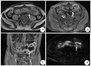
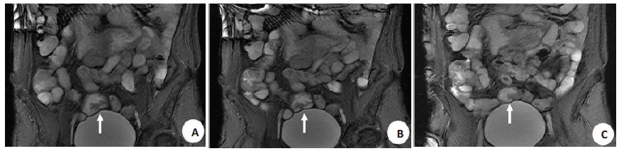
图 2 男,50岁,临床诊断为空肠末段、回肠起始段克罗恩病
A:T2WI,空肠末段、回肠起始段肠壁明显增厚,呈高信号(箭);B~C:增强扫描,病变区明显不均匀强化(箭);D:弥散加权成像(DWI B=800),病变区呈高信号,弥散受限(箭).
Figure 2. Male, 50 years old, the clinical diagnosis was Crohn's disease at the end of jejunum and the beginning of ileum.

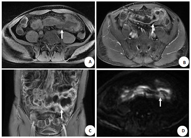
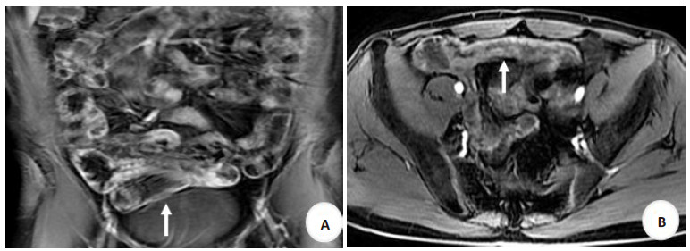
图 3 男,33岁,临床诊断为回肠末端克罗恩病
A~C:稳态进动快速成像(FIESTA),回肠远段呈克罗恩病改变,电影扫描动态观察,其动力较健康肠段动力减低(箭).
Figure 3. Male, 33 years old, the clinical diagnosis was Crohn disease at the end of ileum.
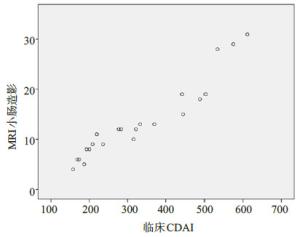
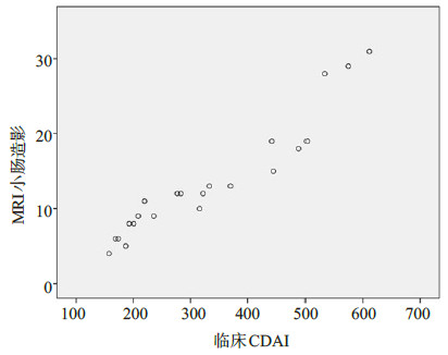
图 4 克罗恩病患者MRE评分结果与临床CDAI值相关性分析散点图
Figure 4. The scatter plot of correlation between MRE score and clinical CDAI value in patients with Crohn's disease.
表 1 CDAI计算法
Table 1. CDAI calculation method
变量 权重 稀便次数(1周) 2 腹痛时间(1周总评,0=无;1=轻;2=中;3=重) 5 一般情况(1周总评,0~3分) 7 肠外表现与并发症(1项1分) 20 阿片类止泻药(0、1分) 30 腹部包块(可疑2分,肯定5分) 10 红细胞比容降低值(正常a:男0.47女0.42) 6 100×(1-体质量/标准体质量) 1 a中国人标准的血细胞比容正常值;取各项分值之和:缓解期CDAI < 150,活动期≥150分;轻度150~220分,中度221~450分,重度 > 450分.  下载: 导出CSV
表 2 MRE克罗恩病活动度评分标准
Table 2. MRE Crohn's disease activity score
项目 缓解期 活动期 0 1 2 3 T2WI信号强度 正常 3~5级 5~7级 7~15级 DWI受限程度 无 5~7级 7~10级 10~15级 增强方式 正常 均匀强化 黏膜强化 分层强化 肠壁厚度(mm) 0~3 3~5 5~9 ≥10 病变累及范围(mm) 0 0~50 50~150 ≥151 病变区肠道动力变化 正常(无动力) 动力降低 动力正常 动力增加 肠系膜区淋巴结(个) 0 0~3 3~7 > 7 梳齿征 无 有 表中范围不包含“~”前的数值且包含“~”后的数值.
下载: 导出CSV
表 3 MRE克罗恩病活动度分级标准
Table 3. MRE Crohn's disease activity classification criteria
无 没有疾病活动迹象 轻度 ①活动迹象,但无并发症;②没有评分为3分的活动迹象;③总得分≤10分 中度 ①活动迹象,但无并发症;②得分11~15分或包含1项评分为3分的活动迹象 重度 ①活动迹象;②总得分≥16分或至少存在1个并发症 克罗恩病并发症:肠管周围蜂窝织炎、肠腔狭窄、瘘管及脓肿.
下载: 导出CSV
表 4 克罗恩病MRE活动度与临床CDAI的相关性
Table 4. Correlation between MRE activity classification standard and clinical CDAI calculation method in Crohn's disease
克罗恩病活动度 r P 轻度 0.873 0.01 中度 0.826 0.006 重度 0.899 0.015 经MRE检查克罗恩病活动性分级后,轻度及重度患者与CDAI具有相关性,中度患者与CDAI显著相关.
下载: 导出CSV
-
[1] Foti PV, Travali M, Farina R, et al. Can conventional and diffusionweighted MR enterography biomarkers differentiate inflammatory from fibrotic strictures in Crohn's disease?[J]. Medicina, 2021, 57 (3): 265. doi: 10.3390/medicina57030265 [2] Chinem ESS, Esberard BC, Moreira ADL, et al. Changes in the management of patients with Crohn's disease based on magnetic resonance enterography patterns[J]. Gastroenterol Res Pract, 2019, 2019: 1-9. http://www.researchgate.net/publication/338010946_Changes_in_the_Management_of_Patients_with_Crohn's_Disease_Based_on_Magnetic_Resonance_Enterography_Patterns [3] Athanasakos A, Mazioti A, Economopoulos N, et al. Inflammatory bowel disease-the role of cross-sectional imaging techniques in the investigation of the small bowel[J]. Insights Imaging, 2015, 6(1): 73-83. doi: 10.1007/s13244-014-0377-6 [4] Chaudhry NA, Riverso M, Grajo JR, et al. A fixed stricture on routine cross-sectional imaging predicts disease-related complications and adverse outcomes in patients with Crohn's disease[J]. Inflamm Bowel Dis, 2017, 23(4): 641-9. doi: 10.1097/MIB.0000000000001054 [5] 肖军华, 贺志龙, 陈冰心, 等. 小肠CT成像对克罗恩病活动性的评估价值[J]. 中国临床研究, 2021, 34(3): 333-6. https://www.cnki.com.cn/Article/CJFDTOTAL-LCFS201511019.htm [6] 王霈, 葸燕燕, 马彩花, 等. CT小肠造影对克罗恩病的临床应用研究. [J]. 影像研究与医学应用, 2020, 4(20): 48-50. doi: 10.3969/j.issn.2096-3807.2020.20.024 [7] 金晓蕾, 孙静, 朱珍, 等. CT影像学检查对克罗恩病患者病情活动度的诊断价值[J]. 临床和实验医学杂志, 2021, 20(1): 85-8. doi: 10.3969/j.issn.1671-4695.2021.01.025 [8] Fang ZN, Li XH, Lin JJ, et al. Magnetisation transfer imaging adds information to conventional MRIs to differentiate inflammatory from fibrotic components of small intestinal strictures in Crohn's disease[J]. Eur Radiol, 2020, 30(4): 1938-47. doi: 10.1007/s00330-019-06594-x [9] 李雪华, 江肖松, 蔡华崧, 等. 磁共振扩散加权成像评估克罗恩病活动性[J]. 中国医学影像技术, 2016, 32(4): 547-51. https://www.cnki.com.cn/Article/CJFDTOTAL-ZYXX201604022.htm [10] Tavares de Sousa H, Estevinho MM, Peyrin-Biroulet L, et al. Transmural histological scoring systems in Crohn's disease: a systematic review with assessment of methodological quality and operating properties[J]. J Crohns Colitis, 2020, 14(6): 743-56. doi: 10.1093/ecco-jcc/jjz178 [11] Pous S, Frasson M, Jiménez R, et al. Relevance of dynamic studies with magnetic resonance enterography in Crohn's disease[J]. Gastroenterol Hepatol, 2020, 43(4): 179-87. doi: 10.1016/j.gastrohep.2019.11.010 [12] Li XH, Mao R, Huang SY, et al. Characterization of degree of intestinal fibrosis in patients with crohn disease by using magnetization transfer MR imaging[J]. Radiology, 2018, 287(2): 494-503. doi: 10.1148/radiol.2017171221 [13] Pendsé DA, Makanyanga JC, Plumb AA, et al. Diffusion-weighted imaging for evaluating inflammatory activity in Crohn's disease: comparison with histopathology, conventional MRI activity scores, and faecal calprotectin[J]. Abdom Radiol, 2017, 42(1): 115-23. doi: 10.1007/s00261-016-0863-z [14] 李璐, 梁宗辉. 炎症性肠病的MRI进展[J]. 国际医学放射学杂志, 2019, 42(4): 454-7. https://www.cnki.com.cn/Article/CJFDTOTAL-GWLC201904018.htm [15] Durayski E, Watte G, Pacini GS, et al. Diffusion-weighted imaging and apparent diffusion coefficient values for evaluating terminal ileitis in patients with Crohn's disease[J]. Radiol Bras, 2019, 52(6): 361-7. doi: 10.1590/0100-3984.2019.0011 [16] Minordi LM, Larosa L, Papa A, et al. A review of Magnetic Resonance Enterography classification and quantitative evaluation of active disease in patients with Crohn's disease[J]. Clin Imaging, 2021, 69: 50-62. doi: 10.1016/j.clinimag.2020.06.006 [17] Biernacka KB, Barańska D, Matera K, et al. The value of magnetic resonance enterography in diagnostic difficulties associated with Crohn's disease[J]. Pjr, 2021, 86(1): 143-50. doi: 10.5114/pjr.2021.104581 [18] Gale HI, Sharatz SM, Taphey M, et al. Comparison of CT enterography and MR enterography imaging features of active Crohn disease in children and adolescents[J]. Pediatr Radiol, 2017, 47(10): 1321-8. doi: 10.1007/s00247-017-3876-z [19] Bruining DH, Bhatnagar G, Rimola J, et al. CT and MR enterography in Crohn's disease: current and future applications[J]. Abdom Imaging, 2015, 40(5): 965-74. doi: 10.1007/s00261-015-0360-9 [20] 任小军, 章士正, 张峭巍, 等. 小肠Crohn病的MRI诊断[J]. 中华放射学杂志, 2004, 38(11): 1201-5. doi: 10.3760/j.issn:1005-1201.2004.11.019 [21] Pesce Lamas Constantino C, Souza Rodrigues R, Araujo Oliveira Neto J, et al. Computed tomography and magnetic resonance enterography findings in Crohn's disease: what does the clinician need to know from the radiologist?[J]. Can Assoc Radiol J, 2014, 65(1): 42-51. doi: 10.1016/j.carj.2012.11.004 [22] Bruining DH, Zimmermann EM, Loftus EV Jr, et al. Consensus recommendations for evaluation, interpretation, and utilization of computed tomography and magnetic resonance enterography in patients with small bowel Crohn's disease[J]. Radiology, 2018, 286 (3): 776-99. doi: 10.1148/radiol.2018171737 [23] Lambrou T, Chaudhry NA, Grajo JR, et al. Small bowel stricture is associated with abnormal motility on the cine MRI sequence in patients with Crohn's disease[J]. Eur J Radiol, 2019, 118: 264-70. doi: 10.1016/j.ejrad.2019.08.001 [24] Cullmann JL, Bickelhaupt S, Froehlich JM, et al. MR imaging in Crohn's disease: correlation of MR motility measurement with histopathology in the terminal ileum[J]. Neurogastroenterol Motil, 2013, 25(9): 749-e577. doi: 10.1111/nmo.12162 [25] Bickelhaupt S, Froehlich JM, Cattin R, et al. Differentiation between active and chronic Crohn's disease using MRI small-bowel motility examinations—Initial experience[J]. Clin Radiol, 2013, 68 (12): 1247-53. doi: 10.1016/j.crad.2013.06.024 [26] Ohtsuka K, Takenaka K, Kitazume Y, et al. Magnetic resonance enterography for the evaluation of the deep small intestine in Crohn's disease[J]. Intest Res, 2016, 14(2): 120. doi: 10.5217/ir.2016.14.2.120 [27] Yu-Tai S, Tien-Yu C. The comb sign of Crohn's disease[J]. QJM: Int J Med, 2020, 113(8): 581. doi: 10.1093/qjmed/hcz285 [28] Guimarães LS, Greer MLC, Dillman JR, et al. Magnetic resonance in Crohn's disease: diagnosis, disease burden, and classification[J]. Magn Reson Imaging Clin N Am, 2020, 28(1): 31-44. doi: 10.1016/j.mric.2019.08.003 -

 点击查看大图
计量
- 文章访问数: 592
- HTML全文浏览量: 252
- PDF下载量: 11
- 被引次数: 0