


Diagnostic analysis and imaging findings of MSCT and MRI in intracranial anaplastic meningioma
-
摘要:
目的探讨颅内间变性室管膜瘤的多层螺旋CT(MSCT)及MRI表现。 方法收集我院2011年1月~2019年12月经手术病理证实的颅内间变性室管膜瘤病例,最终共收集到符合标准的患者17例,其中男15例,女2例;年龄1~67岁,其中 < 10岁的患者7例, > 50岁的患者7例,呈两极分布;主要收集病例的颅脑MSCT平扫、MRI的T1WI、T2WI、FLAIR及增强检查,分析其形态特征及影像学特点。 结果本组病例幕上16例,幕下1例;形态分2型,囊实性11例,实质性6例。囊实性肿瘤形状多不规则,囊性部分与实性部分比例差别较大,肿瘤边界不清或欠清,瘤周水肿较轻;实质性肿瘤呈类圆形或分叶状,边界较清,多伴有轻或中度瘤周水肿。MSCT平扫示囊实性肿瘤实性成分及实质性肿瘤主要呈等或稍高密度,而囊实性肿瘤囊性成分呈脑脊液样密度改变;MRI扫描示囊实性肿瘤的实性成分及实质性肿瘤于T1WI呈稍低或低信号,T2WI及FLAIR呈等或稍高信号,MRI增强扫描示囊实性肿瘤的实性成分及实质性肿瘤明显不均匀强化,囊实性肿瘤的囊壁明显环形强化,囊内无强化。 结论颅内间变性室管膜瘤的MSCT及MRI表现有一定的特征性,结合患者性别、发病年龄及临床病史,对其诊断及鉴别诊断有较高的临床意义。 Abstract:ObjectiveTo analyze MSCT and MRI findings of intracranial anaplastic meningioma, compare with the pathological results, and improve the diagnosis of the disease. MethodsWe collected cases of intracranial anaplastic meningioma confirmed by pathology in our hospital from January 2011 to December 2019. Seventeen patients were collected, including 15 males and 2 females. Aged from 1 to 67 years old, with a bipolar distribution, the patients included 7 patients under 10 years old and 7 patients over 50 years old. The imaging data of intracranial anaplastic meningioma cases were collected, including MSCT scan, T1WI, T2WI, FLAIR and enhanced examination of MRI. Their imaging characteristics were analyzed. ResultsThere were 16 tumors mainly located in the supra, 1 case was under the curtain, the main body was located in the fourth ventricle. The morphology of this group was divided into 2 types, 11 cases were cystic-solid and 6 cases were solid. The shape of cystic-solid tumors was mostly irregular. The ratio of cystic parts to solid parts was quite different. The tumor borders were unclear, and the peritumoral edema was light. Solid tumors were mainly round or lobulated, with clear borders, often accompanied by light or moderate peritumoral edema. The MSCT scan showed that the solid components of cystic-solid tumors and solid tumors were mainly of equal or slightly higher density. The cystic components of cystic-solid tumors showed cerebrospinal fluid-like density changes. MRI scans showed solid components of cystic-solid tumors and solid tumors with slightly lower or lower signals at T1WI, equal or slightly higher signals at T2WI and FLAIR, and enhanced MRI scans showed solid components of cystic-solid tumors and solid tumors were significantly unevenly enhanced, and the cyst wall of the cystic-solid tumors was obviously strengthened in a ring shape. There was no enhancement in the cyst. ConclusionMSCT and MRI manifestations of intracranial anaplastic meningioma had certain characteristics. Combining with the patient's gender, age and clinical history, it has higher clinical significance for its diagnosis and differential diagnosis. -
Key words:
- anaplastic ependymal tumor /
- tomography /
- X-ray computer /
- magnetic resonance imaging
-
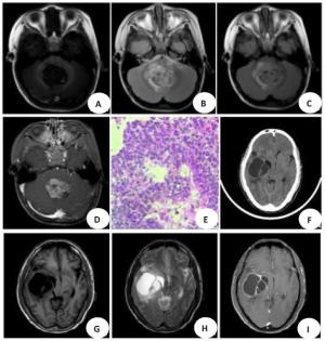
图 1 患者颅内间变性室管膜瘤的影像学表现
A~E: 患者第四脑室内见混杂信号肿块; A~D: TIWI呈稍低信号,其内见点状高信号(出血灶),T2WI及FLAIR呈等或稍高信号,其内见少许点片状短T2信号影,肿块累及双侧小脑半球,周围可见轻度水肿带影,增强扫描不均匀明显强化;E: 病理示瘤细胞呈团片状,部分形成假菊型团结构,细胞排列密集,核大(HE×200)。F~I: MRI示肿瘤TIWI呈低信号,T2WI呈高信号,其内可见分隔样改变,并与右侧侧脑室关系密切,增强扫描肿瘤囊壁明显强化,囊内无强化.
Figure 1. Imaging of intracranial anaplastic ependymal tumor of the patient.
-
[1] Evenson M, Cai CY, Hucthagowder V, et al. Utility of copy number variants in the classification of intracranial ependymoma[J]. Cancer Genet, 2020, 240: 66-72. doi: 10.1016/j.cancergen.2019.11.003 [2] 陈灵朝, 姚瑜, 汪洋, 等. 93例室管膜瘤的临床特点及预后分析[J]. 中华神经外科杂志, 2013, 29(11): 1087-9. doi: 10.3760/cma.j.issn.1001-2346.2013.11.004 [3] Kharosekar H, Bhide A, Velho V, et al. Pediatric isolated cortical (ectopic) anaplastic ependymoma[J]. Asian J Neurosurg, 2018, 13 (1): 144-6. doi: 10.4103/1793-5482.181133 [4] 任翔, 吴于淳, 杨秀军. DWI和最小ADC值鉴别诊断儿童颅内间变性室管膜瘤与室管膜瘤[J]. 中国医学影像技术, 2019, 35(1): 46-9. https://www.cnki.com.cn/Article/CJFDTOTAL-ZYXX201901015.htm [5] 郑瑞平, 张勇, 程敬亮, 等. 增强MRI全域直方图分析在鉴别间变型与非间变型室管膜瘤中的价值[J]. 临床放射学杂志, 2019, 38(4): 589-92. https://www.cnki.com.cn/Article/CJFDTOTAL-LCFS201904005.htm [6] Louis DN, Perry A, Reifenberger G, et al. The 2016 world health organization classification of tumors of the central nervous system: a summary[J]. Acta Neuropathol, 2016, 131(6): 803-20. doi: 10.1007/s00401-016-1545-1 [7] Chao MM, Packer RJ, Myseros JS, et al. Isolated extracranial recurrence of anaplastic ependymoma[J]. Pediatr Blood Cancer, 2011, 56(2): 317-8. doi: 10.1002/pbc.22764 [8] St Jeor JD, Thacker PG, Benson JC, et al. Anaplastic ependymoma metastases though a ventriculoperitoneal shunt[J]. Radiol Case Rep, 2020, 15(6): 650-4. doi: 10.1016/j.radcr.2020.02.036 [9] 徐焱, 姚振威, 韩芳. 幕上脑实质室管膜瘤影像表现及病理对照分析[J]. 放射学实践, 2014, 29(9): 1031-4. https://www.cnki.com.cn/Article/CJFDTOTAL-FSXS201409019.htm [10] 王凯, 张姝, 施露, 等. 2016年世界卫生组织中枢神经系统肿瘤分类概述[J]. 磁共振成像, 2016, 7(12): 881-96. doi: 10.12015/issn.1674-8034.2016.12.001 [11] 张国晋, 马莉, 王丹, 等. DWI对脑实质间变性室管膜瘤与多形性胶质母细胞瘤的鉴别价值[J]. 磁共振成像, 2017, 8(11): 812-6. doi: 10.12015/issn.1674-8034.2017.11.003 [12] Lavrador JP, Oliveira E, Teixeira JC, et al. Adult supratentorial extraventricular anaplastic ependymoma: therapeutic approach and clinical review[J]. Asian J Neurosurg, 2018, 13(1): 105-9. doi: 10.4103/1793-5482.181121 [13] 马玲, 吴靖雯, 于昊, 等. 脑实质内室管膜瘤的影像学诊断[J]. 山东医药, 2016, 56(43): 7-10. doi: 10.3969/j.issn.1002-266X.2016.43.003 [14] 李小会, 黄仲奎. 颅内脑室外室管膜瘤的MRI诊断[J]. 实用放射学杂志, 2012(3): 342-4, 416. doi: 10.3969/j.issn.1002-1671.2012.03.005 [15] 白玉贞, 韩晓东, 牛广明. 脑实质间变性室管膜瘤的MRI表现[J]. 放射学实践, 2012, 27(12): 1304-7. doi: 10.3969/j.issn.1000-0313.2012.12.006 [16] McGuire CS, Sainani KL, Fisher PG. Incidence patterns for ependymoma: a surveillance, epidemiology, and end results study[J]. J Neurosurg, 2009, 110(4): 725-9. doi: 10.3171/2008.9.JNS08117 [17] Sayegh ET, Aranda D, Kim JM, et al. Prognosis by tumor location in adults with intracranial ependymomas[J]. J Clin Neurosci, 2014, 21(12): 2096-101. doi: 10.1016/j.jocn.2014.05.011 [18] Yadav YR, Neha, Chandrakar SK. Pure cortical supratentorial extraventricular ependymoma[J]. Neurol India, 2009, 57(2): 213-5. doi: 10.4103/0028-3886.51301 [19] 王杨灵犀, 孙凤娇, 王翀, 等. 颅内间变性室管膜瘤10例临床分析[J]. 广东医学, 2019, 40(21): 3074-9. https://www.cnki.com.cn/Article/CJFDTOTAL-GAYX201921021.htm [20] Amirian ES, Armstrong TS, Gilbert MR, et al. Predictors of survival among older adults with ependymoma[J]. J Neurooncol, 2012, 107 (1): 183-9. doi: 10.1007/s11060-011-0730-2 [21] 管英, 李继兵, 邹萍. 多层螺旋CT与MRI诊断甲状腺结节的价值比较[J]. 分子影像学杂志, 2020, 43(4): 697-700. doi: 10.12122/j.issn.1674-4500.2020.04.32 -

 下载:
下载:
 点击查看大图

图(1)
计量
- 文章访问数: 666
- HTML全文浏览量: 281
- PDF下载量: 13
- 被引次数: 0