


Evaluation value of HR-MRI detection of core types of vulnerable carotid plaque
-
摘要:
目的通过颈动脉易损斑块核心类型高分辨率MRI检测的评估价值,为进一步为临床预防、诊断脑梗死提供有价值的线索。 方法选取2017年6月~2020年6月于我院接受治疗的150例急性脑梗死患者为观察组,随机选取同期接受体检的自愿者30例为健康对照组。MRI检查采用GE Discovery MR750 3.0T超导型磁共振仪,8通道相控阵表面线圈成像方法,扫描层厚4 mm,层间距1 mm检查。所有患者均行常规横断位T1WI、T2WI及T2液体衰减反转恢复(FLAIR),矢状位T1WI检查。 结果观察组和健康对照组两组对象性别、年龄及TC、TG水平比较无统计学意义(P>0.05),观察组LDL-C、FIB均较健康对照组更高(P < 0.05),而HDL-C较健康对照组更低(P < 0.05)。观察组血清游离脂肪酸(FFA)、oxLDL-C水平均明显高于健康对照组(P < 0.05)。观察组患者根据MRI检查判定得到的颈动脉粥样硬化性斑块稳定性情况可分为无斑块组(n=35)、稳定斑块组(n=50)和不稳定板块组(n=65),3组组间血清FFA、oxLDL-C水平比较有统计学意义(P < 0.05)。经Spearman相关分析,急性脑梗死患者颈动脉粥样硬化性斑块稳定性与血清FFA、oxLDL-C水平均具有正相关性(P < 0.05)。 结论颈动脉易损斑块核心类型高分辨率MRI检测的评估价值良好。 Abstract:ObjectiveTo evaluate the value of HR-MRI detection of the core types of vulnerable carotid plaques to provide valuable clues for further clinical prevention and diagnosis of cerebral infarction. MethodsA total of 150 ACI patients who were treated in our hospital from June 2017 to June 2020 were selected as the observation group. Thirty volunteers who received physical examinations during the same period were randomly selected as the healthy control group. MRI examination adoptted GE Discovery MR750 3.0 T superconducting magnetic resonance instrument, -channel phased array surface coil imaging method, scanning slice thickness 4 mm, slice spacing 1mm inspection. All patients were performed with conventional transverse T1WI, T2WI and T2 fluid attenuation inversion recovery (FLAIR), sagittal T1WI examination. ResultsThe differrences of gender, age, TC and TG levels of the observation group and the healthy control group were not significant (P>0.05). The observation group had higher LDL-C and FIB than the healthy control group (P < 0.05). HDL-C was lower in the healthy control group (P < 0.05). Serum FFA and oxLDL-C levels in the observation group were significantly higher than those in the healthy control group (P < 0.05). Patients in the observation group were divided into non-plaque group (n=35), stable plaque group (n=50) and unstable plate group (n=35) based on the stability of carotid atherosclerotic plaque determined by MRI examination (n=65). The differrences of serum FFA and oxLDL-C levels between the three groups were significant (P < 0.05). According to Spearman correlation analysis, the stability of carotid atherosclerotic plaque in ACI patients was positively correlated with serum FFA and oxLDL-C levels (P < 0.05). ConclusionThe evaluation value of HR-MRI for the core type of carotid artery vulnerable plaque is good. -
Key words:
- vulnerable carotid plaque /
- core type /
- HR-MRI /
- evaluation value
-

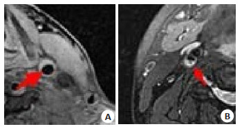
图 1 颈动脉易损斑块影像学结果
A: 坏死核心MRI(男, 67岁); B: 脂质核心(男, 42岁)
Figure 1. Imaging results of carotid artery vulnerable plaque
表 1 患者一般资料比较
Table 1. Comparison of general information of subjects (Mean±SD)
组别 性别[n(%)] 年龄(岁) TG(mmol/L) TC(mmol/L) HDL-C(mmol/L) LDL-C(mmol/L) FIB(g/L) 男 女 观察组(n=150) 84(56.00) 66(44.00) 62.56±4.15 1.96±0.42 5.43±0.58 1.06±0.14 3.75±0.36 3.76±0.74 健康对照组(n=30) 17(56.67) 13(43.33) 62.25±4.36 1.82±0.26 5.46±0.42 1.38±0.15 3.06±0.27 3.30±0.56 t/χ2 0.005 0.370 1.757 0.269 11.293 9.944 3.222 P 0.946 0.712 0.081 0.788 0.000 0.000 0.002 TG: 三酰甘油; TC: 总胆固醇; LDL-C: 低密度脂蛋白胆固醇; HDL-C: 高密度脂蛋白胆固醇; FIB: 纤维蛋白原.  下载: 导出CSV
表 2 两组血清FFA、oxLDL-C水平比较
Table 2. Comparison of serum FFA and oxLDL-C levels between the two groups (Mean±SD)
组别 FFA(µmol/L) oxLDL-C(µg/L) 观察组(n=150) 467.18±107.41 581.67±109.23 健康对照组(n=30) 125.89±31.47 393.26±71.58 t 17.221 9.056 P 0.000 0.000 FFA: 游离脂肪酸; oxLDL-C: 氧化低密度脂蛋白.
下载: 导出CSV
表 3 不同硬化斑块组血清FFA、oxLDL-C水平比较
Table 3. Comparison of serum FFA and oxLDL-C levels in different plaque groups (Mean±SD)
组别 FFA(µmol/L) oxLDL-C(µg/L) 无斑块组(n=35) 353.73±72.51 457.70±92.08 稳定斑块组(n=50) 465.69±89.45a 579.32±103.38a 不稳定斑块组(n=65) 529.41±132.81ab 650.22±121.44ab F 30.370 35.315 P 0.000 0.000 aP < 0.05 vs无斑块组; bP < 0.05 vs稳定斑块组.
下载: 导出CSV
-
[1] 田瑞. 高分辨MRI与超声检查诊断颈动脉粥样硬化斑块的临床价值比较[J]. 临床医学工程, 2020, 27(7): 853-4. doi: 10.3969/j.issn.1674-4659.2020.07.0853 [2] 宋微, 杨帆, 张国富, 等. 血浆Lp-PLA2、幽门螺杆菌感染对颈动脉粥样硬化的影响分析[J]. 临床和实验医学杂志, 2021, 20(6): 613-7. [3] 杨梅, 周丹. 颈动脉易损斑块磁共振特征与临床危险因素评分的相关性分析[J]. 实用放射学杂志, 2020, 36(11): 1759-62. doi: 10.3969/j.issn.1002-1671.2020.11.014 [4] 朱文卿, 柳琦, 陈晓. 血管紧张素Ⅱ诱导产生兔颈动脉不稳定斑块及其高分辨率MRI [J]. 实用放射学杂志, 2020, 36(1): 139-43. doi: 10.3969/j.issn.1002-1671.2020.01.035 [5] 查晴, 于晨溪, 刘亚, 等. miR-16在冠状动脉粥样硬化斑块进展中的作用及初步机制研究[J]. 诊断学理论与实践, 2021, 20(1): 82-7. [6] 吴静静, 贾琳, 王云玲, 等. 磁共振血管壁成像对症状性大脑中动脉粥样硬化斑块的定量评估[J]. 临床放射学杂志, 2020, 39(11): 2152-5. [7] 梁群娣, 吴文军, 佘子瑜. 颈动脉粥样硬化斑块患者淋巴细胞功能相关抗原-1、可溶性CD40、组织蛋白酶K水平分析[J]. 心脑血管病防治, 2021, 21(1): 93-4, 98. doi: 10.3969/j.issn.1009-816x.2021.01.023 [8] 齐炳才, 靳琦文, 胡杰, 等. 颈动脉粥样硬化斑块内新生血管的研究现状及进展[J]. 中国动脉硬化杂志, 2021, 29(4): 359-62, 368. https://www.cnki.com.cn/Article/CJFDTOTAL-AHYX201211064.htm [9] 戴婷, 何文明, 麦一峰. 麝香保心丸对小鼠动脉粥样硬化斑块形成的影响及相关机制探讨[J]. 心电与循环, 2021, 40(2): 139-44, 148, 227. [10] 周立平, 孙钦亮, 王鸿凤, 等. 颈动脉粥样硬化斑块内新生血管超声造影分级与血脂水平的相关性[J]. 临床超声医学杂志, 2021, 23(3): 183-6. doi: 10.3969/j.issn.1008-6978.2021.03.008 [11] 张颖怡, 刘金波, 刘欢, 等. 全身动脉粥样硬化斑块与脑梗死的关系: 北京血管病变评价研究结果[J]. 心血管病学进展, 2021, 42(3): 277-84. https://www.cnki.com.cn/Article/CJFDTOTAL-CGZC201701004.htm [12] 李雯, 陈朔华, 赵剑秋, 等. 颈动脉斑块和低踝臂指数联合作用增加老年人缺血性心脑血管事件的发生风险[J]. 中华心血管病杂志, 2021, 49(3): 263-8. doi: 10.3760/cma.j.cn112148-20200401-00272 [13] 陶飞, 赵旺, 琚双五. 血小板与淋巴细胞比值、中性粒细胞与淋巴细胞比值与急性脑梗死颈动脉粥样硬化斑块的相关性研究[J]. 临床和实验医学杂志, 2021, 20(6): 606-9. https://www.cnki.com.cn/Article/CJFDTOTAL-ZFSJ201709012.htm [14] 黄宜华. 大数据机器学习系统研究进展[J]. 大数据, 2015, 1(1): 35-54. https://www.cnki.com.cn/Article/CJFDTOTAL-DSJU201501005.htm [15] 王彬, 韦玉新, 徐常清, 等. 高分辨率MRI评价颈动脉斑块稳定性的优势及临床分析[J]. 中国实用医药, 2020, 15(24): 78-80. https://www.cnki.com.cn/Article/CJFDTOTAL-ZSSA202024033.htm [16] 孟庆国. 高分辨MRI与超声检查应用于颈动脉粥样硬化斑块诊断的效果对比分析[J]. 保健文汇, 2020, (17): 178-9. doi: 10.3969/j.issn.1671-5217.2020.17.134 [17] 葛东泉, 曲刚成, 崔福进. 3.0T MRI多序列扫描在颈动脉斑块分析中的运用价值研究[J]. 影像研究与医学应用, 2020, 4(6): 46-7. https://www.cnki.com.cn/Article/CJFDTOTAL-YXYY202006026.htm [18] 远奇, 马玉波. 18F-FDG PET/CT和MRI评估颈动脉粥样硬化斑块炎症活动程度的价值分析[J]. 中国CT和MRI杂志, 2020, 18(9): 24-7. doi: 10.3969/j.issn.1672-5131.2020.09.008 [19] 孙培育, 吴娇艳, 刘梦秋, 等. 应用高分辨MRI评估颈动脉斑块与体质指数的相关性[J]. 医学影像学杂志, 2020, 30(2): 182-6. https://www.cnki.com.cn/Article/CJFDTOTAL-XYXZ202002004.htm [20] 巴建, 苗红, 张艾红. 磁共振高分辨技术在脑梗死复发患者颈动脉斑块评估中的应用观察[J]. 癫痫与神经电生理学杂志, 2020, 29(1): 17-20. https://d.wanfangdata.com.cn/periodical/lcsjdslxzz202001005 [21] 王霞, 塔娜, 勉丽, 等. 高分辨MRI与超声检查对颈动脉粥样硬化斑块的诊断价值[J]. 影像研究与医学应用, 2019, 3(1): 95-6. doi: 10.3969/j.issn.2096-3807.2019.01.059 [22] 张振, 夏纪筑, 彭雪莲, 等. 速度向量成像技术评价维持性血液透析患者颈动脉弹性的应用价值[J/OL]. 解放军医学院学报, 2021: 1-5. http://kns.cnki.net/kcms/detail/10.1117.R.20210318.0946.006.html. https://www.cnki.com.cn/Article/CJFDTOTAL-JYJX202102008.htm [23] 陈绍琦, 杜西亚, 姚中铿, 等. 超声造影评估大动脉粥样硬化型缺血性卒中风险的初步研究[J]. 中国超声医学杂志, 2021, 37(3): 244-7. [24] 熊云志, 井斌, 李慧, 等. 颈动脉内膜增厚和斑块发生率及其相关因素分析[J]. 中国城乡企业卫生, 2021, 36(3): 188-91. -

 点击查看大图
计量
- 文章访问数: 663
- HTML全文浏览量: 310
- PDF下载量: 10
- 被引次数: 0