


Correlation between the apical morphology and cardiac function of left ventricular and plaque vulnerability in patients with coronary heart disease
-
摘要:
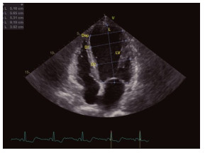
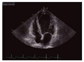
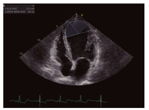
目的探讨冠心病患者左心室心尖形态及功能与斑块易损性的相关性。 方法选取我院2018年1月~2020年1月收治的53例冠心病患者为研究对象,所有患者均行超声心动图及冠状动脉CT造影检查。根据CT造影检测结果分为稳定斑块组(n=28)、易损斑块组(n=25)。比较两组左室收缩末期内径(LVESD)、左室舒张末期内径(LVEDD)、射血分数、心尖部舒张末期左右径(Dap)、心尖球形指数(Siap)、舒张末期心尖角度、收缩末期心尖角度。采用logistics回归分析冠心病患者左心室心尖形态及功能与斑块易损性的相关性。 结果稳定斑块组心功能指标LVEDD、LVESD明显低于易损斑块组(P < 0.05),射血分数明显高于易损斑块组(P < 0.05);稳定斑块组左心室Dap、舒张末期心尖夹角、收缩末期心尖夹角明显低于易损斑块组(P < 0.05),Siap明显高于易损斑块组(P < 0.05);斑块易损性是影响左心室Dap、舒张末期心尖夹角、收缩末期心尖夹角、LVEDD、LVESD、射血分数的独立危险因素(P < 0.05)。 结论冠心病患者左室心尖形态及功能和斑块易损性具有显著相关性。 Abstract:ObjectiveTo explore the correlation between the apical morphology and cardiac function of left ventricular and plaque vulnerability in patients with coronary heart disease. Methods53 patients with coronary heart disease in our hospital from January 2018 to January 2020 were enrolled. Echocardiography and coronary CT angiography (CTA) were performed in all patients. According to CTA results, patients were divided into stable plaque group (n=28) and vulnerable plaque group (n= 25). Various indexes were compared between groups, including left ventricular end systolic diameter (LVESD), left ventricular end diastolic diameter (LVEDD), left ventricular ejection fraction (LVEF), anterior to posterior diameter (DAP), apical spherical index (Siap), end diastolic apex angle and end systolic apex angle. Correlation between the apical morphology and cardiac function of left ventricular and plaque vulnerability was analyzed using Logistic regression analysis. ResultsLVEDD and LVESD in stable plaque group were significantly lower than those in vulnerable plaque group (P < 0.05), and LVEF was significantly higher than that in vulnerable plaque group (P < 0.05). The left ventricular Dap, end-diastolic apex Angle and endsystolic apex Angle in stable plaque group were significantly lower than those in vulnerable plaque group (P < 0.05), and Siap was significantly higher than that in vulnerable plaque group (P < 0.05). Plaque vulnerability was an independent risk factor affecting left ventricular Dap, end-diastolic apex angle, end-systolic apex angle, LVEDD, LVESD, and LVEF (P < 0.05). ConclusionThe morphology and function of left ventricular apex in patients with coronary heart disease have significant correlation with plaque vulnerability. -
Key words:
- coronary heart disease /
- apical morphology /
- cardiac function /
- plaque vulnerability
-
表 1 心功能指标比较
Table 1. Comparison of cardiac function indicators (Mean±SD)
组别 LVEDD(mm) LVESD(mm) LVEF(%) 稳定斑块组(n=28) 50.60±3.52 40.11±2.65 56.77±6.31 易损斑块组(n=25) 54.86±4.28 43.97±3.22 48.03±5.04 t 3.984 4.784 5.257 P < 0.001 < 0.001 < 0.001 LVEDD: 左心室舒张末期内径; LVESD: 左室收缩末期内径; LVEF: 左心室射血分数.  下载: 导出CSV
表 2 左心室心尖心态比较
Table 2. Comparison of left ventricular apical morphology (Mean±SD)
组别 Dap(mm) Siap 舒张末期心尖夹角(°) 收缩末期心尖夹角(°) 稳定斑块组(n=28) 30.59±2.66 1.35±0.20 74.11±6.34 60.21±5.39 易损斑块组(n=25) 33.98±3.41 1.19±0.13 78.35±7.29 69.51±6.28 t 4.058 3.407 2.246 5.75 P < 0.001 0.001 0.029 < 0.001 Dap: 心尖部舒张末期左右径; Siap: 心尖球形指数.
下载: 导出CSV
表 3 冠心病患者左心室心尖形态及功能与斑块易损性的logistics回归分析
Table 3. Logistic regression analysis of correlation between the apical morphology and cardiac function of left ventricular and plaque vulnerability
影响因素 β SE Wald P OR(95%CI) LVEDD 0.508 0.17 8.931 0.003 1.662(1.249-2.432) LVESD 0.363 0.165 4.86 0.027 1.438(1.160-2.213) LVEF 0.419 0.185 5.14 0.023 1.521(1.310-2.705) Dap 0.524 0.198 7.005 0.008 1.688(1.446-3.140) Siap 0.552 0.197 7.816 0.005 1.736(1.526-3.307) 舒张末期心尖夹角 0.801 0.276 8.395 0.004 2.228(1.730-5.114) 收缩末期心尖夹角 0.893 0.282 10.004 0.002 2.442(1.767-5.343)
下载: 导出CSV
-
[1] 黄国虹, 陈艳, 游卫华, 等. 慢性充血性心力衰竭患者左室射血分数与心脏变时不良的相关性[J]. 广东医学, 2017, 38(S1): 60-1. https://www.cnki.com.cn/Article/CJFDTOTAL-GAYX2017S1020.htm [2] Yang G, Yao XW, Liang L, et al. Correlation between Cys-C, β2-MG levels and left ventricular structure and function in patients with coronary heart disease[J]. Chin J Evid Based Cardiovasc Med, 2017, 16(1): 173-9. http://en.cnki.com.cn/Article_en/CJFDTOTAL-PZXX201706037.htm [3] 韩艳, 杨朝宽, 高传玉, 等. ApoB/A1比值与稳定型心绞痛患者左主干斑块易损性的相关性分析[J]. 中华医学杂志, 2017, 97(27): 2101-6. doi: 10.3760/cma.j.issn.0376-2491.2017.27.005 [4] 韩艳, 杨朝宽, 高传玉, 等. 脂蛋白(a)浓度与稳定型心绞痛患者左主干斑块性质的相关性分析[J]. 中华医学杂志, 2019, 99(19): 1490-3. doi: 10.3760/cma.j.issn.0376-2491.2019.19.011 [5] 郑刚. 指导临床实践的新指南: 2007年冠心病诊治指南[J]. 华夏医学, 2008, 21(3): 575-7. doi: 10.3969/j.issn.1008-2409.2008.03.108 [6] 田菊荣. 平板运动试验结果与冠心病患者左心室整体收缩功能及冠状动脉病变程度的相关性研究[J]. 空军医学杂志, 2019, 35(2): 149-52. https://www.cnki.com.cn/Article/CJFDTOTAL-ZJZY201902020.htm [7] 李丹, 张杰. 老年冠状动脉粥样硬化性心脏病患者三维超声心动图特征及与左室收缩功能的相关性研究[J]. 国际心血管病杂志, 2020, 47(1): 59-61. doi: 10.3969/j.issn.1673-6583.2020.01.015 [8] Gumauskiene B, Padervinskiene L, Vaskelyte JJ, et al. Left ventricular morphology and function as a determinant of pulmonary hypertension in patients with severe aortic Stenosis: cardiovascular magnetic resonance imaging study[J]. Medicina (Kaunas), 2019, 55(10): E711. doi: 10.3390/medicina55100711 [9] 柳春霞, 朱琳, 李雅菁. 冠心病患者颈动脉斑块易损性与脂代谢、炎症反应、蛋白酶活性的相关性研究[J]. 海南医学院学报, 2018, 24(12): 1147-50. https://www.cnki.com.cn/Article/CJFDTOTAL-HNYY201812003.htm [10] 麦兴盛, 王娟利, 李亚峰, 等. 老年冠心病患者超声三维空间运动指标及其与左心室收缩功能的关系[J]. 海南医学, 2019, 30(14): 1841-3. doi: 10.3969/j.issn.1003-6350.2019.14.022 [11] 王辉, 徐磊, 贺毅, 等. 心脏磁共振评价心尖肥厚型心肌病患者左心房室结构、功能及其相关性研究[J]. 磁共振成像, 2019, 10(6): 415-9. https://www.cnki.com.cn/Article/CJFDTOTAL-CGZC201906007.htm [12] Pramushinta L, Pikir BS, Pranawa, et al. Correlation between arterial stiffness with left ventricular mass index and diastolic function in patients with stage 3 and 4 of non-diabetic chronic kidney disease [J]. IOP Conf Ser Earth Environ Sci, 2020, 441(1): 121. http://www.researchgate.net/publication/339468701_Correlation_between_Arterial_Stiffness_with_Left_Ventricular_Mass_Index_and_Diastolic_Function_in_Patients_with_Stage_3_and_4_of_Non-Diabetic_Chronic_Kidney_Disease [13] 于海奕, 莫小丽, 王新宇, 等. 应用心脏核磁共振成像评价高血压相关收缩性心力衰竭患者左心室形态改变[J]. 中国介入心脏病学杂志, 2019, 27(2): 64-9. doi: 10.3969/j.issn.1004-8812.2019.02.002 [14] 卢飞, 张伟, 李茂琴, 等. 左心室应变和扭转对冠心病患者心脏功能水平的关系研究[J]. 中国心血管病研究, 2019(2): 143-6. doi: 10.3969/j.issn.1672-5301.2019.02.012 [15] 唐军. 偶然发现的左室心尖部"铲形"FDG摄取可能提示心尖肥厚性心肌病[J]. 中华核医学与分子影像杂志, 2018, 38(1): 72-3. doi: 10.3760/cma.j.issn.2095-2848.2018.01.104 [16] Vancheri F, Vancheri S, Henein M. Relationship between QRS measurements and left ventricular morphology and function in asymptomatic individuals [J]. Echocardiography, 2018, 35(3): 301-7. doi: 10.1111/echo.13782 [17] 王一超, 王莺, 周振堰, 等. 多排螺旋CT评估老年冠心病患者的冠状动脉病变严重性与左心室收缩功能[J]. 分子影像学杂志, 2020, 43(4): 651-4. doi: 10.12122/j.issn.1674-4500.2020.04.21 [18] Otsuka K, Nakanishi K, Shimada K, et al. Associations of sensitive cardiac troponin-Ⅰ with left ventricular morphology, function and prognosis in end-stage renal disease patients with preserved ejection fraction[J]. Heart Vessels, 2018, 33(11): 1334-42. doi: 10.1007/s00380-018-1192-7 [19] 高亚坤, 张玉辉, 刘颖, 等. 三维超声心动技术评估老年冠心病患者心脏三维空间运动及其与左心室收缩功能的关系[J]. 中国老年学杂志, 2018, 38(10): 2329-31. doi: 10.3969/j.issn.1005-9202.2018.10.011 [20] Goto TK. Diagnosis of the morphology and function in patients with mandibular laterognathism by 3DMRI[J]. Jpn J Jaw Deform, 2017, 27(2): 57-9. doi: 10.5927/jjjd.27.57 [21] 郑嘉荣, 邓剑玲, 邢超, 等. 超声心动图对冠心病心衰患者左室心尖形态与功能的分析[J]. 中国超声医学杂志, 2020, 36(4): 336-8. doi: 10.3969/j.issn.1002-0101.2020.04.016 [22] Šarić S, Cvetković T, Petrović D, et al. Correlation between oxidative stress parameters and left ventricular geometry in patients with chronic heart failure[J]. Acta Fac Medic Naissensis, 2020, 37(3): 241-51. https://www.cnki.com.cn/Article/CJFDTOTAL-ZHHL201503035.htm [23] 李靖, 高敬. 二尖瓣环自动追踪技术评价冠状动脉粥样硬化性心脏病病人左心室收缩功能[J]. 首都医科大学学报, 2017, 38(3): 386-90. doi: 10.3969/j.issn.1006-7795.2017.03.012 [24] 周洁舲, 孙书菊. 三维斑点追踪显像对冠心病患者左室运动功能评价效果的研究[J]. 中国心血管杂志, 2020, 25(5): 442-6. doi: 10.3969/j.issn.1007-5410.2020.05.008 -

 点击查看大图
图(3) / 表(3)
计量
- 文章访问数: 482
- HTML全文浏览量: 214
- PDF下载量: 5
- 被引次数: 0