


Effect factors of the measurement results of Laser Speckle Imaging Analyzer
-
摘要:
目的通过模拟不同的使用条件,找出影响激光散斑衬比成像(LSCI)结果值的影响因素。 方法在暨南大学第一附属医院的疼痛门诊模拟各种使用条件。采用武汉迅微光电子技术有限公司的商用仪器(Laser Speckle Blood Flow Imager Ⅱ),利用仿皮肤硅胶模型作为被测物体,探索4种使用条件下散斑成像系统结果值的变化:包括肤色、绿、黄、蓝、红、黑6种物体颜色的不同的曝光时间(1~15 ms)、不同的光线条件(遮光、室内灯光及无额外光照)、不同的探头距被测物体距离(15、20、25、30、35、40、45、50 cm)以及不同的探头激光束与被测物体夹角(90°、85°、80°、75°、70°、65°)。 结果曝光时间影响:6种被测物体颜色的散斑成像测量结果值都会随着曝光时间的增加而减少;仅当被测物体表面是黑色时,测量结果值减少的变化规律和其他5种颜色的变化规律不一致。光线条件影响:被测物体表面颜色为红色时,3种光线条件下的散斑成像测量结果值差异无统计学意义;当被测物体表面颜色为黑色时,仅室内灯光和无光条件下的散斑成像测量结果值差异无统计学意义。探头距被测物体距离的影响:被测物体表面颜色为红色时,在20~40 cm探头距被测物体距离范围内,散斑成像测量结果值差异无统计学意义;当被测物体表面颜色为黑色时,在20~35 cm探头距被测物体距离范围内,散斑成像测量结果值无明显统计学差异。探头激光束与被测物体夹角的影响:被测物体表面颜色为红色,曝光时间为5 ms时,6个探头激光束与被测物体夹角下的散斑成像测量结果值差异均无统计学意义;曝光时间为10 ms时,在80°~90°和65°~85°两个探头激光束与被测物体夹角范围内,散斑成像测量结果值差异无统计学意义。当被测物体表面颜色为黑色,曝光时间为5 ms、角度在80°~90°及65°~75°范围内时,散斑成像测量结果值差异无统计学意义;曝光时间为10 ms、角度在85°~90°及65°~80°范围内时,散斑成像测量结果值差异无统计学意义。 结论曝光时间、光线条件、探头距被测物体距离及激光束与被测物体夹角均是影响激光散斑成像仪测量结果值的影响因素。除此之外,被测物体表面颜色是黑色时的测量结果值和其他任何颜色都不一致,我们应该重新审视不同被测者的测量结果值的可比性。 Abstract:ObjectiveTo find out the factors that affect the measurement results of laser speckle contrast imaging (LSCI) by simulating using conditions. MethodsWe simulated using conditions in the pain clinic of The First Affiliated Hospital of Jinan University. The LSCI device we used is a commercial product from SIM Opto-Technology Co., Wuhan, China (Laser Speckle Blood Flow Imager Ⅱ). Using a silicon-imitated skin model, we collected the results changes of Laser Speckle Blood Flow Imager Ⅱ data in four using conditions, including different exposure times (1-15 ms), different lighting situations (shading condition, lamplight condition, and no light condition), different distances between the LSCI device and object (15, 20, 25, 30, 35, 40, 45, 50 cm), and different angles between the LSCI device laser beam and object (90°, 85°, 80°, 75°, 70°, 65°). ResultsThe influence of the exposure time: The measurement results of the six colors area of the object decreased when the setting of exposure time increased, and particularly showed different change pattern when the object color was black, compared to the other five colors. The influence of the light condition: When the detected area was red, the laser speckle results showed no significant difference in three light conditions; when the detected area was black, the laser speckle results showed no significant difference only in lamplight conditions and no light conditions. The influence of the distance of the device and object: When the detected area was red, and the distances were between 20-40 cm, the laser speckle results showed no significant difference; when the detected area was black, and the distances were between 20-35 cm, the laser speckle results showed no significant difference. The influence of the angle between the device laser beam and the object: When the detected area was red, and the exposure time was 5 ms, the laser speckle results showed no significant difference in all six angles between the device laser beam and object; and when the exposure time was 10ms, the laser speckle results showed no significant difference in two ranges of 80°-90° and 65°-85°. When the detected area was black, and the exposure time was 5 ms, the laser speckle results showed no significant difference in two ranges of 80°-90° and 65°-75°; when the exposure time was 10 ms, the laser speckle results showed no significant difference in two ranges of 85°-90° and 65°-80°. ConclusionExposure time, lighting condition, the distance between the LSCI device and object, the angle between the device laser beam and the object are all factors that affect the measurement results. In addition, compared to other colors, measurement results varied from the other results when the color of object is black, we should reconsider the comparability of LSCI blood flow value between different people. -
Key words:
- laser speckle contrast imaging /
- affecting factors /
- using condition
-

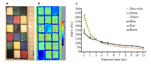
图 2 被测物彩色图和散斑伪彩图以及不同曝光时间下的PBFV
PBFV: 伪血流灌注值; A: 硅胶皮肤模型彩色图; B: 硅胶皮肤模型散斑伪彩图; C: 不同曝光时间下的PBFV.
Figure 2. The color image, speckle pseudo color image and PBFV values under different exposure time

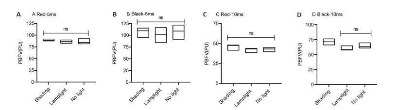
图 3 不同光线条件下的PBFV
红色表面、曝光时间5 ms (A), 黑色表面、曝光时间5 ms (B),红色表面、曝光时间10 ms (C), 黑色表面、曝光时间10 ms (D)时不同光线条件下的PBFV.
Figure 3. The PBFV of different light conditions
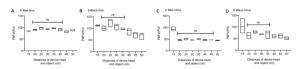
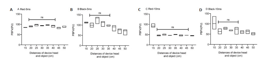
图 4 不同探头距被测物体距离下的PBFV
红色表面、曝光时间5 ms(A), 黑色表面、曝光时间5 ms (B), 红色表面、曝光时间10 ms (C), 黑色表面、曝光时间10 ms (D)时不同激光探头距被测物距离的PBFV.
Figure 4. The PBFV of different distances of the device and object
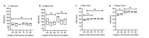
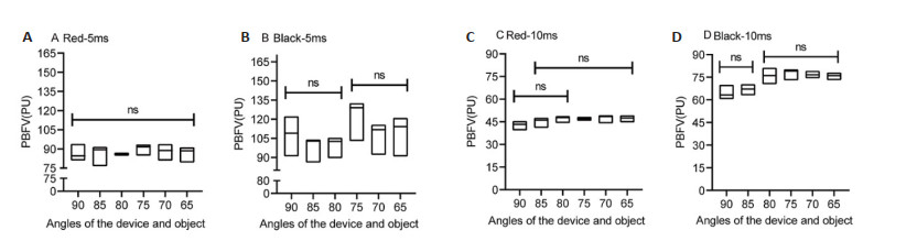
图 5 不同探头距被测物体角度下的PBFV
红色表面、曝光时间5 ms(A), 黑色表面、曝光时间5 ms (B), 红色表面、曝光时间10 ms (C), 黑色表面、曝光时间10 ms (D)时不同激光探头距被测物距离的PBFV.
Figure 5. The PBFV of different angles of the device and object
表 1 不同颜色区域及曝光时间下的伪血流灌注值
Table 1. The PBFV of different color areas and exposure times (Mean±SD)
曝光时间(ms) 肤色 绿色 黄色 蓝色 红色 黑色 1 408.4±28.8* 398.1±11.1* 417.8±6.8* 411.9±8.3* 408±16.7* 215.7±46.5 2 205±8.5* 200.8±7.2* 209.5±3.2* 204.7±3.4* 203.1±7.8* 155.7±21.4 3 137.9±5.6 137.3±6.6 143.7±4.6 141.9±4.7 140.1±5.7 137.8±16.7 4 102.9±3.2 100.7±3.2* 106±2.1 105.2±1.0 102.9±4.4 114±9.5 5 84.3±2.9* 82.7±3.8* 85.3±2.0* 85±2.1* 84.2±5.1* 101.1±8.9 6 70.9±2.2* 70.4±3.2* 71.4±1.4* 70.2±0.9* 71.1±3.7* 88.5±4.3 7 61±2.0* 62±3.2* 61.3±1.8* 60.1±1.0* 61.7±2.2* 79.6±2.6 8 53.6±1.2* 54.5±2.7* 53.3±1.9* 52.8±0.5* 54±1.6* 72.8±2.3 9 48.2±2.1* 49±2.2* 49.4±2.8* 48.5±1.6* 47.8±2.9* 69.5±5.1 10 43.1±1.2* 43.7±1.6* 45±2.6* 44.5±1.3* 43.1±2.7* 65.2±4.7 15 28.9±0.3* 29.7±0.9* 32.6±2* 31.4±1.3* 29.6±1.4* 49.4±3.4 *P < 0.05 vs黑色区域; PBFV: 伪血流灌注值.  下载: 导出CSV
-
[1] Briers JD, Webster S. Laser speckle contrast analysis (LASCA): a nonscanning, full-field technique for monitoring capillary blood flow [J]. J Biomed Opt, 1996, 1(2): 174-9. doi: 10.1117/12.231359 [2] Briers JD, Richards G, He XW. Capillary blood flow monitoring using laser speckle contrast analysis (LASCA)[J]. J Biomed Opt, 1999, 4(1): 164-75. doi: 10.1117/1.429903 [3] Li YY, Liu R, Wang Y, et al. Detecting relative speed changes of moving objects through scattering medium by using wavefront shaping and laser speckle contrast analysis[J]. Opt Express, 2016, 24 (8): 8382-90. doi: 10.1364/OE.24.008382 [4] Cutolo M, Vanhaecke A, Ruaro B, et al. Is laser speckle contrast analysis (LASCA) the new kid on the block in systemic sclerosis? A systematic literature review and pilot study to evaluate reliability of LASCA to measure peripheral blood perfusion in Scleroderma patients[J]. Autoimmun Rev, 2018, 17(8): 775-80. doi: 10.1016/j.autrev.2018.01.023 [5] Roustit M, Giai J, Gaget O, et al. On-demand sildenafil as a treatment for raynaud phenomenon: a series of n-of-1 trials[J]. Ann Intern Med, 2018, 169(10): 694-703. doi: 10.7326/M18-0517 [6] Lindahl F, Tesselaar E, Sjöberg F. Assessing paediatric scald injuries using Laser Speckle Contrast Imaging[J]. Burns, 2013, 39(4): 662-6. doi: 10.1016/j.burns.2012.09.018 [7] Reyal J, Lebas N, Fourme E, et al. Post-occlusive reactive hyperemia in basal cell carcinoma and its potential application to improve the efficacy of solid tumor therapies[J]. Tohoku J Exp Med, 2012, 227 (2): 139-47. doi: 10.1620/tjem.227.139 [8] Klijn E, Hulscher HC, Balvers RK, et al. Laser speckle imaging identification of increases in cortical microcirculatory blood flow induced by motor activity during awake craniotomy[J]. J Neurosurg, 2013, 118(2): 280-6. doi: 10.3171/2012.10.JNS1219 [9] Wu X, Li J, Joypaul K, et al. Blood flow index as an indicator of successful sciatic nerve block: a prospective observational study using laser speckle contrast imaging[J]. Br J Anaesth, 2018, 121(4): 859-66. doi: 10.1016/j.bja.2018.05.065 [10] 中国微循环学会周围血管疾病专业委员会糖尿病足学组. 糖尿病足创面修复治疗专家共识[J]. 中华糖尿病杂志, 2018, 10(5): 305-9. doi: 10.3760/cma.j.issn.1674-5809.2018.05.001 [11] Secomb TW, Pries AR. Microvascular plasticity: angiogenesis in health and disease: preface[J]. Microcirculation, 2016, 23(2): 93-4. doi: 10.1111/micc.12262 [12] Houben AJHM, Martens RJH, Stehouwer CDA. Assessing microvascular function in humans from a chronic disease perspective[J]. J Am Soc Nephrol, 2017, 28(12): 3461-72. doi: 10.1681/ASN.2017020157 [13] Beckman JA, Duncan MS, Damrauer SM, et al. Microvascular disease, peripheral artery disease, and amputation[J]. Circulation, 2019, 140(6): 449-58. doi: 10.1161/CIRCULATIONAHA.119.040672 [14] Roustit M, Millet C, Blaise S, et al. Excellent reproducibility of laser speckle contrast imaging to assess skin microvascular reactivity[J]. Microvasc Res, 2010, 80(3): 505-11. doi: 10.1016/j.mvr.2010.05.012 [15] van Vuuren TM, van Zandvoort C, Doganci S, et al. Prediction of venous wound healing with laser speckle imaging[J]. Phlebology, 2017, 32(10): 658-64. doi: 10.1177/0268355517718760 [16] 汪洋. 激光散斑衬比成像血流定量检测问题研究[D]. 武汉: 华中科技大学, 2018. [17] Mahé G, Haj-Yassin F, Rousseau P, et al. Distance between laser head and skin does not influence skin blood flow values recorded by laser speckle imaging[J]. Microvasc Res, 2011, 82(3): 439-42. doi: 10.1016/j.mvr.2011.06.014 -

 点击查看大图
计量
- 文章访问数: 702
- HTML全文浏览量: 266
- PDF下载量: 8
- 被引次数: 0