


Application value of bi-plane DSA in cerebral angiography
-
摘要:
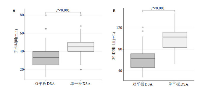
目的探讨双平板数字减影血管造影(DSA)机在全脑血管造影中的应用价值。 方法将拟行脑血管造影的患者,随机分为实验组(n=50,采用双平板DSA)和对照组(n=59,采用单平板DSA),对比两组患者的手术时间、对比剂用量、电影序列数、摄影帧数、剂量面积乘积以及空气比释动能。 结果实验组手术时间[33.50(25.00,40.50)min vs 45.00(40.00,50.00)min,P < 0.001]、对比剂用量(62.68±22.40 mL vs 100.46±20.91 mL,P < 0.001)、以及电影序列数[10.00(8.00,13.25)vs 14.00(12.00, 16.00),P < 0.001]均低于对照组,差异有统计学意义。两组电影序列数均与手术时间(r=0.586,P < 0.001)、术中对比剂用量(r=0.637,P < 0.001)成正相关。两组患者在剂量面积乘积、空气比释动能、摄影帧数的差异无统计学意义(P>0.05)。 结论采用双平板DSA机造影的辐射剂量与常规采用单平板DSA无明显差别,但使用双平板DSA造影,手术时间以及术中对比剂用量明显减少,有益于患者。 -
关键词:
- 脑血管造影 /
- 双平板数字减影血管造影机 /
- 碘对比剂 /
- 造影期并发症 /
- 辐射剂量
Abstract:ObjectiveTo analyze the application values of bi- plane digital subtraction angiography (DSA) in whole cerebral angiography. MethodsThe patients who underwent cerebral angiography were randomly divided into the experimental group (n=50, using bi-plane DSA) and the control group (n=59, using single-plane DSA). And the operation time, the amount of contrast agent, the number of movie sequences, the unmber of photographic frames, the dose area product (DAP) and air kerma (AK) of the two groups were compared. ResultsThe operation time of the experimental group [33.50 (25.00, 40.50) min vs 45.00 (40.00, 50.00) min, P < 0.001], the amount of contrast agent (62.68 ± 22.40 mL vs 100.46 ± 20.91 mL, P < 0.001), and the number of movie sequences [10.00 (8.00, 13.25) vs 14.00 (12.00, 16.00), P < 0.001] were lower than the control group. The difference is statistically significant. The two groups of patients have difference in dose area product, air kerma, and number of photographic frames. The difference was not statistically significant (P>0.05). The number of movie sequences in both groups was positively correlated with operation time (r=0.637, P < 0.001) and intraoperative contrast agent dosage (r=0.586, P < 0.001). ConclusionThere is no significant difference between the radiation dose of bi- plane DSA angiography and conventional single-plate DSA. However, the use of bi-plane DSA angiography significantly reduces the operation time and the amount of contrast agent during the operation, which is beneficial to patients. -
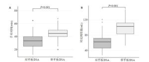
图 1 两组手术时间及对比剂用量对比
A: 手术时间; B: 对比剂用量.
Figure 1. Comparison of operation time and contrast agent dosage between the two groups
表 1 患者基本信息
Table 1. Basic information of patients
组别 男性[n (%)] 年龄(岁) BMI (kg/m2) 实验组(n=50) 23 (46.0) 42.72±14.11 22.53 (20.85, 24.70) 对照组(n=59) 26 (44.1) 50.73±14.82 23.44 (20.81, 26.44) χ2/t/Z 0.041 -2.885 -1.016 P 0.849 0.005 0.310  下载: 导出CSV
表 2 辐射剂量相关参数的比较
Table 2. Comparison of radiation dose related parameters
组别 实验组(n=50) 对照组(n=59) t/Z P 电影序列数 10.00 (8.00,13.25) 14.00(12.00,16.00) -4.480 0.001 摄影帧数(帧) 521.00 (446.75,666.25) 534.00 (459.00,630.00) -0.049 0.961 AK (mGy) 307.38 (233.85,467.57) 294.75 (230.76,339.82) -1.618 0.106 DAP (mGy·cm2) 77987.00 (57240.00,121153.50) 77110.00 (39565.00,95249.00) -1.909 0.056 AK: 空气比释动能; DAP: 剂量面积乘积.
下载: 导出CSV
-
[1] 中华医学会神经病学分会脑血管病学组缺血性脑卒中二级预防指南撰写组. 中国缺血性脑卒中和短暂性脑缺血发作二级预防指南2010 [J]. 中国医学前沿杂志: 电子版, 2011, 3(3): 84-93. https://www.cnki.com.cn/Article/CJFDTOTAL-ZLYS201111028.htm [2] 叶瑞东, 孙文, 刘新峰. 脑血管造影术操作规范中国专家共识[J]. 中华神经科杂志, 2018, 51(1): 7-13. doi: 10.3760/cma.j.issn.1006-7876.2018.01.003 [3] 朱栋梁, 卢建华, 陈胜利, 等. 脑血管介入造影检查中患者X射线辐射评价与剂量控制方法[J]. 生物医学工程与临床, 2012, 16(3): 246-9. https://www.cnki.com.cn/Article/CJFDTOTAL-SGLC201203018.htm [4] 卢贤贵. 全脑血管造影检查中患者X射线辐射评价与剂量控制方法[J]. 系统医学, 2016, 1(9): 68-70. https://www.cnki.com.cn/Article/CJFDTOTAL-XTYX201609024.htm [5] 彭方强, 檀书斌, 刘旻谛, 等. 西门子双大平板DSA机在神经介入治疗中的应用[J]. 介入放射学杂志, 2019, 28(12): 1215-8. https://www.cnki.com.cn/Article/CJFDTOTAL-JRFS201912023.htm [6] 马廉亭. 对《脑血管造影术操作规范中国专家共识》的评价与建议[J]. 中华神经科杂志, 2018, 51(9): 774-6. doi: 10.3760/cma.j.issn.1006-7876.2018.09.022 [7] Smith K, Crowhurst J, Walters D, et al. Bi-plane and single plane angiography: a study to compare contrast usage and radiation doses for adult cardiac patients in diagnostic studies[J]. Br J Radiol, 2019, 92(1093): 20180367. http://www.ncbi.nlm.nih.gov/pubmed/30209953 [8] Friedrich B, Maegerlein C, Lobsien D, et al. Endovascular stroke treatment on single-plane vs. Bi-plane angiography suites: technical considerations and evaluation of treatment success[J]. Clin Neuroradiol, 2019, 29(2): 303-9. doi: 10.1007/s00062-017-0655-z [9] 宋宾, 姜宏, 乔林, 等. 60例脑血管疾病患者脑血管造影的诊断及介入治疗[J]. 南昌大学学报: 医学版, 2016, 56(1): 70-3. https://www.cnki.com.cn/Article/CJFDTOTAL-JXYB201601018.htm [10] Alakbarzade V, Pereira AC. Cerebral catheter angiography and its complications[J]. Pract Neurol, 2018, 18(5): 393-8. doi: 10.1136/practneurol-2018-001986 [11] 杨清, 曾强军, 章东映, 等. 1123例脑血管造影中并发症分析[J]. 华西医学, 2008, 23(6): 1255-6. https://www.cnki.com.cn/Article/CJFDTOTAL-HXYX200806010.htm [12] Artico M, Spoletini M, Fumagalli L, et al. Egas Moniz: 90 years (1927-2017) from cerebral angiography[J]. Front Neuroanat, 2017, 11(9): 81. http://pubmedcentralcanada.ca/pmcc/articles/PMC5610728/ [13] Leffers AM, Wagner A. Neurologic complications of cerebral angiography[J]. Acta Radiol, 2000, 41(3): 204-10. doi: 10.1080/028418500127345299 [14] 徐剑峰, 曾令勇, 蒋正方, 等. 脑血管造影术的并发症临床分析[J]. 中国实用医药, 2011, 6(13): 19-21. https://www.cnki.com.cn/Article/CJFDTOTAL-ZSSA201113009.htm [15] American College of Radiology. ACR manual on contrast mediaversion 10.3[EB/OL]. [2019- 09- 12]. https: //www. acr. org/-/media/ ACR/Files/ClinicalResources/Contrast_Media.pdf. [16] van der Molen AJ, Reimer P, Dekkers IA, et al. Post-contrast acute kidney injury-Part 1: Definition, clinical features, incidence, role of contrast medium and risk factors[J]. Eur Radiol, 2018, 28(7): 2845- 55. doi: 10.1007/s00330-017-5246-5 [17] 朱俊, 杜倩. 碘对比剂引起急性肾损伤的相关危险因素分析[J]. 药物评价研究, 2020, 43(4): 760-4. https://www.cnki.com.cn/Article/CJFDTOTAL-YWPJ202004033.htm [18] Biondi-Zoccai G, Lotrionte M, Thomsen HS, et al. Nephropathy after administration of Iso-osmolar and low-osmolar contrast media: evidence from a network meta-analysis[J]. Int J Cardiol, 2014, 172 (2): 375-80. http://www.ncbi.nlm.nih.gov/pubmed/24502883 [19] 滕树恩. 造影剂用量与肾小球滤过率比值用于预测PCI术后造影剂肾病发生的临床意义[D]. 广州: 南方医科大学, 2016. [20] Krasinski Z, Krasińska B, Olszewska M, et al. Acute Renal Failure/ Acute Kidney Injury (AKI) Associated with Endovascular Procedures[J]. Diagnostics (Basel), 2020, 10(5): 274. http://www.researchgate.net/publication/341127514_Acute_Renal_FailureAcute_Kidney_Injury_AKI_Associated_with_Endovascular_Procedures [21] 黄卓, 范瑶华, 岳保荣, 等. 辅助防护设施对降低介入职业人员眼晶状体受照剂量的影响[J]. 中华放射医学与防护杂志, 2017, 37(6): 456-60. [22] 张文龙, 董硕, 白玫, 等. 双平板数字减影血管造影投照角度对介入医师辐射剂量影响的研究[J]. 中国医学装备, 2020, 17(3): 22-6. https://www.cnki.com.cn/Article/CJFDTOTAL-YXZB202003006.htm [23] 阿浣, 黄艺峰, 张乾营, 等. HR-VW-MRI与数字减影血管造影检查在评估脑动脉狭窄病变中的价值比较[J]. 分子影像学杂志, 2020, 43 (4): 731-4 doi: 10.12122/j.issn.1674-4500.2020.04.40 -

 点击查看大图
图(1) / 表(2)
计量
- 文章访问数: 786
- HTML全文浏览量: 248
- PDF下载量: 7
- 被引次数: 0