


Transvaginal ultrasound monitoring the development of follicles and endometrium can guide the treatment of infertility
-
摘要:
目的分析不孕症患者卵泡及子宫内膜的发育特点,为临床治疗提供准确依据。 方法选取128例不孕症患者设为观察组,128例成功妊娠女性设为对照组。比较两组月经周期第8、10、12、14天卵泡大小,同时测量并比较观察组与对照组卵泡个数、扁卵泡个数、最大卵泡长与宽度,宫体、宫颈大小及子宫内膜厚度,监测结局。 结果观察组排卵期卵泡个数、卵泡最大直径、卵泡每日增长直径及内膜厚度均低于对照组,差异有统计学意义(P < 0.05);观察组排卵正常型4例,明显低于对照组126例,差异有统计学意义(P < 0.05);观察组卵泡黄体化不破裂型、扁卵泡及无优势卵泡型分别为42例、64例及有84例均明显高于对照组2例、6例及1例,差异有统计学意义(P < 0.05)。观察组B型内膜的患者中,排卵正常型和排卵异常型分别有1例和18例,明显差于对照组125例和3例,差异有统计学意义(P < 0.05);观察组无优势卵泡的患者中,A型内膜和C型内膜分别有76例和7例,明显高于对照组0例和0例,差异有统计学意义(P < 0.05);观察组卵泡黄体化不破裂型的患者中,A型内膜和C型内膜分别有11例和15例,明显高于对照组0例和0例,差异有统计学意义(P < 0.05)。 结论不孕症患者卵泡及子宫内膜发育状况异常,经阴道超声动态监测使不孕症患者实现“早发现,早诊断,早治疗”,有效提高受孕率。 Abstract:ObjectiveTo analyze the developmental characteristics of follicles and endometrium in patients with infertility and provide precise dependence for clinical treatment. MethodsA total of 128 patients with infertility were selected and regarded as the observation group, and 128 women with successful pregnancy were regarded as the control group. The monitoring cycle was from the 8th day of the menstrual cycle to the ovulation period for 6 consecutive cycles. The differences in the monitoring results between the two groups were compared. ResultsCompared with the control group, the number of ovulation follicles, the maximum diameter of follicles, the daily growth diameter of follicles and the thickness of endometrium were all lower in the observation group, and the differences were statistically significant (P < 0.05). In the control group, there were 4 cases with normal ovulation type, which were significantly lower than 126 cases in the control group, and the differences were statistically significant (P < 0.05). In the observation group, there were 42 cases, 64 cases and 84 cases with luteinized unruptured follicles, flat follicles and non-dominant follicles respectively, which were significantly higher than 2 cases, 6 cases and 1 case in the control group, and the differences were statistically significant (P < 0.05). Among patients with type B endometrium in the observation group, there were 1 case and 18 cases with normal ovulation type and abnormal ovulation type, which were significantly lower than 125 cases and 3 cases in the control group, and the difference was statistically significant (P < 0.05). Among the patients with non-dominant follicles in the observation group, there were 76 cases and 7 cases with type A endometrium and type C endometrium, which were significantly higher than 0 cases and 0 cases in the control group, and the difference was statistically significant (P < 0.05); Among the patients with luteinized unruptured follicle type in the observation group, there were 11 cases and 15 cases with type A endometrium and type C endometrium, which were significantly higher than 0 cases and 0 cases in the control group, and the differences were statistically significant (P < 005). ConclusionThe developmental characteristics of follicles and endometrium in patients with infertility are abnormal. Realizing "early detection, early diagnosis, and early treatment" by transvaginal ultrasound dynamic monitoring for patients with infertility can effectively increase the pregnancy rate. -
Key words:
- transvaginal ultrasound /
- infertility /
- follicles /
- endometrium
-
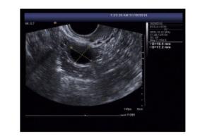
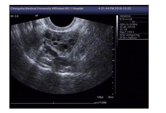
图 1 成熟卵泡超声表现图
对照组患者, 女, 30岁, 可见优势卵泡, 大小为19.7 mm×17.2 mm, 卵泡壁薄, 光滑, 张力好, 呈圆形无回声区, 形态饱满.
Figure 1. The ultrasonographic images of mature follicles
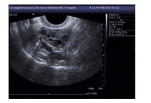
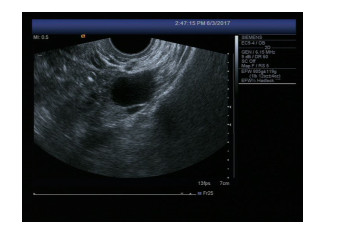
图 2 无优势卵泡型超声表现图
对照组患者, 女, 33岁, 左侧卵巢内最大卵泡, 大小为10 mm×6 mm, 呈小圆形无回声区, 卵泡壁厚且不规则, 张力偏低.
Figure 2. The ultrasonographic images of immature follicles
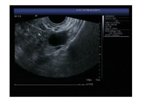
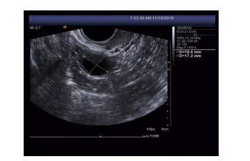
图 3 卵泡黄体化不破裂型超声表现图
观察组患者, 女, 32岁, 排卵期后优势卵卵泡存在, 呈椭圆形无回声区, 壁薄, 张力大, 大小为37 mm×34 mm, 子宫直肠陷凹未见液性暗区.
Figure 3. The ultrasonographic images of Luteinized Unruptured Follicles
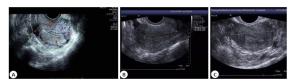
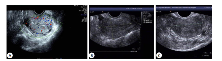
图 4 A型内膜(A)、B型内膜(B)及C型内膜(C)的超声表现图
Figure 4. The ultrasonographic images of membrane of Type A(A), membrane of Type B (B) and membrane of Type C (C).
表 1 两组卵泡发育情况比较
Table 1. Comparison of normal and abnormal follicle development between groups (n=128, Mean±SD)
组别 排卵正常型 卵泡黄体化不破裂型 扁卵泡型 无优势卵泡型 卵泡每日增长直径(mm) 卵泡个数 卵泡最大直径(mm) 观察组 4 42 64 84 0.84±0.05 4.88±0.32 14.35±2.34 对照组 126 2 6 1 2.35±0.07 5.92±0.55 20.29±3.28 t/χ2 232.619 43.911 66.143 121.334 198.594 8.392 16.679 P 0.000 0.000 0.000 0.000 0.000 0.002 0.000  下载: 导出CSV
表 2 两组子宫内膜厚度和子宫大小比较
Table 2. Comparison of endometrial thickness and uterine size between two groups (mm, Mean±SD)
组别 子宫内膜厚度 子宫大小 观察组 8.54±1.32 46.24±8.34 对照组 6.32±1.03 55.35±8.63 t 5.385 2.192 P 0.032 0.073
下载: 导出CSV
表 3 两组卵泡情况综合内膜分型的比较
Table 3. Comparison of Follicle Condition and Endometrial Type between two groups
组别 排卵正常型(B型内膜) 排卵异常(B型内膜) 无优势卵泡型(A型内膜) 无优势卵泡型(C型内膜) 卵泡黄体化不破裂型(A型内膜) 卵泡黄体化不破裂型(C型内膜) 观察组 1 18 76 7 11 15 对照组 125 3 0 0 0 0 χ2 240.309 11.672 108.089 7.197 11.494 15.934 P 0.000 0.001 0.000 0.014 0.001 0.000
下载: 导出CSV
-
[1] Practice Committee of the american society for reproductive medicine. Definitions of infertility and recurrent pregnancy loss: a committee opinion[J]. Fertil Steril, 2013, 99(1): 63. doi: 10.1016/j.fertnstert.2012.09.023 [2] Casu G, Gremigni P. Screening for infertility-related stress at the time of initial infertility consultation: psychometric properties of a brief measure[J]. JAdv Nurs, 2016, 72(3): 693-706. doi: 10.1111/jan.12830 [3] 张怡. 经阴道超声监测卵泡发育在不孕患者中应用的价值研究[J]. 影像研究与医学应用, 2019, 3(6): 22-3. doi: 10.3969/j.issn.2096-3807.2019.06.012 [4] 冯敏芝, 伍诗媚, 李建聪. 超声在妊娠合并卵巢肿瘤患者的诊断价值[J]. 分子影像学杂志, 2019, 42(4): 439-43. doi: 10.12122/j.issn.1674-4500.2019.04.04 [5] 熊敏, 周志红. 经阴道超声卵泡监测在治疗不孕不育中的价值分析[J]. 母婴世界, 2019, 2(16): 44. https://www.cnki.com.cn/Article/CJFDTOTAL-ZYCX201816039.htm [6] 柴小霞. 经阴道彩超在不孕症患者卵泡监测中的应用价值[J]. 影像研究与医学应用, 2020, 4(2): 134-5. https://www.cnki.com.cn/Article/CJFDTOTAL-YXYY202002087.htm [7] Boivin J, Bunting L, Collins JA, et al. International estimates of infertility prevalence and treatment-seeking: potential need and demand for infertility medical care[J]. Hum Reprod, 2007, 22(6): 1506-12. doi: 10.1093/humrep/dem046 [8] 高蜀君, 隋龙. 宫腔病变与不孕症的关系及诊治进展[J]. 国际生殖健康/计划生育杂志, 2016, 35(3): 237-40. https://www.cnki.com.cn/Article/CJFDTOTAL-GWJS201603016.htm [9] 林小琼, 观志强, 谢瑞娜. 评估经阴道超声检查对女性不孕症患者卵泡发育及排卵的价值[J]. 广州医科大学学报, 2016, 44(1): 68-70. https://www.cnki.com.cn/Article/CJFDTOTAL-GZXI201601018.htm [10] 易芳, 周天志. 经腹部联合经阴道彩色多普勒超声可提高子宫肌瘤诊断的准确率[J]. 分子影像学杂志, 2020, 43(3): 512-5. doi: 10.12122/j.issn.1674-4500.2020.03.30 [11] Yue G, Deng Y, Yang S, et al. Early Doppler ultrasound in the superior mesenteric artery and the prediction of necrotizing enterocolitis in preterm neonates[J]. J Ultrasound Med, 2019, 38 (12): 3283-9. doi: 10.1002/jum.15064 [12] 何楠. 经阴道超声监测卵泡发育在不孕症治疗中的指导价值分析[J]. 中国冶金工业医学杂志, 2015, 32(6): 671-2. https://www.cnki.com.cn/Article/CJFDTOTAL-ZGYJ201506042.htm [13] Titus S, Szymanska KJ, Musul B, et al. Individual-oocyte transcriptomic analysis shows that genotoxic chemotherapy depletes human primordial follicle reserve in vivo by triggering proapoptotic pathways without growth activation[J]. Sci Rep, 2021, 11(1): 407. doi: 10.1038/s41598-020-79643-x [14] 王海英. 经阴道彩色超声检查对监测卵泡生长及指导不孕症治疗的临床价值分析[J]. 中国当代医药, 2020, 27(34): 114-7. doi: 10.3969/j.issn.1674-4721.2020.34.034 [15] 徐颖, 许小凤. 卵泡发育障碍的中西医研究进展[J]. 实用医学杂志, 2013, 29(6): 1009-11. doi: 10.3969/j.issn.1006-5725.2013.06.059 [16] 张苗, 汪彩英, 李霞, 等. 子宫内膜分型与厚度对不孕患者促排卵治疗后妊娠率的影响[J]. 中国性科学, 2016, 25(3): 118-20. https://www.cnki.com.cn/Article/CJFDTOTAL-XKXZ201603042.htm [17] Ruan X, Li M, Mueck AO. Why does polycystic ovary syndrome (PCOS) need long-term management?[J]. Curr Pharm Des, 2018, 24 (39): 4685-92. http://www.researchgate.net/publication/330787335_why_polycystic_ovary_syndrome_pcos_needs_long-term_management [18] Costello MF, Garad RM, Hart R, et al. A review of second- and thirdline infertility treatment s and supporting evidence in women with policy sticovary syndrome[J]. MedSci(Basel), 2019, 7(7): 75. http://www.researchgate.net/publication/334043886_A_Review_of_Second-_and_Third-line_Infertility_Treatments_and_Supporting_Evidence_in_Women_with_Polycystic_Ovary_Syndrome [19] Senturk LM, Erel CT. Thinen domet riuminassis tedreprod uctivete chnology[J]. Curr Opin Obstet Gynecol, 2009, 20(3): 221-8. [20] Kunicki M, Łukaszuk K, Woclawek-Potocka I, et al. Evaluation of granulocyte colony-stimulating factor effects on treatment-resistant thin endometrium in women undergoing in vitro fertilization[J]. Biomed Res Int, 2014, 2014: 913235. http://europepmc.org/articles/pmc3944906/ [21] Bakas P, Hassiakos D, Grigoriadis C, et al. Role of hysteroscopy prior to assisted reproduction techniques[J]. J Minim Invasive Gynecol, 2014, 21(2): 233-7. doi: 10.1016/j.jmig.2013.07.023 -

 点击查看大图
计量
- 文章访问数: 1135
- HTML全文浏览量: 409
- PDF下载量: 7
- 被引次数: 0