


Up-regulation factors of BI-RADS 4 breast nodules by two-dimensional gray-scale ultrasound
-
摘要:
目的分析二维灰阶超声对BI-RADS 4类乳腺结节评估上调的原因,提高超声识别乳腺良性结节的能力,减少过度诊断与治疗。 方法回顾性分析我院2019年12月~2020年11月因乳腺结节住院患者380例,依据病理结果将超声BI-RADS 4类乳腺结节分为2组,结节上调组及结节一致组。将乳腺结节为良性但超声BI-RADS 4类结节认为结节上调组,其中女性68例,男性2例,年龄26~76岁(48.43±11.52岁);将乳腺结节为恶性且超声BI-RADS 4类结节认为结节一致组,其中女性310例,男性0例,年龄26~87岁(52.84±11.28岁)。分析两组声像图征象,总结超声对BI-RADS 4类乳腺结节评估上调的原因,进行鉴别诊断。 结果380例患者中,结节上调组70例(18.42%),结节一致组310例(81.58%)。结节上调组中乳腺纤维腺瘤13例(18.57%),导管内乳头状瘤12例(17.14%),乳腺腺病7例(10.00%),非哺乳期乳腺炎5例(7.14%),良性叶状肿瘤3例(4.29%),男性乳腺发育2例(2.86%),乳腺小叶萎缩1例(1.43%),2种以上良性结节并发27例(38.57%);结节一致组中乳腺癌310例(100%)。超声征象:结节上调组中边界清晰,纵横比 < 1,边缘光整或呈大分叶状,粗大钙化多见,结节一致组中边界不清晰,纵横比>1,边缘成角或微小分叶状,微钙化多见,2组超声征象之间差异有统计学意义(P < 0.05)。 结论正确识别超声乳腺结节良、恶性征象,减少主观性诊断,能有效提高超声诊断良性结节准确率,提高诊断自信。 Abstract:ObjectiveTo explore the reasons of up-regulation of BI- RADS 4 breast nodules by two-dimensional gray-scale ultrasound and improve the ability of ultrasound to identify benign breast nodules so as to reduce over-diagnosis and treatment. MethodsA total of 380 cases of breast nodules hospitalized in our hospital from December 2019 to November 2020 were analyzed retrospectively. According to the pathological results, the ultrasound BI-RADS 4 breast nodules were divided into two groups: the nodule up-regulation group and the nodule consistent group. Breast nodules are benign, but ultrasound BI-RADS 4 nodules were considered as up-regulation group, including 68 females and 2 males, aged from 26 to 76 years, with an average age of 48.43±11.52 years. The breast nodules were considered as malignant and the ultrasound BI-RADS 4 types were consistent, including 310 females and 0 males, aged from 26 to 87 years, with an average of 52.84 ± 11.28 years. The ultrasonographic features of the two groups were analyzed, and the reasons for the up-regulation of ultrasound evaluation of BI-RADS 4 breast nodules were summarized for differential diagnosis. ResultsFrom December 2019 to November 2020, there were 380 patients (100.00%) with Bi-RADS type 4 nodules, including 70 patients (18.42%) with up-regulated nodules and 310 patients (81.58%) with consistent nodules. There were 13 breast fibroadenomas (18.57%), 12 intraductal papillomas (17.14%), 7 breast adenopathy (10.00%), 5 non-lactation mastitis (7.14%), 3 benign phyllodes tumors (4.29%), 2 male breast development (2.86%), and 1 breast lobular atrophy(1.43%), 27 cases with more than two kinds of benign nodules. A total of 310 cases (100%) of breast cancer were in the same nodule group. Ultrasonographic signs: in the up-regulated nodule group, the boundary was clear, the aspect ratio was less than 1, the edge was smooth or lobulated, and coarse calcification was more common. In the consistent nodule group, the boundary was unclear, the aspect ratio was more than 1, the edge was angular or lobulated, and microcalcification was more common. There was significant difference between the two groups (P < 0.05). ConclusionCorrect identification of benign and malignant signs of ultrasound breast nodules and reduction of subjective diagnosis can effectively improve the accuracy of ultrasound benign nodules and improve diagnostic confidence. -
Key words:
- breast /
- benign nodules /
- ultrasonic sign
-
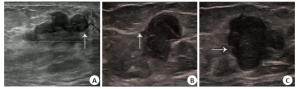
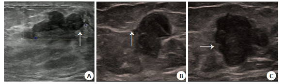
图 1 结节上调组及结节一致组超声征象
A: 纤维腺瘤呈大分叶状; B: 乳腺癌成角; C: 乳腺癌呈微小分叶.
Figure 1. Ultrasound signs of up-regulated nodules and consistent nodules.
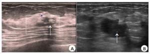
图 2 结节上调组及结节一致组超声征象
A: 纤维腺瘤伴有粗大钙化; B: 乳腺癌伴有微钙化.
Figure 2. Ultrasound signs of up-regulated nodules and consistent nodules.
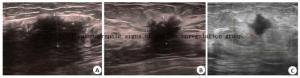
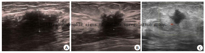
图 3 结节上调组超声征象
A: 乳腺小叶萎缩边界不清; B: 乳腺腺病边界不清; C: 乳腺炎边界不清.
Figure 3. Ultrasonographic signs of nodular upregulation group.
表 1 结节上调组病理结果例数(例)及占比
Table 1. The number of pathological results and the proportion in the up-regulation group of nodules
结节上调组病理结果 例数 占比(%) 导管内乳头状瘤 12 17.14 乳腺腺病 7 10.00 乳腺纤维腺瘤 13 18.57 乳腺小叶萎缩 1 1.43 非哺乳期乳腺炎 5 7.14 良性叶状肿瘤 3 4.29 男性乳腺发育 2 2.86 2种以上良性结节并发 27 38.57 总数 70 100.00  下载: 导出CSV
表 2 结节上调组及结节一致组声像图征象例数
Table 2. Number of Sonographic Signs in Nodule Up-regulated Group and Nodule Consistent Group (n)
超声征象 结节上调组(n=70) 结节一致组(n=310) χ2 P 边界 139.74 < 0.001 清晰 46 20 不清晰 24 290 纵横比 31.03 < 0.001 > 1 5 206 < 1 65 104 边缘 200.99 < 0.001 大分叶 18 84 微小分叶及(或)成角 7 220 光整 45 6 钙化 26.52 < 0.001 粗大钙化 17 33 微钙化 3 100 无钙化 50 177
下载: 导出CSV
-
[1] Feng RM, Zong YN, Cao SM, et al. Current cancer situation in China: good or bad news from the 2018 Global Cancer Statistics?[J]. Cancer Commun, 2019, 39(1): 22. doi: 10.1186/s40880-019-0368-6 [2] 何明艳, 朱碧琪, 钟媛, 等. 2005—2013年中国女性乳腺癌发病及死亡趋势分析[J]. 中华疾病控制杂志, 2019, 23(1): 10-4. https://www.cnki.com.cn/Article/CJFDTOTAL-JBKZ201901003.htm [3] 蒋天安, 陈文, 罗葆明, 等. 乳腺超声若干临床常见问题专家共识(2018版)[ J]. 中国超声医学杂志, 2018, 34(10): 865-70. doi: 10.3969/j.issn.1002-0101.2018.10.001 [4] D'Orsi CJ, Sickles EA, Mendelson EB, et al. ACR BI-RADS atlas, breast imaging reporting and data system. 5th ed[M]. Reston: American College of Radiology, 2013. [5] 刘晶焰, 彭玉兰. 乳腺超声若干临床常见问题专家共识(2018版)解读[J]. 西部医学, 2019, 31(9): 1319-23. doi: 10.3969/j.issn.1672-3511.2019.09.002 [6] 钟建宏, 张春, 罗海兴. 二维及彩超在乳腺良恶性肿块鉴别诊断中的价值评价[J]. 影像研究与医学应用, 2018, 2(11): 169-70. doi: 10.3969/j.issn.2096-3807.2018.11.110 [7] 李易凤, 陈武, 刘晓芳, 等. 剪切波弹性成像在乳腺结节BI-RADS 3、4a类中的应用价值[J]. 中国超声医学杂志, 2020, 36(7): 613-6. doi: 10.3969/j.issn.1002-0101.2020.07.013 [8] 贾怡婷. 超声造影在乳腺良恶性结节鉴别诊断中的应用[J]. 影像研究与医学应用, 2020, 4(3): 70-1. https://www.cnki.com.cn/Article/CJFDTOTAL-YXYY202003041.htm [9] 叶细容, 刘佳. BI-RADS联合超声弹性成像对非哺乳期乳腺炎及乳腺癌的鉴别价值[J]. 医疗卫生装备, 2021, 42(2): 56-9, 77. https://www.cnki.com.cn/Article/CJFDTOTAL-YXQY201807011.htm [10] 王小燕, 韦海明, 蓝春勇, 等. 乳腺导管乳头状瘤的超声图像与超声造影对照分析[J]. 中国超声医学杂志, 2011, 27(3): 213-6. doi: 10.3969/j.issn.1002-0101.2011.03.008 [11] Cheng L, Reddy V, Solmos G, et al. Mastitis, a radiographic, clinical, and histopathologic review[J]. Breast J, 2015, 21(4): 403-9. doi: 10.1111/tbj.12430 [12] 贾晓红, 詹维伟, 周建桥, 等. 非哺乳期乳腺炎超声和MRI表现特征[J]. 中华医学超声杂志: 电子版, 2019, 16(12): 943-8. doi: 10.3877/cma.j.issn.1672-6448.2019.12.011 [13] 朱束华, 范晓芳, 李卫民. 纵横比大于1乳腺良、恶性病灶的超声、病理及临床特征对比[J]. 中外医学研究, 2020, 18(35): 76-8. https://kns.cnki.net/KCMS/detail/detail.aspx?dbcode=CJFD&filename=YJZY202035029 [14] 陈园园, 韩宝三, 王丽君, 等. 无乳头溢液导管内乳头状瘤的超声图像特征分析[J]. 临床超声医学杂志, 2021, 23(2): 152-4. doi: 10.3969/j.issn.1008-6978.2021.02.024 [15] 左晓林, 陈成生, 胡伟, 等. 彩色多普勒超声联合乳腺钼靶对乳腺导管原位癌诊断的临床应用价值[J]. 影像研究与医学应用, 2020, 4 (23): 244-6. doi: 10.3969/j.issn.2096-3807.2020.23.132 [16] 栾丽霞. 乳腺钙化对超声诊断乳腺良恶性病变的临床意义[J]. 临床医药文献电子杂志, 2018, 5(86): 174. doi: 10.3877/j.issn.2095-8242.2018.86.149 [17] 康莉, 郁玉文, 李卫民, 等. 超声在乳腺内钙化灶诊断中的作用[J]. 北华大学学报: 自然科学版, 2019, 20(3): 358-60. https://www.cnki.com.cn/Article/CJFDTOTAL-ZLYY201903016.htm -

 点击查看大图
计量
- 文章访问数: 850
- HTML全文浏览量: 452
- PDF下载量: 7
- 被引次数: 0