


Correlation of dietary inflammatory index and carotid plaque enhancement by contrastenhanced ultrasonography
-
摘要:
目的探讨超声造影对膳食炎症指数(DII)与颈动脉斑块内增强特征的相关性。 方法选取2015年3月~2019年10月在我院行颈动脉斑块复查的77例患者作为研究对象。采用食物频率问卷表调查患者的日常饮食数据,应用膳食营养软件计算食物的膳食成分/营养素的DII指数。根据患者DII指数分组,即DII指数为正数纳入促炎饮食组(n=35),DII指数为负数纳入抗炎饮食组(n=42);并根据患者DII指数划分5个等级,即≤-1.05、-1.05~-0.33、-0.33~0.38、0.38~1.22、≥1.22。采用彩色多普勒超声检测患者颈动脉斑块,对其斑块厚度、回声类型、斑块造影剂分布特征进行分析。双变量相关性分析采用Kendall相关系数法检验。 结果促炎饮食组35例患者共41枚斑块,抗炎饮食组42例患者共47枚斑块。促炎饮食组的斑块厚度较抗炎饮食组的增厚,差异有统计学意义(P < 0.05);促炎饮食组超声造影增强多于抗炎饮食组,差异有统计学意义(P < 0.05)。DII指数与颈动脉斑块内膜有无增强呈显著性正相关(Kendall相关系数为0.659,P < 0.001)。 结论促炎饮食患者的颈动脉斑块较厚,DII指数越高,颈动脉斑块斑块内增强特征越明显。 Abstract:ObjectiveTo explore the correlation between dietary inflammatory index (DII) and enhancement characteristics of carotid plaque by contrast-enhanced ultrasound. MethodsFrom March 2015 to October 2019, 77 patients with carotid plaque reexamined in our hospital were selected as the study objects. Food frequency questionnaire was used to collect the data of patients' daily diet, The DiI index of dietary components/nutrients of food was calculated by dietary nutrition software. According to the DiI index of patients, the positive DiI index (35 cases) was included in the anti-inflammatory diet group, and the negative DiI index (42 cases) was included in the anti-inflammatory diet group. According to the DiI index of patients, 5 grades were divided, That is ≤-1.05, -1.05~-0.33, -0.33~0.38, 0.38~ < 1.22 and ≥1.22. Carotid plaque was detected by color Doppler ultrasound. The plaque thickness, echo type and the distribution of plaque contrast agent were analyzed. Kendall correlation coefficient method was used to test the bivariate correlation analysis. ResultsThere were 41 plaques in 35 patients in the pro-inflammatory diet group and 47 plaques in 42 patients in the anti-inflammatory diet group. Inflammation promoting diet group of plaque thickness relatively anti-inflammatory diet group of thickening. The difference was statistically significant (P < 0.05). Inflammation promoting diet group contrast enhanced ultrasound enhance many to antiinflammatory diet group. The difference was statistically significant (P < 0.05). There was a significant positive correlation between DiI index and carotid plaque intima enhancement (Kendall correlation coefficient 0.659, P < 0.001). ConclusionsCarotid plaques are quite thick in patients with pro-inflammatory diet. The higher the DiI index, the more obvious the enhancement characteristics of carotid plaque. -
Key words:
- contrast-enhanced ultrasound /
- dietary inflammation index /
- carotid plaque
-

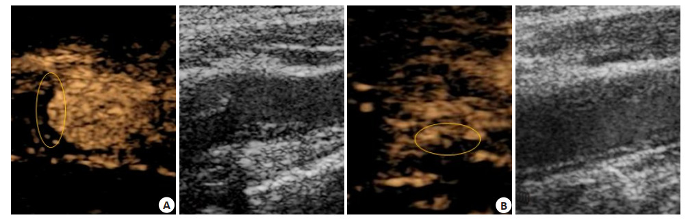
图 1 颈动脉斑块CEUS图像
A: 促炎饮食组患者, 男, 66岁, 颈动脉分叉处斑块造影2级增强, 斑块内弥漫性增强(圈处); B:抗炎饮食组患者, 男, 67岁, 颈动脉分叉处斑块造影1级 增强, 近基底部点状增强(圈处)
Figure 1. Contrast-enhanced ultrasound image of carotid plaque
表 1 促炎饮食组与抗炎饮食组的一般资料对比[n(%), Mean±SD]
Table 1. Comparison of general data between the pro-inflammatory diet group and the anti-inflammatory diet group
组别 性别(男/女) 年龄(岁) BMI(kg/m2) 血糖(mmol/L) 舒张压(mmHg) 收缩压(mmHg) 促炎饮食组(n=35) 30/5 62.89± 9.17 24.58土2.09 5.31±0.76 76.98± 7.47 121.47±6.98 抗炎饮食组(n=42) 34/8 64.79± 8.96 24.63± 2.12 5.16 ± 0.73 77.45土7.22 122.75±7.16 t 0.308 0.916 0.103 0.881 0.280 0.790 P 0.579 0.362 0.917 0.381 0.780 0.432  下载: 导出CSV
表 2 两组斑块厚度及回声类型比较[n(%)]
Table 2. Comparison of patch thickness and echo type between two groups
组别 斑块数(n) 斑块厚度(mm,Mean±SD) 回声类型 混合回声 等回声 低回声 促炎饮食组(n=35) 41 4.12±1.03 27(65.85) 9(21.95) 5(12.20) 抗炎饮食组(n=42) 47 3.69±0.95 26(55.32) 17(36.17) 4(8.51) t/χ2 2.037 2.193 P 0.045 0.334
下载: 导出CSV
表 3 两组CEUS检查结果比较[n(%)]
Table 3. Comparison of contrast-enhanced ultrasonography results between two groups
组别 斑块数(n) 0级 1级 2级 促炎饮食组(n=35) 41 2(4.88) 17(41.46) 22(53.66) 抗炎饮食组(n=42) 47 7(14.89) 27(57.45) 13(27.66) t/χ2 2.392 2.238 6.179 P 0.122 0.135 0.013
下载: 导出CSV
表 4 DII指数与颈动脉斑块内增强的相关性分析(n)
Table 4. CorrelationAnalysis of DiI index and carotid plaque internal enhancement
DII指数等级(分) 颈动脉斑块内增强 合计 + - ≤-1.05 2 26 28 -1.05~-0.33 4 13 17 -0.33~0.38 5 10 15 0.38~1.22 11 3 14 ≥1.22 13 1 14 合计 35 53 88
下载: 导出CSV
-
[1] 阚艳敏, 何文, 宁彬, 等.颈动脉斑块内钙化分布特征对斑块稳定性的影响[J].中国动脉硬化杂志, 2020, 28(2): 128-33. http://www.wanfangdata.com.cn/details/detail.do?_type=perio&id=zgdmyhzz202002008 [2] Zhou MG, Wang HD, Zeng XY, et al. Mortality, morbidity, and risk factors in China and its Provinces, 1990-2017: a systematic analysis for the Global Burden of Disease Study 2017[J]. Lancet, 2019, 394 (10204): 1145-58. https://pubmed.ncbi.nlm.nih.gov/31248666/ [3] 张广俊, 黄圣奇, 宋秀莲.超声微血管成像与超声造影评价颈动脉斑块内新生血管的比较[J].中国超声医学杂志, 2019, 35(12): 1066-9. http://med.wanfangdata.com.cn/Paper/Detail?id=PeriodicalPaper_zgcsyxzz201912004 [4] Wang WZ, Jiang B, Sun HX, et al. Prevalence, incidence, and mortality of stroke in China: results from a nationwide populationbased survey of 480 687 adults[J]. Circulation, 2017, 135(8): 759-71. http://europepmc.org/abstract/MED/28052979 [5] Howard DP, van Lammeren GW, Rothwell PM, et al. Symptomatic carotid atherosclerotic disease: correlations between plaque composition and ipsilateral stroke risk[J]. Stroke, 2015, 46(1): 182-9. https://www.ncbi.nlm.nih.gov/pmc/articles/PMC4285579/ [6] Ricordi C, Garcia-Contreras M, Farnetti S. Diet and inflammation: possible effects on immunity, chronic diseases, and life span[J]. J Am Coll Nutr, 2015, 34(Suppl 1): 10-3. doi: 10.1080/07315724.2015.1080101 [7] 黄海彬, 赵秋革, 方晓涛, 等.膳食纤维及常见危险因素对广州社区脑梗死发病的影响[J].热带医学杂志, 2019, 19(1): 60-4, 2. http://www.cnki.com.cn/Article/CJFDTotal-RDYZ201901015.htm [8] 宁彬, 何文, 张东, 等.斑块内新生血管分布特征的超声造影与病理对照[J].中国医学影像技术, 2015, 31(5): 655-8. http://www.cnki.com.cn/Article/CJFDTotal-ZYXX201505006.htm [9] 黄莉莉, 罗小铭, 谈晔, 等.广州地区食物频率问卷信度和效度研究[J].中华疾病控制杂志, 2013, 17(8): 711-4. http://med.wanfangdata.com.cn/Paper/Detail/PeriodicalPaper_jbkzzz201308017 [10] 《营养学报》编辑部.《中国食物成分表》标准版第6版第一二册出版[J].营养学报, 2019, 41(5): 426. [11] Shivappa N, Steck SE, Hurley TG, et al. Designing and developing a literature-derived, population-based dietary inflammatory index[J]. Public Health Nutr, 2014, 17(8): 1689-96. https://pubmed.ncbi.nlm.nih.gov/23941862/ [12] Kuk M, Wannarong T, Beletsky V, et al. Volume of carotid artery ulceration as a predictor of cardiovascular events[J]. Stroke, 2014, 45 (5): 1437-41. doi: 10.1161/strokeaha.114.005163 [13] 王萍, 周成礼.颈动脉超声相关指标及动态动脉硬化指数在冠心病风险预测中的意义[J].分子影像学杂志, 2017, 40(3): 284-7. doi: 10.3969/j.issn.1674-4500.2017.03.10 [14] 王言憬, 勇强, 刘欣, 等.冠状动脉病变程度与颈动脉粥样硬化斑块易损性相关性研究[J].中国超声医学杂志, 2019, 35(7): 601-4. http://www.cnki.com.cn/Article/CJFDTotal-ZGCY201907008.htm [15] 金鑫, 侯秀昆, 杨光.超声联合超声造影评价颈动脉斑块稳定性的价值研究[J].大连医科大学学报, 2019, 41(6): 487-91. http://www.cnki.com.cn/Article/CJFDTotal-DLYK201906004.htm [16] 刘莉, 叶鹏, Bondonno CP, 等.老年女性蔬菜硝酸盐的摄入与颈动脉粥样硬化和缺血性脑血管病的相关性[J].中华高血压杂志, 2017, 25(9): 887. http://www.cnki.com.cn/Article/CJFDTOTAL-ZGGZ201709030.htm [17] 张静波, 武志远, 刘相佟, 等.北京市功能单位在职人员饮食行为对血脂异常的影响研究[J].中国全科医学, 2018, 21(24): 2993-6. http://www.cqvip.com/QK/91169X/201824/7000768225.html [18] 李华, 卢吴柱, 程林, 等.饮食习惯对高血压患者动脉粥样硬化的影响[J].分子影像学杂志, 2018, 41(2): 264-7. doi: 10.3969/j.issn.1674-4500.2018.02.30 [19] Giebe S, Cockcroft N, Hewitt K, et al. Cigarette smoke extract counteracts atheroprotective effects of high laminar flow on endothelial function[J]. Redox Biol, 2017, 12: 776-86. https://www.sciencedirect.com/science/article/pii/S2213231717301064 [20] Behradmanesh S, Nasri P. Serum cholesterol and LDL-C in association with level of diastolic blood pressure in type 2 diabetic patients[J]. J Renal Inj Prev, 2012, 1(1): 23-6. https://www.ncbi.nlm.nih.gov/pubmed/25340098 [21] 薛万华, 杨婷.氧化应激与自噬在动脉粥样硬化发生发展中的研究进展[J].分子影像学杂志, 2019, 42(1): 95-8. doi: 10.12122/j.issn.1674-4500.2019.01.22 [22] 陈丽, 王凤娇, 薛雅卓, 等.颈动脉粥样硬化影响内皮祖细胞功能的研究进展[J].中国动脉硬化杂志, 2018, 26(4): 419-23. http://www.cqvip.com/QK/98074X/201804/675385550.html [23] Rafailidis V, Charitanti A, Tegos T, et al. Contrast-enhanced ultrasound of the carotid system: a review of the current literature[J]. J Ultrasound, 2017, 20(2): 97-109. doi: 10.1007%2Fs40477-017-0239-4 [24] Wang Q, Liu LH, Li YY, et al. Hypoxic preconditioning enhances biological function of endothelial progenitor cells via notch-Jagged1 signaling pathway[J]. Med Sci Monit, 2017, 23: 4665-7. https://www.ncbi.nlm.nih.gov/pubmed/28959004 [25] Huang RQ, Abdelmoneim SS, Ball CA, et al. Detection of carotid atherosclerotic plaque neovascularization using contrast enhanced ultrasound: a systematic review and meta- analysis of diagnostic accuracy studies[J]. JAm Soc Echocardiogr, 2016, 29(6): 491-502. https://www.sciencedirect.com/science/article/pii/S0894731716001267 [26] Medina-Remón A, Casas R, Tressserra-Rimbau A, et al. Polyphenol intake from a Mediterranean diet decreases inflammatory biomarkers related to atherosclerosis: a substudy of the PREDIMED trial[J]. Br J Clin Pharmacol, 2017, 83(1): 114-28. doi: 10.1111/bcp.12986 -

 点击查看大图
计量
- 文章访问数: 577
- HTML全文浏览量: 265
- PDF下载量: 3
- 被引次数: 0