


Value of endoscopic ultrasound in the selection of early gastric cancer invasion depth and guiding treatment methods
-
摘要:
目的探讨内镜超声对早期胃癌浸润深度及指导治疗方法选择中的应用价值。 方法选取2019年1月~2020年8月在我院接受内镜超声检查的早期胃癌患者作为观察组(n=80),同时选取未接受内镜超声检查的早期胃癌患者作为对照组(n=60),比较内镜超声与病理结果情况,分析内镜超声诊断Tis/T1a、T1b的价值,比较观察组和对照组治愈性切除比例的差异。 结果观察组Tis/T1a、T1b比例分别为86.23%和13.75%,与对照组差异无统计学意义(P>0.05);内镜超声与病理结果一致性Kappa值为0.634(P < 0.05),内镜超声诊断准确率为90.00%,其中有2例T1b误诊为Tis/T1a,有6例Tis/T1a误诊为T1b,内镜超声诊断Tis/T1a的灵敏性、特异性、阳性预测值和阴性预测值分别为91.30%、81.82%、96.92%和60.00%;内镜超声诊断T1b的灵敏性、特异性、阳性预测值和阴性预测值分别为81.82%、91.30%、60.00%和96.92%;观察组治愈性切除比例高于对照组,差异有统计学意义(P < 0.05)。 结论内镜超声在早期胃癌浸润深度诊断中有较高的价值,有助于指导医师对切除范围的了解,提高治疗效果。 Abstract:ObjectiveTo investigate the application value of endoscopic ultrasonography in the depth of invasion and treatment of early gastric cancer. MethodsEighty patients with early gastric cancer who underwent endoscopic ultrasonography in our hospital from January 2019 to August 2020 were selected as the observation group, and 60 patients with early gastric cancer who did not receive endoscopic ultrasonography were selected as control group. The results of endoscopic ultrasonography and pathology were compared, the value of endoscopic ultrasonography in the diagnosis of tis/T1a and T1b were analyzed. And the difference of curative incision were compared between the observation group and the control group. ResultsThe ratio of tis/T1a and T1b in the observation group was 86.23% and 13.75%, respectively. Comparing with the control group, the difference was not statistically significant (P>0.05). The Kappa value of endoscopic ultrasonography consistent with the pathologic results was 0.634 (P < 0.05); The diagnostic accuracy of endoscopic ultrasonography was 90.00%, among which 2 cases of T1b were misdiagnosed as Tis/T1a and 6 cases of Tis/T1a were misdiagnosed as T1b. The sensitivity, specificity, positive predictive value and negative predictive value of endoscopic ultrasonography in Tis/T1a diagnosis were 91.30%, 81.82%, 96.92% and 60.00%, respectively. The sensitivity, specificity, positive predictive value and negative predictive value of endoscopic ultrasonography in T1b diagnosis were 81.82%, 91.30%, 60.00% and 96.92%, respectively. The curative resection ratio in the observation group was significantly higher than that in the control group (P < 0.05). ConclusionEndoscopic ultrasonography is of great value in the diagnosis of invasion depth of early gastric cancer, which is helpful to guide doctors to understand the scope of resection and improve the therapeutic effect. -
Key words:
- endoscopic ultrasonography /
- early gastric cancer /
- depth of invasion /
- treatment /
- application value
-

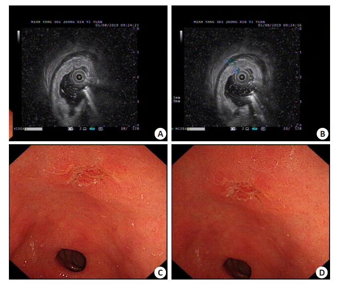
图 1 患者男,77岁,超声内镜及胃镜影像表现
A~B: 超声内镜下提示胃窦对应病灶见黏膜层呈低回声增厚, 黏膜下层, 固有肌层, 外膜层显影清 晰, 连续; C~D: 普通胃镜下提示胃窦小弯侧近幽门处可见一大小约1.5 cm IIa+IIc型病灶, 表面可见糜烂.
Figure 1. Endoscopic ultrasound and gastroscope image of a 77-year-old male patient.
表 1 两组患者一般资料比较[n(%)]
Table 1. Comparison of general data between the two groups
组別 男/女 年龄(岁,Mean±SD) 病变部位 内镜分型 上1/3 中1/3 下1/3 隆起型 平坦型 凹陷型 观察组(n=80) 51/29 56.60±4.93 2(26.25) 33(41.25) 26(32.50) 18(22.50) 39(48.75) 23(28.75) 对照组(n=60) 32/18 55.12±5.03 17(28.33) 28(46.67) 15(25.00) 16(26.67) 30(50.00) 14(23.33) t/χ2 0.001 1.743 0.944 0.637 p 0.977 0.084 0.624 0.727  下载: 导出CSV
表 2 两组病理浸润深度情况比较[n(%)]
Table 2. Comparison of pathological infiltration depth between the two groups
组別 Tis/T1a T1b 观察组(n=80) 69(86.25) 1(13.75) 对照组(n=60) 48(80.00) 12(20.00) χ2 0.975 P 0.323
下载: 导出CSV
表 3 内镜超声与病理结果比较(n)
Table 3. Comparison of endoscopic ultrasonography and pathological results
内镜超声 病理 Kappa P Tis/T1a T1b Tis/T1a 63 2 0.634 0.000 T1b 6 9
下载: 导出CSV
表 4 两组治愈性切除比较[n(%)]
Table 4. Comparison of curative resection between the two groups
组別 治愈性切除 χ2 P 观察组(n=80) 7(88.75) 22.118 0.000 对照组(n=60) 32(53.33)
下载: 导出CSV
-
[1] Spolverato G, Pawlik TM. Clinicopathological evaluation of recurrence in early gastric cancer[J].Am J Surg, 2019, 157(3): 202-7. https://academic.oup.com/jjco/article/33/5/209/861965 [2] Luo MC, Li LF. Clinical utility of miniprobe endoscopic ultrasonography for prediction of invasion depth of early gastric cancer: a meta-analysis of diagnostic test from PRISMA guideline [J]. Medicine (Baltimore), 2019, 98(6): e14430. https://www.ncbi.nlm.nih.gov/pmc/articles/PMC6417529/ [3] Suzuki T, Kitagawa Y, Nankinzan R, et al. Early gastric cancer diagnostic ability of ultrathin endoscope loaded with laser light source[J]. World J Gastroenterol, 2019, 25(11): 1378-86. https://pubmed.ncbi.nlm.nih.gov/30918430/ [4] Park JC, Choi SI, Kim EH, et al. Su1164 predictive model for endoscopic ultrasonography accuracy of invasion depth in early gastric cancer[J]. Gastrointest Endosc, 2019, 89(6): 295-6. https://www.researchgate.net/publication/333590872_Su1164_PREDICTIVE_MODEL_FOR_ENDOSCOPIC_ULTRASONOGRAPHY_ACCURACY_OF_INVASION_DEPTH_IN_EARLY_GASTRIC_CANCER [5] 苏有盛, 吴封.超声内镜联合NBI在胃早癌中的诊断价值[J].临床医学工程, 2018, 25(1): 9-10. http://www.cqvip.com/QK/98448X/201801/674348948.html [6] Kuroki K, Oka S, Tanaka S, et al. Clinical significance of endoscopic ultrasonography in diagnosing invasion depth of early gastric cancer prior to endoscopic submucosal dissection[J]. Gastric Cancer, 2020, 23(1): 1-11. doi: 10.1007/s10120-020-01100-5 [7] Takamaru H, Yoshinaga S, Takisawa H, et al. Endoscopic ultrasonography miniature probe performance for depth diagnosis of early gastric cancer with suspected submucosal invasion[J]. Gut Liver, 2020, 14(5): 581-8. https://gut.bmj.com/content/52/8/1220 [8] Cheng JY, Wu X, Yang AM, et al. Model to identify early-stage gastric cancers with deep invasion of submucosa based on endoscopy and endoscopic ultrasonography findings[J]. Surg Endosc, 2018, 32(2): 855-63. doi: 10.1007/s00464-017-5754-z [9] 林言, 郑祺, 黄蓉, 等.内镜超声联合传统内镜对早期胃癌浸润深度的预测及临床价值研究[J].中华消化内镜杂志, 2018, 35(12): 895-900. http://www.wanfangdata.com.cn/details/detail.do?_type=perio&id=zhxhnjzz98201812007 [10] Lee MW, Kim GH, Kim KB, et al. Digital image analysis-based scoring system for endoscopic ultrasonography is useful in predicting gastrointestinal stromal tumors[J]. Gastric Cancer, 2019, 22(5): 980-7. doi: 10.1007/s10120-019-00928-w.pdf [11] Zhao B, Zhang J, Zhang J, et al. Risk factors associated with lymph node metastasis for early gastric cancer patients who underwent noncurative endoscopic resection: a systematic review and meta-analysis [J]. J Gastrointest Surg, 2019, 23(7): 1318-28. https://www.ncbi.nlm.nih.gov/pubmed/30187319 [12] Muto M, Saito S, Muto M, et al. Effect of adding X-ray examination to endoscopy in the assessment of the invasion depth of early gastric cancer[J]. Jpn J Gastro-Enterol, 2019, 116(6): 506-14. https://www.ncbi.nlm.nih.gov/pubmed/31178580 [13] 王炘, 陈南云, 詹志刚.小探头超声内镜对早期胃癌浸润深度的评估价值[J].临床消化病杂志, 2019, 31(3): 173-4. http://www.cnki.com.cn/Article/CJFDTotal-LCXH201903015.htm [14] 林言, 郑祺, 闫昆, 等.超声内镜联合64排双源计算机断层扫描检查对胃癌局部临床分期和腹膜转移的预测价值[J].中华消化杂志, 2018, 38(2): 98-104. http://d.wanfangdata.com.cn/periodical/zhxhzz201802008 [15] 周振玉, 洪明.胃窗超声造影和高频小探头超声内镜在早期胃癌术前T分期中的应用价值[J].实用癌症杂志, 2018, 33(12): 2023-5, 2029. http://www.wanfangdata.com.cn/details/detail.do?_type=perio&id=syazzz201812033 [16] 阮江, 殷云勤, 高宇辉, 等.早期胃癌浸润深度判断的研究进展[J].中华消化病与影像杂志:电子版, 2018, 8(6): 267-70. http://www.cqvip.com/QK/71537X/201806/7000982499.html [17] 朱敏, 张澍田.内镜技术预测早期胃癌浸润深度的研究进展[J].中华消化内镜杂志, 2019, 36(1): 58-61. http://www.wanfangdata.com.cn/details/detail.do?_type=perio&id=zhxhnjzz98201901016 [18] 王会丰, 刘鑫, 张锦, 等.小探头超声内镜用于早期胃癌分期判断的影响因素[J].中国医学物理学杂志, 2019, 36(12): 1449-52. http://www.cnki.com.cn/Article/CJFDTotal-YXWZ201912016.htm [19] 徐瑶, 王志洋, 罗凌玉, 等.超声内镜对早期胃癌浸润深度的诊断价值及影响因素分析[J].实用临床医学, 2020, 21(4): 5-9. http://d.wanfangdata.com.cn/periodical/sylcyx202004002 [20] 徐涛, 王拥军, 张澍田.超声内镜对食管早癌浸润深度诊断价值的Meta分析[J].中华消化内镜杂志, 2018, 35(2): 126-32. http://www.wanfangdata.com.cn/details/detail.do?_type=perio&id=zhxhnjzz98201802010 [21] 程捷瑶, 吴晰, 杨爱明, 等.超声内镜对浅表胃癌诊断及治疗决策的影响[J].中华消化内镜杂志, 2016, 33(10): 663-6. http://med.wanfangdata.com.cn/Paper/Detail/PeriodicalPaper_zhxhnjzz98201610004 [22] 张家璐, 王贵齐.超声内镜联合放大内镜窄带成像对早期结直肠癌的诊断价值[J].肿瘤研究与临床, 2018, 30(7): 456-9. http://d.wanfangdata.com.cn/periodical/zlyjylc201807006 [23] 李峰.超声内镜检查在早期胃癌术前分期的应用分析[J].临床医药文献电子杂志, 2017, 4(81): 15969-70. http://www.cnki.com.cn/Article/CJFDTotal-LCWX201781071.htm [24] 夏晨梅, 陈霞, 李倩倩, 等.超声内镜对胃癌术前T、N分期准确率的评估及其影响因素分析[J].浙江医学, 2018, 40(3): 255-7, 265. https://www.zhangqiaokeyan.com/academic-journal-cn_zhejiang-medical-journal_thesis/0201254651711.html [25] 朱志超, 樊荣荣, 徐宝宏, 等. EUS判断胃癌术前病理学特点的应用价值[J].现代消化及介入诊疗, 2019, 24(1): 29-31, 35. http://www.cnki.com.cn/Article/CJFDTotal-XDXH201901008.htm -

 点击查看大图
计量
- 文章访问数: 577
- HTML全文浏览量: 340
- PDF下载量: 4
- 被引次数: 0