


Comparison on application value of MSCT and MRI in the differential diagnosis of thyroid nodules
-
摘要:
目的探讨多层螺旋CT(MSCT)与MRI在甲状腺结节鉴别诊断的应用价值。 方法选取2018年3月~2019年10月于我院收治的甲状腺结节患者并经手术后取得病理诊断结果的94例患者(94个结节)作为研究对象,对入组患者均行MSCT和MRI检查,以病理组织学检查为金标准,比较两种检查方法的诊断结果,计算两种检查方法的敏感度、特异度、准确率、阳性预测值、阴性预测值和kappa值。 结果94例甲状腺结节患者中,73例(77.66%)诊断为良性结节,21例(22.34%)为恶性结节。MSCT诊断甲状腺结节良恶性的灵敏度、特异度、准确率、阳性预测值、阴性预测值与MRI检查相比差异均无统计学意义(P>0.05),但MSCT诊断与病理诊断的Kappa值为0.746,大于MRI诊断与病理诊断的Kappa值为0.737。 结论MSCT与MRI诊断甲状腺结节良恶性的灵敏度、特异度、准确率均较高,与病理学诊断结果均具有较高的一致性,具有较高的临床应用价值,而MSCT在诊断甲状腺恶性结节钙化方面更具优势。 Abstract:ObjectiveTo explore the application value of multi-slice spiral CT (MSCT) and MRI in the differential diagnosis of thyroid nodules. Methods94 thyroid nodules patients (94 nodules) admitted to the hospital from March 2018 to October 2019 and obtained pathological diagnosis results after surgery were enrolled as the research objects. All patients underwent MSCT and MRI examination. Taking histopathological examination as the golden standard, the diagnosis results of the two examination methods were compared. The sensitivity, specificity, accuracy, positive predictive value, negative predictive value and kappa value of the two examination methods were calculated. ResultsIn 94 cases of thyroid nodules, 73 cases (77.66%) were diagnosed as benign nodules, 21 cases (22.34%) as malignant nodules. There were no significant differences in sensitivity, specificity, accuracy, positive predictive value and negative predictive value between MSCT and MRI for diagnosis of benign and malignant thyroid nodules (P>0.05). However, the kappa value of MSCT diagnosis and pathological diagnosis was 0.746, which was higher than that of MRI diagnosis and pathological diagnosis (0.737). ConclusionThe sensitivity, specificity and accuracy of both MSCT and MRI are high in the diagnosis of benign and malignant thyroid nodules. They are highly consistent with the pathological diagnosis results and have high clinical application value. However, MSCT is more advantageous in diagnosis of malignant thyroid nodules calcification. -
Key words:
- multislice spiral CT /
- MRI /
- thyroid nodule /
- differential diagnosis /
- accuracy
-
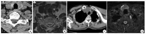
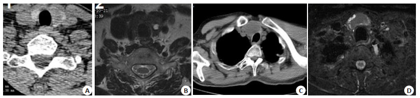
图 1 MSCT与MRI影像学表现的对比
A~B:结甲囊变患者, 女, 57岁. A: MSCT横断面图像; B: MRI T2WI横断面图像. C~D:结节性甲状腺肿患者, 男, 68岁; C: MSCT横断面图像; D: MRI T2WI横断面压脂图像, 可见甲状腺肿大, 密度减低.
Figure 1. Comparison of MSCT and MRI image.
表 1 MSCT诊断结果和病理学诊断的比较(n)
Table 1. Comparison on the diagnosis results of MSCT and pathology
方法 病理学珍断(金标准) 合计 阳性 阴性 MSCT 阳性 70 5 75 阴性 3 16 19 合计 73 21 94  下载: 导出CSV
表 2 MRI诊断结果和病理学诊断的比较(n)
Table 2. Comparison on the diagnosis results of MRI and pathology
方法 病理学珍断(金标准) 合计 阳性 阴性 MRI 阳性 67 3 70 阴性 6 18 24 合计 73 21 94
下载: 导出CSV
表 3 两种检查方法对肿瘤诊断结果比较(%, n=94)
Table 3. Comparison on the diagnosis results of tumors by the two methods
检杳方法 灵敏度 特异度 准确率 阳性预测值 阴性预测值 Kappa值 MSCT 95.89(70/73) 76.19(16/21) 91.49(86/94) 93.33(70/75) 84.21(16/19) 0.746 MRI 91.78(67/73) 85.71(18/21) 90.43(85/94) 95.71(67/70) 75.00(18/24) 0.737 χ2 0.474 0.154 0.065 0.070 0.130 P 0.491 0.694 0.799 0.792 0.719
下载: 导出CSV
-
[1] Zhao CK, Chen SG, Alizad A, et al. Three-dimensional shear wave elastography for differentiating benign from malignant thyroid nodules[J]. J Ultrasound Med, 2018, 37(7): 1777-88. doi: 10.1002/jum.14531 [2] Farihah AG, Nurismah MI, Husyairi H, et al. Reliability of the ultrasound classification system of thyroid nodules in predicting malignancy[J]. Med J Malaysia, 2018, 73(1): 9-15. [3] 袁文平, 马步云.多普勒超声联合多排螺旋CT在甲状腺结节良恶性鉴别中的应用价值[J].中国CT和MRI杂志, 2019, 17(9): 33-5. [4] 王洪序, 郑岩岩, 付荣湛, 等.甲状腺结节良恶性质应用CT和MRI的鉴别诊断价值比较研究[J].医学影像学杂志, 2018, 28(6): 917-9, 924. [5] 蒲玉洁, 李玉杰, 毛泽庆.超声、CT、MRI在甲状腺良恶性结节鉴别诊断中的应用价值分析[J].内科, 2018, 13(5): 753-5, 814. [6] Li MH, Liu JT. Screening of benign and malignant thyroid nodules in 5 196 physical examination population[J]. Zhonghua Zhong Liu Za Zhi, 2018, 40(2): 151-4. doi: 10.3760/cma.j.issn.0253-3766.2018.02.014 [7] Chang N, Zhang XC, Wan WJ, et al. The preciseness in diagnosing thyroid malignant nodules using shear-wave elastography[J]. Med Sci Monit, 2018, 24: 671-7. [8] Watanabe K, Igarashi T, Ashida H, et al. Diagnostic value of ultrasonography and TI-201/Tc-99m dual scintigraphy in differentiating between benign and malignant thyroid nodules[J]. Endocrine, 2019, 63(2): 301-9. doi: 10.1007/s12020-018-1768-0 [9] Pei SF, Cong SZ, Zhang B, et al. Diagnostic value of multimodal ultrasound imaging in differentiating benign and malignant TIRADS category 4 nodules[J]. Int J Clin Oncol, 2019, 24(6): 632-9. http://www.ncbi.nlm.nih.gov/pubmed/30825007 [10] 赵立群, 张祥林. CT能谱成像在甲状腺结节良恶性鉴别诊断中的应用价值[J].陕西医学杂志, 2019, 48(5): 614-6, 623. [11] 王岩, 陈国栋. CT及MRI对甲状腺结节的临床诊断价值[J].中国医药指南, 2019, 17(17): 93-4. [12] Hou HJ, Xu ZS, Zhang HS, et al. Combination diagnosis of multislice spiral computed tomography and secretary phospholipase A2- Ⅱa for solitary pulmonary nodules[J]. J Clin Lab Anal, 2018, 32(2): e22250. DOI: 10.1002/jcla.22250. [13] 刘明标, 傅晓琴, 郭威, 等.多层螺旋CT检查在甲状腺良恶性结节诊断及鉴别诊断中的应用价值[J].中国现代医生, 2018, 56(33): 114-6. [14] 黄玉娃, 朱新进, 靳仓正, 等.钙化征在CT诊断甲状腺良、恶性结节中的价值[J].分子影像学杂志, 2018, 41(4): 448-51. doi: 10.12122/j.issn.1674-4500.2018.04.07 [15] 黄敬忠, 程晓燕.甲状腺结节应用螺旋CT的诊断价值分析[J].影像研究与医学应用, 2018, 2(7): 148-9. [16] 肖连宏, 黄璞.螺旋CT在甲状腺良恶性结节鉴别诊断中的应用[J].中国CT和MRI杂志, 2019, 17(3): 44-6. [17] He ZH, Lv F, Cao ZF, et al. Value of multi-slice spiral CT in diagnosis of malignant gastrointestinal stromal tumors[J]. Chin-Ger J Clin Oncol, 2009, 8(8): 443-6. [18] Sebag F, Vaillant-Lombard J, Berbis J, et al. Shear wave elastography: a new ultrasound imaging mode for the differential diagnosis of benign and malignant thyroid nodules[J]. J Clin Endocrinol Metab, 2010, 95(12): 5281-8. http://www.ncbi.nlm.nih.gov/pubmed/20881263 [19] 陈广银, 陈悦熙.多层螺旋CT诊断结节性甲状腺肿合并甲状腺癌[J].分子影像学杂志, 2018, 41(2): 175-7. doi: 10.3969/j.issn.1674-4500.2018.02.10 -

 点击查看大图
计量
- 文章访问数: 638
- HTML全文浏览量: 278
- PDF下载量: 5
- 被引次数: 0