


Comparison of the application of CEUS and MSCT in the diagnosis and surgical assessment of primary clear carcinoma cell of the liver
-
摘要:
目的观察超声造影(CEUS)与多层螺旋CT扫描(MSCT)在肝透明细胞型癌(PCCCL)诊断及手术疗效评估的应用效果。 方法选取2016年2月~2019年3月本院收治的经手术病理确诊的PCCCL患者100例,另选取同期肝脏局灶性结节增生患者42例,分别行CEUS、MSCT肝脏检查,以手术病理结果为金标准,比较CEUS、MSCT对PCCCL患者不同直径病灶、腹腔积液、后腹膜淋巴结肿大、边缘不清的诊断效果;统计CEUS、MSCT检测的灵敏度、特异度、漏诊率、误诊率;术后1年复诊时采用CEUS、MSCT结果对PCCCL患者的手术疗效(肿瘤复发、转移)进行评价。 结果MSCT对直径 < 1 cm的病灶、腹腔积液、后腹膜淋巴结肿大、边缘不清检出情况优于CEUS,差异有统计学意义(P < 0.05);MSCT对PCCCL的诊断的灵敏度、特异度高于CEUS(P < 0.05),误诊率、漏诊率低于CEUS(P < 0.05);MSCT对PCCCL患者术后肿瘤复发、转移的检出率高于CEUS(P < 0.05)。 结论CEUS及MSCT对于PCCCL均有一定的诊断价值,但MSCT对于微小病灶及异常影像学表现的诊断效果更好,灵敏度、特异度更高,对手术疗效的评估效果更优。 Abstract:ObjectiveTo observe the application effect of contrast-enhanced ultrasound (CEUS) and multi-slice spiral computed tomography (MSCT) in the diagnosis and surgical assessment of primary clear carcinoma cell of the liver (PCCCL). Methods100 patients with PCCCL admitted in the hospital and 42 patients with focal nodular hyperplasia of the liver from February 2016 to March 2019 were enrolled in this study. CEUS and MSCT were performed respectively.The diagnostic effects of CEUS and MSCT on different diameter lesions, peritoneal effusion, retroperitoneal lymph node enlargement and unclear margin in PCCCL patients were compared with surgical pathological results as the gold standard; Sensitivities, specificities, missed diagnosis rates and misdiagnosis rates of CEUS and MSCT were statistically analyzed. The results of CEUS and MSCT were used to evaluate the surgical efficacy (tumor recurrence and metastasis) of PCCCL patients at 1 year after operation. ResultsMSCT was superior to CEUS in detecting lesions smaller than 1 cm in diameter, hydrops abdominis, posterior peritoneal lymphadenopathy and unclear margins and the difference was statistically significant (P < 0.05). MSCT had higher sensitivity and specificity than CEUS in the diagnosis of PCCCL (P < 0.05), but the misdiagnosis rate and missed diagnosis rate were lower than those of CEUS (P < 0.05). The detection rates of postoperative tumor recurrence and metastasis in patients with PCCCL by MSCT was higher than those by CEUS (P < 0.05). ConclusionBoth CEUS and MSCT can be used for diagnosis of PCCCL, but MSCT is better for diagnosing small lesions and abnormal imaging findings, with higher sensitivity and specificity. Besides, it is better for surgical effect evaluation. -
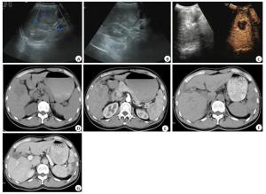
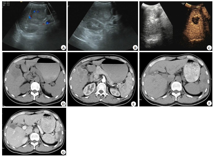
图 1 肝透明细胞癌手术前后影像学图片
A~B:术前超声图像, 肝右叶稍高回声肿块影, 病灶部分边界欠清,未见明显血流信号, 增强后轻度强化, 内见无强化区; C:术后超声图像, 肝右叶不均质回声影, 病灶边界欠清, 未见明显血流信号, 增强后无明显强化; D~E:术前CT, 肝右前叶下段见片团状稍低密度区, 形态不规则, 边界欠清晰, 其内密度尚均匀, 增强后动脉期病灶周边明显不均匀强化, 门脉期及延迟期轻度强化, 中心见片状无强化区; F~G:术后图像, 肝右前叶下段片状低密度区, 形态不规则, 边界欠清晰, 其内密度不均匀, 见散在多发高密度影,增强后动脉期病灶边缘不均匀强化, 门脉期及延迟期轻度强化.
Figure 1. Images of PCCCL before and after surgery.
表 1 CEUS与MSCT对不同尺寸病灶检出情况比较[n(%)]
Table 1. Comparison between CEUS and MSCT in detecting different sizes of lesions
检查方法 病灶尺寸(cm) < 1 (n=32) 1~5 (n=58) > 5(n=20) CEUS 2(65.63) 5(87.93) 18(90.00) MSCT 30(93.75) 56(96.55) 20(100.00) χ2 7.824 1.927 0.526 P 0.005 0.165 0.468 CEUS: Contrast-enhanced ultrasound; MSCT: Multi-slice spiral computed tomography.  下载: 导出CSV
表 2 PCCCL患者影像学表现结果分析[n=100, n(%)]
Table 2. Imaging findings of patients with PCCCL
检查方法 腹腔积液 后腹膜淋巴结肿大 边缘不清 CEUS 11(11.00) 10(10.00) 17(17.00) MSCT 22(22.00) 23(23.00) 42(42.00) χ2 4.392 6.135 15.034 P 0.036 0.013 0.000 PCCCL: Primary clear carcinoma cell of the liver.
下载: 导出CSV
表 3 CEUS、MSCT对PCCCL的诊断分析(n)
Table 3. Diagnosis of PCCCL by CEUS and MSCT
组别 手术病理诊断结果 灵敏度 特异度 PCCCL(n=100) 肝脏局灶性结节增生(n=42) CEUS组 71/100(71.00) 29/42(69.05) 阳性 71 13 阴性 29 29 MSCT组 85/100(85.00) 38/42(90.47) 阳性 85 4 阴性 15 38 χ2 5.712 5.168 P 0.017 0.023
下载: 导出CSV
表 4 CEUS、MSCT对PCCCL的漏诊率和误诊率比较[n(%)]
Table 4. Comparison of missed diagnosis rates and misdiagnosed rates of CEUS and MSCT for PCCCL
组别 漏诊率 误诊率 CEUS组 29/142(20.42) 13/142(9.15) MSCT组 15/142(10.56) 4/142(2.82) χ2 5.274 5.076 P 0.022 0.024
下载: 导出CSV
表 5 CEUS、MSCT对PCCCL患者手术疗效的评价[n=100, n(%)]
Table 5. Surgical assessment of patients with PCCCL by CEUS and MSCT
检测方法 肿瘤复发 转移 CEUS 3(3.00) 1(1.00) MSCT 12(12.00) 8(8.00) χ2 4.613 4.189 P 0.032 0.041
下载: 导出CSV
-
[1] Laino ME, Ragucci M, Klimstra DS, et al.Hepatobiliary and Pancreatic:Primary acinar cell carcinoma of the liver showing good response to chemotherapy[J].Journal of Gastroenterol Hepatol, 2018, 33(5):977. [2] Li ZY, Bi XY, Zhao JJ, et al.Clinicopathological and prognostic analysis of primary clear cell carcinoma of the liver[J].Chin J Cancer, 2013, 35(2):140-3. [3] Chen ZS, Zhu SL, Qi LN, et al.Long-term survival and prognosis for primary clear cell carcinoma of the liver after hepatectomy[J].Onco Ther, 2016, 9:4129-35. [4] Fanqing, Meng, Jun.B-Mode ultrasound based diagnosis of liver cancer with CEUS images as privileged information[J].Conf Proc IEEE Eng Med Biol Soc, 2018, 11(8):3124-7. [5] Ma AD, Zhang Y, Xue Z, et al.Angiogenesis of hepatocellular carcinoma under multislice spiral CT plain scan and enhanced scan[J].J Biol Regul HomeostAgents, 2015, 29(4):895-903. [6] Kothadia JP, Kaur N, Arju R, et al.Primary clear cell carcinoma of the non-cirrhotic liver presenting as an acute abdomen:a case report and review of the literature[J].J Gastrointest Cancer, 2017, 48(2):211-6. [7] 马秀华, 薛鹏, 肖智博, 等.原发性肝脏透明细胞癌的影像学诊断与临床应用价值[J].中华肝胆外科杂志, 2013, 19(7):491-4. [8] Zhou HY, Lv L, Ding XH.Primary clear cell carcinoma of the liver with intracerebral hemorrhage as first presentation:case report[J].Onkologie, 2011, 34(1/2):51-3. [9] Wang HY, Tan BY, Zhao B, et al.CT findings of primary clear cell carcinoma of liver:with analysis of 19 cases and review of the literature[J].Abdom Imaging, 2014, 39(4):736-43. [10] 张娜, 许万博.原发性肝脏透明细胞癌的影像学特征[J].实用放射学杂志, 2019, 35(2):321-3. [11] 王德玲, 李卉, 耿志君, 等.原发性肝透明细胞癌的CT、MRI表现[J].中国医学影像技术, 2013, 29(4):587-90. [12] 钟景云, 梁满球, 聂悦富, 等.肝硬化并发原发性肝癌合并肝囊肿患者的影像学特征[J].分子影像学杂志, 2018, 41(2):165-8. [13] 康霞, 张斯佳.肝细胞肝癌的多层螺旋CT征象与肿瘤血管生成指标的相关性探讨[J].癌症进展, 2018, 16(4):442-4, 491. [14] 张慧, 周庭永, 吕发金, 等.64-MSCT重建肝门静脉左支的临床解剖学研究[J].解剖学杂志, 2019, 42(2):173-6. [15] 刘力, 金梅, 曹大勇, 等.128层MSCT测量肝脏体积在肝巨大血管瘤精准切除中的应用研究[J].医学影像学杂志, 2018, 28(7):1130-3. [16] 周文华, 徐大伟, 王立军, 等.多层螺旋CT及动态增强磁共振成像血流参数在肝良恶性结节鉴别诊断中应用价值探究[J].中华生物医学工程杂志, 2019, 25(5):518-22. [17] 宋杰峰, 张亚珍, 聂忠仕, 等.原发性透明细胞型肝癌患者应用磁共振成像诊断的效果及其临床价值分析[J].中国医学装备, 2019, 16(7):91-4. [18] 黄雅琴, 饶圣祥, 曾蒙苏, 等.原发性肝透明细胞癌的CT及MRI表现[J].实用放射学杂志, 2014, 8(13):226-30. [19] Xiong JJ, He D, Hu WM, et al.Retroperitoneal and intrahepatic metastasis from primary clear cell carcinoma of the liver[J].Medicine, 2017, 96(12):e6452. [20] 胡井泉, 王彦辉.MSCT评估肝癌伴肝硬化患者肝脏储备功能中的价值分析[J].标记免疫分析与临床, 2018, 25(1):21-5, 49. -

 点击查看大图
计量
- 文章访问数: 527
- HTML全文浏览量: 248
- PDF下载量: 3
- 被引次数: 0