


Diagnostic value of abdominal color Doppler flow imaging and CT in acute pancreatitis
-
摘要:
目的探讨腹部彩色多普勒超声(CDFI)与CT对急性胰腺炎诊断价值。 方法回顾性分析2013年1月~2020年6月在本院就诊的81例急性胰腺炎患者的病例资料。将入组患者CDFI与CT检查结果进行比较,探讨CDFI、CT在急性胰腺炎患者诊断中的影像学特征,比较二者对急性胰腺炎的诊断准确率,及其对水肿型、坏死出血型胰腺炎的分类诊断价值。 结果总结81例急性胰腺炎患者CT、CDFI检查的影像学特征,发现CDFI检查检出胰管扩张率25.93%高于CT检查的12.35%,CDFI检出胰内小灶液化、胰外片状高密度率均为12.35%,分别低于CT检查的30.86%、27.16%,差异有统计学意义(P < 0.05);两种检查方式对胰外脓肿、胆总管结石、脂肪层模糊、实质不均的检出率比较,差异无统计学意义(P>0.05)。以临床综合诊断结果为金标准,CDFI检查漏诊16例、CT检查漏诊9例,CDFI、CT对急性胰腺炎的诊断符合率分别为80.25%(65/81)、88.89%(72/81),差异无统计学意义(P>0.05)。CDFI对水肿型胰腺炎、坏死出血型胰腺炎的诊断符合率分别为78.79%、86.67%与CT检查87.88%、93.33%比较,差异无统计学意义(P>0.05)。 结论腹部CDFI可以为急性胰腺炎患者提供较为准确的判断,对胰管扩张的判断上具有天然优势,可以作为临床诊治的首选检查方式之一。 Abstract:ObjectiveTo explore the diagnostic value of color Doppler flow imaging (CDFI) and CT for acute pancreatitis. Methods81 patients with acute pancreatitis admitted in the hospital from January 2013 to June 2020 were retrospectively analyzed. The examination results of CDFI and CT were compared among patients. The imaging features of CDFI and CT were explored in the diagnosis of acute pancreatitis patents. The diagnostic accuracy rate of acute pancreatitis, and diagnostic valu by the two methods were compared in oedematous and necrotic hemorrhagic pancreatitis. ResultsBy summarizing the imaging features of CT and CDFI in 81 patients with acute pancreatitis, The results showed that the expansion rate of pancreatic duct was 25.93% higher than that of CT 12.35%. The rates of small intrapancreatic liquefaction and extrapancreatic high density detected by CDFI were 12.35%, which were lower than 30.86% and 27.16% of CT examination respectively, and the difference was statistically significant (P < 0.05). There were no significant differences in the detection rates of extrapancreatic abscess, common bile duct stones (CBDS), fat layer blur and parenchyma unevenness between the two examination methods (P>0.05). Taking the clinical comprehensive diagnosis results as the gold standard, CDFI missed 16 cases diagnosis and CT missed 9 cases. The diagnostic coincidence rates of CDFI and CT for acute pancreatitis were 80.25% (65/81) and 88.89% (72/81), respectively, with no significant difference (P>0.05). The diagnostic coincidence rates of CDFI for oedematous pancreatitis and necrotic hemorrhagic pancreatitis were 78.79% and 86.67%, without significant difference compared with those of CT (87.88%, 93.33%) (P>0.05). ConclusionAbdominal CDFI can provide more accurate judgment for patients with acute pancreatitis. It has natural advantages in the judgment of pancreatic duct dilatation, and can be applied as one of the first choice for clinical diagnosis and treatment. -
Key words:
- Acute pancreatitis /
- Color Doppler flow imaging /
- CT /
- Clinical value
-
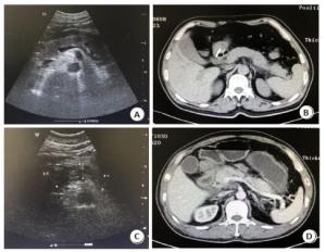
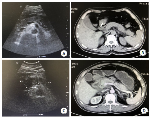
图 1 水肿型、坏死出血型胰腺炎患者的CT、CDFI图像
A~B:患者男,52岁,确诊为水肿型胰腺炎;A: CDFI图像可见胰腺呈弥漫性增大、边缘清晰、形态较规则; B: CT图可见胰腺呈均匀性强化、肿大; C~D:患者女, 50岁, 确诊为坏死出血型胰腺炎; C: CDFI图像可见胰周有渗出液、胰腺形态不规则、边缘欠清晰、内部呈强回声; D: CT图可见胰腺内部出现低密度灶, 轮廓不清晰.
Figure 1. CT and CDFI images of patients with oedematous and necrotic hemorrhagic pancreatitis.
表 1 急性胰腺炎患者CT、CDFI检查的影像学特征[n=81, n(%)]
Table 1. Imaging features of CT and CDFI in patients with acute pancreatitis
检查方法 胰外脓肿 胆总管结石 胰内小灶液化 脂肪层模糊 实质不均 胰管扩张 胰外片状高密度 CDFI 12(14.81) 7(8.64) 10(12.35) 13(16.05) 20(24.69) 2(25.93) 10(12.35) CT 15(18.52) 3(3.70) 25(30.86) 10(12.35) 19(23.46) 10(12.35) 22(27.16) χ2 0.400 1.705 8.200 0.456 0.034 4.827 5.608 P 0.527 0.192 0.004 0.499 0.854 0.028 0.018  下载: 导出CSV
表 2 急性胰腺炎患者CT、CDFI检查诊断符合率比较[n=81, n(%)]
Table 2. Comparison on diagnostic coincidence rates of CT and CDFI in patients with acute pancreatitis
检查方法 水肿型胰腺炎(n=66) 坏死出血型胰腺炎(n=15) 合计 CDFI 52(78.79) 13(86.67) 65(80.25) CT 58(87.88) 14(93.33) 72(88.89) χ2 1.964 0.370 2.318 P 0.161 0.543 0.128
下载: 导出CSV
-
[1] 孙备, 李冠群.重症急性胰腺炎局部并发症外科干预策略[J].中华消化外科杂志, 2020, 19(4):379-83. [2] Liu MW, Wei R, Su MX, et al.Effects of Panax notoginseng saponins on severe acute pancreatitis through the regulation of mTOR/Akt and caspase-3 signaling pathway by upregulating miR-181b expression in rats[J].BMC Complement Altern Med, 2018, 18(1):51. [3] Li B, Han X, Ye X, et al.Substance P-regulated leukotriene B4 production promotes acute pancreatitis-associated lung injury through neutrophil reverse migration[J].Int Immunopharmacol, 2018, 57:147-56. [4] 牛美红, 郭继慧, 赵丹.老年重症急性胰腺炎患者并发急性肾损伤的影响因素[J].中国老年学杂志, 2020, 40(9):1859-62. [5] Vege SS, DiMagno MJ, Forsmark CE, et al.Initial medical treatment of acute pancreatitis:American gastroenterological association institute technical review[J].Gastroenterology, 2018, 154(4):1103-39. [6] 郭晓梅, 吾红光, 毛春英, 等.超早期肠内营养支持对重症急性胰腺炎患者血淀粉酶尿淀粉酶恢复时间的影响[J].中国药物与临床, 2020, 20(7):1176-7. [7] Naqvi R.Acute kidney injury in association with acute pancreatitis[J].Pak J Med Sci, 2018, 34(3):606-9. [8] 王斌, 纪仁浩, 贺启龙.超声与多层螺旋CT在诊断急性胰腺炎中的应用比较[J].浙江临床医学, 2018, 20(8):1441-2. [9] 中华医学会消化病学分会胰腺疾病学组, 中华胰腺病杂志编委会, 中华消化杂志编委.中国急性胰腺炎诊治指南(2019年, 沈阳)[J].临床肝胆病杂志, 2019, 35(12):2706-11. [10] 卢磊, 周舒, 陈刘成, 等.比较磁共振成像与CT对急性胰腺炎诊断价值的荟萃分析[J].实用放射学杂志, 2020, 36(5):759-63, 787. [11] Kiss L, Fűr G, Mátrai P, et al.The effect of serum triglyceride concentration on the outcome of acute pancreatitis:systematic review and meta-analysis[J].Sci Rep, 2018, 8(1):14096. [12] Palestino-Dominguez M, Pelaez-Luna M, Lazzarini-Lechuga R, et al.Recombinant human hepatocyte growth factor provides protective effects in cerulein-induced acute pancreatitis in mice[J].J Cell Physiol, 2018, 233(12):9354-64. [13] 木塔里甫·买合木提, 郭峻氚, 肖东.超声影像在重症胰腺炎病情评估中的应用价值[J].中国医师杂志, 2020, 22(4):517-20. [14] Huang PJ, Liu AL, Ren H, et al.Color Doppler flow imaging of retrobulbar ocular blood flow changes in retinal artery occlusions caused by cosmetic facial filler injections[J].Ophthalmic Plast Reconstr Surg, 2019, 35(3):227-31. [15] Ahmed SU, Rana SS, Ahluwalia J, et al.Role of thrombophilia in splanchnic venous thrombosis in acute pancreatitis[J].Gastroenterology, 2017, 152(5):S288. [16] Holm TL, Murati MA, Hoggard E, et al.Liver Doppler findings in pediatric patients after total pancreatectomy and islet autotransplantation[J].J Ultrasound Med, 2018, 37(11):2595-601. [17] Fung C, Svystun O, Fouladi DF, et al.CT imaging, classification, and complications of acute pancreatitis[J].Abdom Radiol (NY), 2020, 45(5):1243-52. [18] Yamamoto S, Mutoh T, Sasaki K, et al.Non-invasive threedimensional power Doppler imaging for the assessment of acute cerebral blood flow alteration in a mouse model of subarachnoid haemorrhage[J].Clin Exp Pharmacol Physiol, 2019, 46(1):99-102. [19] Feng F, Tan HN, Li XY, et al.Incidence and risk factors of acute pancreatitis after scoliosis surgery:a prospective study[J].Spine, 2018, 43(9):630-6. [20] 姚公志, 何嘉辉, 姜惠悦, 等.急性胰腺炎患者采用CDFI检查的影像特征及诊断价值[J].中国现代普通外科进展, 2019, 22(2):149-50, 168. [21] 毕超.110例急性胰腺炎患者的彩超诊断分析[J].中国实验诊断学, 2019, 23(2):256-7. [22] 王俊, 周婷.多层螺旋CT、超声联合血淀粉酶检测在ICU中重症急性胰腺炎诊断中的应用价值探讨[J].中国CT和MRI杂志, 2019, 17(5):110-2. -

 点击查看大图
计量
- 文章访问数: 787
- HTML全文浏览量: 314
- PDF下载量: 5
- 被引次数: 0