


Value of ultrasound GI-RADS classification and 16-slice spiral CT in differential diagnosis of benign and malignant ovarian tumors
-
摘要:
目的探讨超声妇科影像报告和数据系统(GI-RADS)分类与16层螺旋CT对良恶性卵巢肿瘤鉴别诊断价值。 方法选取2015年1月~2019年8月在我院就诊的卵巢肿瘤患者85例为研究对象,分别进行超声及16层螺旋CT检查,采用GI-RADS系统评价超声声像图表现,并检测其癌胚抗原(CEA)水平。比较超声GI-RADS系统、16层螺旋CT联合CEA联合检查结果与病理学检查结果的一致性;以病理学检查结果为金标准,比较超声GI-RADS系统、16层螺旋CT联合CEA检查诊断鉴别良恶性卵巢肿瘤的灵敏度、特异度、阳性预测值、阴性预测值及诊断准确率,并采用ROC曲线分析超声GI-RADS系统、16层螺旋CT联合CEA检查对良恶性卵巢肿瘤的诊断鉴别价值。 结果超声GI-RADS系统联合CEA检查结果与病理学检查结果的一致性(Kappa=0.791)大于16层螺旋CT联合CEA(Kappa=0.487);超声GI-RADS系统、16层螺旋CT联合CEA联合检查诊断良恶性卵巢肿瘤的灵敏度、特异度、恶性预测值、良性预测值对比,差异无统计学意义(P>0.05);超声GI-RADS系统联合CEA诊断良恶性卵巢肿瘤的准确率高于16层螺旋CT联合CEA(P < 0.05);经ROC曲线分析得,超声GI-RADS系统联合CEA诊断良恶性卵巢肿瘤的AUC大于16层螺旋CT联合CEA(P < 0.05)。 结论超声GI-RADS系统联合CEA检测对良恶性卵巢肿瘤具有较高的诊断价值,且诊断准确率较高。 Abstract:ObjectiveTo explore value of ultrasound gynecological image report and data system (GI-RADS) classification and 16- slice spiral CT in differential diagnosis of benign and malignant ovarian tumors. MethodsA total of 85 patients with ovarian tumors treated in the hospital from January 2015 to August 2019 were enrolled as study objects. All underwent ultrasound and 16-slice spiral CT examination. GI-RADS was applied to evaluate performances of ultrasonic images, and to detect the CEA level. The pathological examination was performed on lesions. The consistency of ultrasound GI-RADS system and 16-slice spiral CT combined with CEA was compared with those of pathological examination. Using pathology test results as the gold standard, the sensitivity, specificity, positive predictive value, negative predictive value and diagnostic accuracy of ultrasonic GI-RADS system, 16 slice spiral CT combined with CEA were compared in the diagnosis of identification of benign and malignant ovarian tumors. ROC curve was used to analyze the diagnostic value of ultrasound gi-rads system, 16 slice spiral CT combined with CEA in the diagnosis of benign and malignant ovarian tumors. ResultsThe consistency of ultrasound GI-RADS combined CEA examination with pathological examination was higher than that of 16-slice spiral CT combined CEA (Kappa: 0.791 vs 0.487). There were no significant differences in sensitivity, specificity, malignant predictive value and benign predictive value of the two methods for diagnosing benign and malignant ovarian tumors (P>0.05). The accuracy of ultrasound GI-RADS combined with CEA in the diagnosis of benign and malignant ovarian tumors was higher than that of 16-slice spiral CT combined with CEA (P < 0.05). ROC curve analysis showed that AUC of ultrasound GI-RADS combined with CEA was greater than that of 16-slice spiral CT combined with CEA for diagnosing benign and malignant ovarian tumors (P < 0.05). ConclusionThe ultrasound GI-RADS with CEA is of relatively high diagnostic value for benign and malignant ovarian tumors, and its diagnostic accuracy is relatively high. -
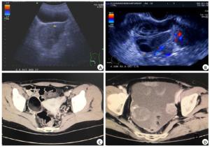
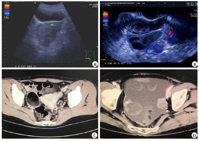
图 1 16层螺旋CT及超声声像图
A~B:超声声像图; C~D: 16层螺旋CT图像; A:右侧卵巢浆液性腺癌; B:右侧卵巢浆液性囊腺瘤; C:盆腔增强CT可见右侧附件区囊性良性病灶占位; D:卵巢囊实性占位,增强扫描可见实性部分中度强化,考虑恶性上皮肿瘤.
Figure 1. Images of 16-slice spiral CT and ultrasound.
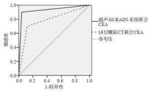
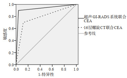
图 2 超声GI-RADS系统、16层螺旋CT联合CEA鉴别卵巢良恶性肿瘤的ROC曲线分析
Figure 2. ROC curves of ultrasound GI- RADS system and 16- slice spiral CT respectively combined with CEA for differential diagnosis of benign and malignant ovarian tumors.
表 1 超声GI-RADS系统、16层螺旋CT联合CEA检查结果与病理学检查结果(n)
Table 1. Examination results of ultrasound GI-RADS system and 16-slice spiral CT respectively combined with CEA and pathology
检查方法 病理学检查结果 合计 恶性(n=10) 良性(n=75) 超声GI-RADS系统联合CEA 恶性 9 3 12 良性 1 72 73 16层螺旋CT联合CEA 恶性 7 9 16 良性 3 66 69 GI-RADS: Gynecological image report and data system.  下载: 导出CSV
表 2 超声GI-RADS系统、16层螺旋CT联合CEA对乳腺良恶性肿瘤的诊断效能比较
Table 2. Comparison on diagnostic efficiency of ultrasound GI-RADS system and 16- slice spiral CT respectively combined with CEAfor benign and malignant breast tumors (n=85, %)
检查方法 灵敏度 特异度 恶性预测值 良性预测值 诊断准确率 超声GI-RADS系统联合CEA 90.00(9/10) 96.00(72/75) 75.00(9/12) 98.63(72/73) 95.29(81/85) 16层螺旋CT联合CEA 70.00(7/10) 88.00(66/75) 43.75(7/16) 95.65(66/69) 85.88(73/85) χ2 0.312 3.261 2.734 0.319 4.415 P 0.576 0.071 0.098 0.572 0.036
下载: 导出CSV
表 3 超声GI-RADS系统、16层螺旋CT联合CEA检查对良恶性卵巢肿瘤的诊断鉴别价值
Table 3. Differential diagnosis value of ultrasound GI-RADS system and 16-slice spiral CT respectively combined with CEAfor benign and malignant ovarian tumors
检查方法 AUC SE值 95%CI 超声GI-RADS系统联合CEA 0.930* 0.051 0.853-0.974 16层螺旋CT联合CEA 0.790 0.078 0.688-0.871 *P < .05 vs 16层螺旋CT.
下载: 导出CSV
-
[1] 李文凯.超声GI-RADS分类法对卵巢良恶性肿瘤的诊断价值[J].实用癌症杂志, 2018, 33(5):756-9. doi: 10.3969/j.issn.1001-5930.2018.05.018 [2] Zhang N, Zeng Z, Li SB, et al.High expression of EZH2 as a marker for the differential diagnosis of malignant and benign myogenic tumors[J].Sci Rep, 2018, 8(1):12331-6. doi: 10.1038/s41598-018-30648-7 [3] Li Z, Yu L, Wang X, et al.Diagnostic performance of mammographic texture analysis in the differential diagnosis of benign and malignant breast tumors[J].Clin Breast Cancer, 2018, 18(4):e621-7. doi: 10.1016/j.clbc.2017.11.004 [4] Hosseinzadeh M, Salmani S, Majles Ara MH, et al.The simple optical methods for early diagnosis of selected benign and malignant brain tumors of human[J].J Nonlinear Optic Phys Mat, 2018, 27(3):1850033. doi: 10.1142/S0218863518500339 [5] 胡长耀, 于世英.肿瘤临床诊疗指南[M].北京:科学出版社, 1999. [6] Qu RF, Guo DR, Chang ZX, et al.Differential diagnosis of benign and malignant breast tumors using apparent diffusion coefficient value measured through diffusion-weighted magnetic resonance imaging[J].J ComputAssist Tomogr, 2015, 39(4):513-22. doi: 10.1097/RCT.0000000000000226 [7] 刘婧, 陈秋月, 吕国荣.超声国际卵巢肿瘤研究组简单法则与妇科影像报告与数据系统分类诊断卵巢肿瘤的比较[J].中国医学影像技术, 2017, 33(5):739-42. doi: 10.13929/j.1003-3289.201610141 [8] 王霞丽, 杨舒萍, 吕国荣, 等.妇科超声影像报告和数据系统联合三维超声造影鉴别诊断卵巢良恶性肿块[J].中国医学影像技术, 2018, 34(6):888-92. doi: 10.13929/j.1003-3289.201707099 [9] Hahn S, Lee YH, Lee SH, et al.Value of the strain ratio on ultrasonic elastography for differentiation of benign and malignant soft tissue tumors[J].J Ultrasound Med, 2017, 36(1):121-7. doi: 10.7863/ultra.16.01054 [10] Sadullahoglu C, Nart D, Veral A.The importance of EZH2 and MOC-31 expression in the differential diagnosis of benign and malignant effusions[J].Diagn Cytopathol, 2017, 45(2):118-24. doi: 10.1002/dc.23653 [11] Cao SB, Hu Y, Gao X, et al.Serum carbohydrate antigen 19-9 in differential diagnosis of benign and malignant pancreatic cystic neoplasms:a meta-analysis[J].PLoS One, 2016, 11(11):e0166406. doi: 10.1371/journal.pone.0166406 [12] Qian H, Li S, Ji M, et al.MRI characteristics for the differential diagnosis of benign and malignant small solitary hypovascular hepatic nodules[J].Eur J Gastroenterol Hepatol, 2016, 28(7):749-56. doi: 10.1097/MEG.0000000000000642 [13] 范维, 刘双双, 脱淑梅, 等.经阴道超声弹性成像在宫颈癌诊断中的价值[J].分子影像学杂志, 2015, 38(2):92-4. doi: 10.3969/j.issn.1674-4500.2015.02.05 [14] 杨舒萍, 王霞丽, 吕国荣, 等.GI-RADS联合3D-CEUS评分系统评价卵巢肿瘤的生物学行为[J].中国超声医学杂志, 2019, 35(7):636-8. doi: 10.3969/j.issn.1002-0101.2019.07.023 [15] Tu Z, Xiao Z, Zheng Y, et al.Benign and malignant skull-involved lesions:discriminative value of conventional CT and MRI combined with diffusion-weighted MRI[J].Acta Radiol, 2019, 60(7):880-6. doi: 10.1177/0284185118773541 [16] 许爱玲, 聂芳, 高峻, 等.超声造影和国际卵巢肿瘤分析组织(IOTA)简单评价法鉴别诊断附件区肿瘤良恶性的价值比较[J].中华超声影像学杂志, 2018, 27(11):986-90. doi: 10.3760/cma.j.issn.1004-4477.2018.11.016 [17] 周玮珺, 曹秋月, 于鹏丽, 等.声触诊组织成像与定量技术联合BIRADS分类标准诊断乳腺良恶性肿瘤的初步研究[J].中华超声影像学杂志, 2017, 26(2):151-4. doi: 10.3760/cma.j.issn.1004-4477.2017.02.016 [18] Iima M, Yamamoto A, Kataoka M, et al.Time-dependent diffusion MRI to distinguish malignant from benign head and neck tumors[J].J Magn Reson Imaging, 2019, 50(1):88-95. doi: 10.1002/jmri.26578 [19] 黄冰冰, 陈秋月, 吕国荣.比较超声妇科影像报告和数据系统分类与恶性风险指数4鉴别卵巢良恶性肿块的价值[J].中国医学影像技术, 2019, 35(4):569-72. doi: 10.13929/j.1003-3289.201809046 [20] 龚时鹏, 陈咏宁, 张雅迪, 等.血清CA125、HE4和哥本哈根指数在卵巢上皮性肿瘤良恶性鉴别诊断中的价值[J].南方医科大学学报, 2017, 37(5):628-32. -

 点击查看大图
计量
- 文章访问数: 575
- HTML全文浏览量: 335
- PDF下载量: 7
- 被引次数: 0