


Clinical significance of MDCT in evaluating the severity of coronary artery disease and left ventricular systolic function in elderly patients with CHD
-
摘要:
目的探讨多排螺旋CT(MDCT)在评估老年冠心病中的应用价值。 方法选取2018年1月~2020年2月在我院治疗的老年冠心病患者110例,在我院行MDCT、超声心动图及冠状动脉造影(CAG)检查,以CAG为“金标准”,分析MDCT诊断价值,比较MDCT和超声心动图测量左室射血分数(LVEF)、左室舒张末期容积(LVEDV)、左室收缩末期容积(LVESV)、左室心肌质量(LVMM)、左室每搏输出量(LVSV)和左室短轴缩短率(LVFS)差异。 结果MDCT诊断灵敏性、特异性、准确性、阳性预测值和阴性预测值分别为93.80%、93.49%、93.73%、97.98%和81.77%;与超声心动图比较,MDCT测量LVEF、LVEDV、LVESV、LVMM、LVSV和LVFS差异无统计学意义(P < 0.05);MDCT测量,高度狭窄患者LVEF、LVSV和LVFS低于轻度狭窄和中度狭窄患者(P < 0.05),而LVEDV、LVESV和LVMM高于轻度狭窄和中度狭窄患者(P < 0.05);Gensini评分与LVEF、LVSV和LVFS呈负相关(r=-0.433、-0.412、-0.422,P < 0.05),与LVEDV、LVESV和LVMM呈正相关(r=0.410、0.366、0.378,P < 0.05)。 结论MDCT在老年冠心病诊断中有较好的价值,其可准确评估左心室功能,与冠状动脉狭窄程度有关。 Abstract:ObjectiveToo discuss the application value of multi- slice spiral CT (MDCT) in the evaluation of elderly coronary heart disease (CHD). Methods110 elderly patients with CHD treated in our hospital from January 2018 to February 2020 were selected. Among them, the MDCT, echocardiography and coronary angiography (CAG) were performed in our hospital. The CAG was used as the "gold standard" to analyze the diagnostic value of MDCT. The left ventricular ejection fraction (LVEF), left ventricular end diastolic volume (LVEDV), left ventricular end systolic volume (LVESV), left ventricular myocardial mass (LVMM), left ventricular stroke volume (LVSV) and left ventricular short axis shortening rate (LVFS) measured by MDCT and echocardiography were compared. ResultsThe sensitivity, specificity, accuracy, positive predictive value and negative predictive value of MDCT diagnosis were 93.80%, 93.49%, 93.73%, 97.98% and 81.77%, respectively. Compared with echocardiography, MDCT showed no significant difference in LVEF, LVEDV, LVESV, LVMM, LVSV and LVFS (P < 0.05). MDCT showed that LVEF, LVSV and LVFS in patients with severe stenosis were significantly lower than those in patients with mild and moderate stenosis (P < 0.05), while LVEDV, LVESV and LVMM were significantly higher than those in patients with mild and moderate stenosis (P < 0.05). Gensini score were negatively correlated with LVEF, LVSV and LVFS (r=-0.433, -0.412 and -0.422, P < 0.05), and positively correlated with LVEDV, LVESV and LVMM (r=0.410, 0.366 and 0.378, P < 0.05). ConclusionMDCT has a good value in the diagnosis of CHD in the elderly, which can accurately evaluate left ventricular function and is related to the degree of coronary artery stenosis. -

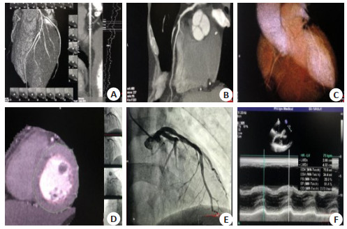
图 1 患者影像学表现
A~B: 320排CT(东芝Aquilion One动态容积MDCT)扫描, 左冠状动脉前降支近段中度狭窄(狭窄约66%); C~D: 320排CT(东芝Aquilion One动态容积MDCT)扫描:左心室功能分析图像; E:西门子(SIEMENS Axiom Artis Zee)全数字血管造影系统-行CAG检查:左冠状动脉前降支近段管腔狭窄; F:(Philips IE)超声诊断仪检查:患者左心室收缩功能减低表现.
Figure 1. Imaging findings of the patient.
表 1 MDCT和超声心动图测量左心室指标比较
Table 1. Comparison of left ventricular indices measured by MDCT and echocardiography (Mean±SD, n=110)
检查方法 LVEF(%) LVEDV(mL) LVESV(mL) LVMM(g) LVSV(mL) LVFS(%) 超声心动图 67.14±3.32 129.15±11.43 42.21±5.82 114.69±15.54 84.40±9.82 34.36±3.12 MDCT 66.70±4.11 130.02±12.10 43.10±6.02 113.02±16.03 86.82±10.12 33.80±3.05 t 0.873 -0.548 -1.115 0.785 -1.800 1.346 P 0.383 0.584 0.266 0.434 0.073 0.180 LVEF: Left ventricular ejection fraction; LVEDV: Left ventricular end diastolic volume; LVESV: Left ventricular end systolic volume; LVMM: Left ventricular myocardial mass; LVSV: Left ventricular stroke volume; LVFS: Left ventricular fraction shortening.  下载: 导出CSV
表 2 MDCT测量不同狭窄患者左心室指标比较
Table 2. Comparison of left ventricular indicators MDCT different stenosis patients (Mean±SD)
组别 LVEF(%) LVEDV(mL) LVESV(mL) LVMM(g) LVSV(mL) LVFS(%) 轻度狭窄(n=37) 71.02±6.02 116.03±11.02 36.82±6.02 105.66±11.20 92.11±9.82 37.82±3.02 中度狭窄(n=47) 67.52±5.56* 128.82±13.01* 42.31±5.83* 112.01±10.43* 86.04±10.11* 33.12±3.42* 高度狭窄(n=26) 60.97±6.04*# 148.21±12.02*# 49.70±6.33*# 125.32±12.21*# 80.70±9.88*# 29.31±3.12*# F 24.493 30.201 25.565 20.332 18.281 15.506 P 0.000 0.000 0.000 0.000 0.000 0.000 *P < 0.05 vs轻度狭窄; #P < 0.05 vs中度狭窄.
下载: 导出CSV
-
[1] 林杰, 曾伟胜, 陈奕州.多层螺旋CT诊断冠心病的应用价值[J].临床医学, 2018, 38(5):50-1. [2] 刘光宇.多层螺旋CT冠脉造影检查诊断冠心病的临床意义[J].饮食保健, 2019, 6(21):266-7. [3] 殷保江, 贾晓辉, 杨星奎.多层螺旋CT与超声心动图在诊断冠心病中的应用比较[J].中国CT和MRI杂志, 2018, 16(8):67-9. [4] 付振杰.64排多层螺旋CT冠状动脉成像的临床应用[J].现代医用影像学, 2018, 27(3):811-2. [5] 王晓港.多层螺旋CT在冠心病临床诊断中的应用价值[J].影像研究与医学应用, 2019, 3(11):177-8. [6] 贾敏, 李璐, 王晶.多层螺旋CT在冠心病的临床诊断中的应用价值研究[J].医药前沿, 2018, 8(2):139-40. [7] 颜红兵, 马长生, 霍勇.临床冠心病诊断与治疗指南[M].北京:人民卫生出版社, 2010:22-4. [8] Wang SQ, Ren FY, Wang JH, et al.Diagnostic value of multislice spiral computed tomography (CT) combined with CT angiography for intra-abdominal undescended testis secondary seminomas[J].Cancer Imaging, 2019, 19(1):24. [9] Social support, unstable angina, and stroke as predictors of depression in patients with coronary heart disease: erratum[J].J Cardiovasc Nurs, 2018, 33(3): 201. [10] 朱彬, 杨飞.多层螺旋CT在冠状动脉狭窄诊断中的应用及其临床价值[J].山西医药杂志, 2018, 47(20):2425-7. [11] 李昌勇.多层螺旋CT诊断冠状动脉狭窄的准确性探讨[J].中国继续医学教育, 2019, 11(12):78-9. [12] 崔楚坤, 李家欢, 曾鹏程, 等.多层螺旋CT血管造影在冠状动脉病变程度诊断中的临床价值[J].海南医学, 2019, 30(7):875-7. [13] Wang M, Wei C, Shi Z, et al.Study on the diagnosis of small hepatocellular carcinoma caused by hepatitis B cirrhosis via multislice spiral CT and MRI[J].Oncol Lett, 2018, 15(1):503-8. [14] 李江华.冠状动脉狭窄的多层螺旋CT诊断分析[J].世界最新医学信息文摘, 2019, 19(11):165-6. [15] Sun MY, Dong Y, Wang Y, et al.Assessment of the left ventricular function in patients with uremia using layer-specific 2-dimensional speckle tracking echocardiography[J].Medicine (Baltimore), 2019, 98(9):e14656. [16] Lukács Krogager M, Skals RK, Appel EVR, et al.Hypertension genetic risk score is associated with burden of coronary heart disease among patients referred for coronary angiography[J].PLoS One, 2018, 13(12):e0208645. [17] Sui YB, Li J, Zou ZX, et al.Comparison of diagnostic value of multi-slice spiral CT and MRI for different pathological stages of prostate cancer[J].Oncol Lett, 2019, 17(6):5505-10. [18] Zhou XC, Chen QL, Huang CQ, et al.The clinical application value of multi-slice spiral CT enhanced scans combined with multiplanar reformations images in preoperative T staging of rectal cancer[J].Medicine (Baltimore), 2019, 98(28):e16374. [19] Blomstrand P, Sjöblom P, Nilsson M, et al.Overweight and obesity impair left ventricular systolic function as measured by left ventricular ejection fraction and global longitudinal strain[J].Cardiovasc Diabetol, 2018, 17(1):113. [20] Ghaleh B, Barthélemy I, Sambin L, et al.Alteration in left ventricular contractile function develops in puppies with Duchenne muscular dystrophy[J].JAm Soc Echocardiogr, 2020, 33(1):120-9. -

 点击查看大图
计量
- 文章访问数: 497
- HTML全文浏览量: 293
- PDF下载量: 2
- 被引次数: 0