


A radiation dose and image quality of coronary artery CT imaging comparison among the GSI model, the traditional front and back door control technique
-
摘要:
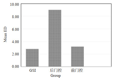
目的对比宝石CT能谱成像(GSI)、传统前门控和后门控技术冠状动脉成像的辐射剂量及图像质量,探讨冠脉成像GSI扫描模式优势及临床应用价值。 方法筛选2019年5月~2020年5月在我院成功完成CT冠状动脉成像患者162例,按照扫描模式分为3组:A组为GSI扫描模式,B组为后门控模式,C组为前门控模式。对比各组图像噪声、对比度、信噪比(SNR),对比噪声比(CNR)及图像优良指数(FOM),并计算比较各组间的有效放射剂量(ED)。 结果图像质量比较:A组图像噪声明显低于后门控组及前门控组,差异具有统计学意义(P < 0.01);A组SNR、CNR及FOM均明显高于后门控组(P < 0.01)及前门控组(P < 0.05);C组SNR、CNR及FOM均高于B组,差异无统计学意义(P>0.05)。有效剂量比较:A组和C组ED均值分别为2.87±0.83、3.34± 2.36 mSv,明显低于B组(9.04±3.06 mSv)(P < 0.01);A组和C组ED较B组分别降低68.25%和63.05%,而A组较C组降低约14.07%。 结论宝石CT冠脉GSI扫描技术与后门控、前门控技术相比,不但能有效大幅度降低患者的辐射剂量,且能显著提高图像的质量,应在临床中积极推广应用。 Abstract:ObjectiveTo explore the advantages and clinical value of coronary angiography in GSI scanning model by comparing the radiation dose and image quality among the GSI model, the traditional front and back door control technique. MethodsA total of 162 patients underwent successfully CT coronary angiography in our hospital from May 2019 to May 2020 were selected as research objects. According to the scanning model, these patients were divided into three groups. Group A was the GSI scanning mode, group B was the backdoor control mode and C group was the front door control mode. The image noise (SD), contrast, SNR (SNR), comparison of noise ratio (CNR), image quality index (FOM), and the effective radiation dose (ED) for these groups were compared. ResultsComparison of image quality: ① SD of A group was significantly lower than that of B group and C group (P < 0.01). ②SNR, CNR and FOM of Group A were significantly higher than B group (P < 0.01) and C group (P < 0.05). ③SNR, CNR and FOM of group C were higher than group B, but without significantly difference (P>0.05). Comparison of ED: ① The mean ED of group A and C were 2.87 ± 0.83 mSv, 3.34 ± 2.36 mSv, respectively, and were both significantly lower than group B (9.04 ± 3.06 mSv) (P < 0.01). ② ED in group A and group C decreased 68.25% and 63.05%, respectively, compared with group B, and ED in group A decreased by about 14.07% compared with group C. ConclusionCompared to the front and back door technique, GSI model scanning technology not only can effectively reduce the radiation dose, but also can significantly improve the quality of image, and it should be popularized in clinic. -
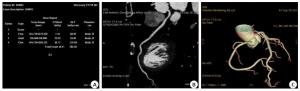
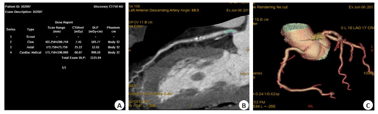
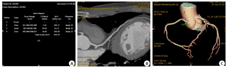
图 1 男,51岁,GSI模式
A:辐射剂量; B: 70 keV单光子图左冠状动脉主干近段轻度狭窄; C: GSI模式显示血管细节更丰富、血管远端更清晰.
Figure 1. Male, 51 years old, GSI model.
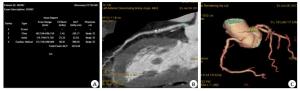
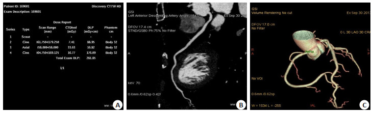
图 2 男,55岁,传统后门控模式
A:辐射剂量; B:左冠状动脉主干近段软硬班块; C:血管侧支显示较少,远端欠清晰.
Figure 2. Male, 55 years old, traditional backdoor control model.
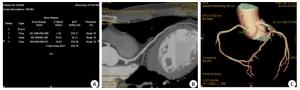
图 3 男,47岁,传统前门控模式
A:辐射剂量; B:左冠状动脉主干未见异常; C:血管侧支显示较多,血管远端清晰.
Figure 3. Male, 47 years old, traditional frontdoor control model.
表 1 各组基本情况比较
Table 1. Comparison of the basic situation of each group (Mean±SD)
组别 BMI(kg/m2) 年龄(岁) 心率(次/min) 心率变化值(次/min) GSI(n=81) 24.17±3.75 55.27±11.50 57.33±5.79 3.56±2.97 后门控(n=52) 25.18±3.13 55.44±12.31 68.92±9.01* 4.04±3.38 前门控(n=29) 24.32±2.82 57.85±11.67 60.31±5.68# 3.76±2.92 F 1.431 0.455 41.387 0.384 P 0.242 0.413 0.000 0.682 GSI: Gemstone spectral imaging; *P < 0.01 vs GSI group; #P < 0.01 vs back door control technique group.  下载: 导出CSV
表 2 各组冠状动脉图像质量比较
Table 2. Comparison of coronary artery image quality of each group (Mean±SD)
组别 SD 对比度 SNR CNR FOM 造影剂所需剂量 GSI(n=81) 21.08±7.02 5.14(E2)±82.04 22.31±7.90 26.85±9.47 10.05±4.03 51.89±10.09 后门控(n=52) 25.68±6.76** 5.09(E2)±94.38 16.61±4.19** 20.12±4.77** 2.45±1.04** 51.02±10.21 前门控(n=29) 26.40±9.23** 5.41(E2)±108.56 19.36±6.69* 22.46±7.92* 8.33±4.22*## 49.88±7.87 F 8.863 1.208 11.586 11.863 80.13 0.477 P 0.000 0.301 0.000 0.000 0.000 0.622 SD: Image noise; SNR: Signal-to- noise ratio; CNR: Cotltrast-to-noise ratio; FOM: Figure of merit; *P < 0.05 vs GSI group, **P < 0.01 vs GSI group; #P < 0.01 vs back door control technique group.
下载: 导出CSV
-
[1] Danad I, Fayad ZA, Willemink MJ, et al. New applications of cardiac computed tomography: dual-energy, spectral, and molecular CT imaging[J]. JACC Cardiovasc Imaging, 2015, 8(6): 710-23. doi: 10.1016/j.jcmg.2015.03.005 [2] de Gonzalez AB, Salotti JA, McHugh K, et al. Relationship between paediatric CT scans and subsequent risk of leukaemia and brain tumours: assessment of the impact of underlying conditions[J]. Br J Cancer, 2016, 114(4): 388-94. doi: 10.1038/bjc.2015.415 [3] 陈俐君, 魏清顺, 杨晓萍.能谱CT的临床应用进展[J].医疗卫生装备, 2017, 38(11): 113-7. http://www.cnki.com.cn/Article/CJFDTotal-YNWS201711030.htm [4] 杨帆, 宋娟, 李勇, 等.宝石能谱CTASiR结合最佳单能量对冠状动脉图像质量的研究[J].实用放射学杂志, 2015, 31(12): 2050-4. doi: 10.3969/j.issn.1002-1671.2015.12.031 [5] Sandfort V, Pourmorteza A, Symons R, et al. Photon-counting CT for coronary artery calcium scoring: Potential for dose reduction in a human population[C]// Radiological Society of North America, 2016. [6] Schirra CO, Brendel B, Anastasio MA, et al. Spectral CT: a technology primer for contrast agent development[J]. Contrast Media Mol Imaging, 2014, 9(1): 62-70. doi: 10.1002/cmmi.1573 [7] Verburg FA, Apitzsch J, Lensing C, et al. Body surface area adapted iopromide 300 mg/ml versus 370 mg/ml contrast medium injection protocol: influence on quantitative and clinical assessment in combined PET/CT[J]. Eur J Radiol, 2013, 82(12): 2348-52. doi: 10.1016/j.ejrad.2013.09.013 [8] 李耀斌, 于秀梅. CT放射中的防护问题分析[J].中国卫生产业, 2016, 13(11): 92-4. http://www.cnki.com.cn/Article/CJFDTotal-WSCY201611038.htm [9] Morford K, Watts LK. Bismuth shielding during CT exams: a literature review[J]. Radiol Manage, 2012, 34(3): 45-7. https://pubmed.ncbi.nlm.nih.gov/22720541/ [10] Han RJ, Sun K, Lu B, et al. Diagnostic accuracy of coronary CT angiography combined with dual-energy myocardial perfusion imaging for detection of myocardial infarction[J]. Exp Ther Med, 2017, 14(1): 207-13. doi: 10.3892/etm.2017.4485 [11] Lu MT, Douglas PS, Udelson JE, et al. Safety of coronary CT angiography and functional testing for stable chest pain in the PROMISE trial: a randomized comparison of test complications, incidental findings, and radiation dose[J]. J Cardiovasc Comput Tomogr, 2017, 11(5): 373-82. doi: 10.1016/j.jcct.2017.08.005 [12] 宋鹏, 李彩英, 宋登浩, 等. CT冠状动脉血管造影前瞻性与回顾性心电门控2种扫描方式的比较[J].河北医科大学学报, 2015, 36(7): 838-40. doi: 10.3969/j.issn.1007-3205.2015.07.028 [13] 刘辉, 王卫, 王玉海, 等.下肢动脉CTA能谱成像的扫描剂量及成像质量研究[J].医疗卫生装备, 2017, 38(8): 91-3, 107. http://www.cnki.com.cn/Article/CJFDTOTAL-YNWS201708025.htm [14] 王良炯, 邓生德, 徐裕, 等. 256层CT冠状动脉前瞻性与回顾性心电门控成像质量和辐射剂量对比分析[J].实用放射学杂志, 2014, 30 (2): 301-5. doi: 10.3969/j.issn.1002-1671.2014.02.026 [15] Mangold S, Cannaó PM, Schoepf UJ, et al. Impact of an advanced image-based monoenergetic reconstruction algorithm on coronary stent visualization using third generation dual-source dual-energy CT: a phantom study[J]. Eur Radiol, 2016, 26(6): 1871-8. doi: 10.1007/s00330-015-3997-4 [16] 柳青, 张智琴, 宗会迁, 等.心率及心律对256层螺旋CT冠状动脉CT血管造影图像质量的影响[J].中国老年学杂志, 2016, 36(8): 1951-3. doi: 10.3969/j.issn.1005-9202.2016.08.076 [17] 潘存雪, 古丽娜·阿扎提, 邢艳, 等.冠状动脉CT单能量成像改善心肌射线硬化伪影的价值[J].中华放射学杂志, 2015, 49(9): 679-84. doi: 10.3760/cma.j.issn.1005-1201.2015.09.009 [18] Hickethier T, Baeßler B, Kroeger JR, et al. Monoenergetic reconstructions for imaging of coronary artery stents using spectral detector CT: In-vitro experience and comparison to conventional images[J]. J Cardiovasc Comput Tomogr, 2017, 11(1): 33-9. doi: 10.1016/j.jcct.2016.12.005 [19] 曲岷.适应性统计迭代重建技术在低剂量CT扫描中的应用进展[J].实用医学影像杂志, 2014, 15(6): 450-2. http://www.cnki.com.cn/Article/CJFDTotal-SYXY201406032.htm [20] Shen HS, Dai GC, Luo MY, et al. Image quality and radiation dose of CT coronary angiography with automatic tube current modulation and strong adaptive iterative dose reduction three-dimensional (AIDR3D)[J]. PLoS One, 2015, 10(11): e0142185. doi: 10.1371/journal.pone.0142185 -

 点击查看大图
计量
- 文章访问数: 579
- HTML全文浏览量: 286
- PDF下载量: 4
- 被引次数: 0