


Coronary computed tomography angiography for the evaluation of congenital absence of the right coronary artery
-
摘要:
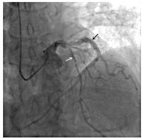
目的总结先天性右冠状动脉缺如患者的临床和冠状动脉CT血管造影(CCTA)影像表现。 方法回顾性分析2014年1月~2019年6月于本院行CCTA检查并经选择性冠状动脉造影(SCA)确诊的13例先天性右冠状动脉缺如患者的临床资料和CCTA影像表现。 结果13例先天性右冠状动脉缺如患者的临床表现缺乏特异性,大多表现为心血管疾病的临床症状。CCTA平扫显示左主干、左前降支和(或)左回旋支走行区内数量不等、形态各异的高密度钙化斑块形成;增强扫描显示左主干、左前降支和(或)左回旋支不同程度变窄,升主动脉和右冠状窦区均未见右冠状动脉发出,左主干、左前降支和左回旋支均不同程度增粗,并左回旋支延伸至右心室背面,进而发出分支供应右心房和右心室。CCTA与SCA表现基本一致。 结论先天性右冠状动脉缺如罕见,其临床表现缺乏特异性,仅凭临床表现往往难以诊断,CCTA联合SCA检查常可用于确诊。 Abstract:ObjectiveTo summarize the clinical characteristics and coronary CT angiography (CCTA) findings of congenital absence of the right coronary arteries (RCA). MethodsThirteen patients with congenital absence of RCA diagnosed by selective coronary angiography (SCA) in our hospital from January 2014 to June 2019 were retrospectively reviewed with emphasis placed upon the patients' clinical manifestations and CCTA findings. ResultsThe clinical manifestations of the 13 patients were of not specific, and most of them showed symptoms of cardiovascular diseases. Non-enhanced CCTA scans showed high density calcified plaques in the left main, left anterior descending and/or left circumflex coronary arteries areas. Enhanced CCTA scans showed different degrees of narrowness in the left main, left anterior descending and/or left circumflex, and no RCA were given off in the ascending aorta or right coronary sinus areas. The left main, left anterior descending and left circumflex were enlarged in different degrees, and the enlarged left circumflex extended to the posterior surface of the right ventricle, where it gave off its branches to supply the right atrium and right ventricle. Besides that, findings on selective coronary angiography (SCA) were almost identical to those of CCTA. ConclusionCongenital absence of the RCA is rare, and it's clinical manifestations are not specific, which make it difficult to diagnosis by the clinical manifestations alone. However, CCTA combined with SCA can be used to confirm the diagnosis. -
Key words:
- coronary artery /
- variation /
- computed tomography /
- X-Ray computed
-
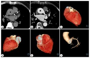
图 1 女,66岁,先天性右冠脉缺如
CCTA平扫轴位(A), 增强扫描轴位(B)以及容积再现(C~F)显示LCA多发钙化斑块形成, 升主动脉或右冠状窦区(黑五角星)未见右冠脉发出, LAD(白箭头)和LCX(黑箭头)均增粗, LCX延伸至右心室背面, 并发出分支供应右心房和右心室.
Figure 1. A sixty-six-year-old female patient, shows an congenital absence of RCA.

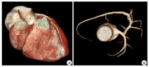
图 3 女,34岁,风湿性心脏病病史(二尖瓣及主动脉瓣狭窄),先天性右冠脉缺如
CCTA VR像(A)显示升主动脉及右冠状窦区(黑箭头)未见右冠脉发出,冠脉树(B)显示LAD(红箭头)和LCX(白箭头)稍增粗,并LCX延伸至右心室背面,发出分支供应右心房和右心室.
Figure 3. A thirty-four-year-old female patient, with a history of rheumatic heart disease (mitral and aortic stenosis), shows an congenital absence of RCA.
-
[1] Yan GW, Bhetuwal A, Yang GQ, et al. Congenital absence of the right coronary artery: a case report and literature review[J]. Medicine (Baltimore), 2018, 97(12): e0187-92. http://europepmc.org/abstract/MED/29561437 [2] 申淑荣, 王兴德, 曾金美, 等.先天性右冠状动脉缺如伴胸前导联ST-T改变一例[J].中国心脏起搏与心电生理杂志, 2018, 32(6): 621-2. http://www.cnki.com.cn/Article/CJFDTotal-ZGXZ201806027.htm [3] 黄佳兵, 周胜华, 唐亮, 等.先天性右冠状动脉缺如合并梗阻性肥厚型心肌病1例[J].临床心血管病杂志, 2016, 32(1): 103-5. http://www.cnki.com.cn/Article/CJFDTotal-LCXB201601028.htm [4] 刘瑞杰, 李自成, 陈家元, 等.先天性右冠状动脉缺如一例并文献复习[J].中国心血管杂志, 2012, 17(5): 364-7. http://www.cnki.com.cn/Article/CJFDTotal-XIXG201205020.htm [5] 姚金龙, 龚艳艳. 64层螺旋CT诊断先天性右冠状动脉缺如一例[J].放射学实践, 2013, 28(4): 419. [6] 中华医学会放射学分会心胸学组, 《中华放射学杂志》心脏冠状动脉多排CT临床应用指南写作专家组.心脏冠状动脉CT血管成像技术规范化应用中国指南[J].中华放射学杂志, 2017, 51(10): 732-43. http://d.wanfangdata.com.cn/Periodical/zhfsx201710004 [7] Lipton MJ, Barry WH, Obrez I, et al. Isolated single coronary artery: diagnosis, angiographic classification, and clinical significance[J]. Radiology, 1979, 130(1): 39-47. http://icvts.oxfordjournals.org/external-ref?access_num=10.1148/130.1.39&link_type=DOI [8] Namgung J, Kim JA. The prevalence of coronary anomalies in a single center of Korea: origination, course, and termination anomalies of aberrant coronary arteries detected by ECG-gated cardiac MDCT[J]. BMC Cardiovasc Disord, 2014, 14: 48. doi: 10.1186/1471-2261-14-48 [9] Türkmen S, Yolcu M, Sertçelik A, et al. Single coronary artery in 215140 patients undergoing coronary angiography[J]. J Am Coll Cardiol, 2013, 62(18): C134-5. http://www.sciencedirect.com/science/article/pii/S0735109713035006 [10] Shriki JE, Shinbane JS, Rashid MA, et al. Identifying, characterizing, and classifying congenital anomalies of the coronary arteries[J]. Radiographics, 2012, 32(2): 453-68. http://www.ncbi.nlm.nih.gov/pubmed/22411942 [11] Agarwal PP, Dennie C, Pena E, et al. Anomalous coronary arteries that need intervention: review of pre- and postoperative imaging appearances[J]. Radiographics, 2017, 37(3): 740-57. http://smartsearch.nstl.gov.cn/paper_detail.html?id=f5d980fc71c3b31cc7a863e618a3cec2 [12] 杨家虎, 王军娜, 韩璐, 等.冠状动脉CTA对先天性单一冠状动脉的诊断价值[J].临床放射学杂志, 2017, 36(1): 148-50. http://d.wanfangdata.com.cn/Periodical/lcfsxzz201701039 [13] Li SH, Ni QQ, Wu HL, et al. Diagnostic accuracy of 320- slice computed tomography angiography for detection of coronary artery stenosis: meta-analysis[J]. Int J Cardiol, 2013, 168(3): 2699-705. http://espace.library.curtin.edu.au/R?func=dbin-jump-full&object_id=193278 [14] Yan GW, Li HW, Yang GQ, et al. Iatrogenic arteriovenous fistula of the iliac artery after lumbar discectomy surgery: a systematic review of the last 18 years[J]. Quant Imaging Med Surg, 2019, 9(6): 1163- 75. http://www.researchgate.net/publication/333395964_Iatrogenic_arteriovenous_fistula_of_the_iliac_artery_after_lumbar_discectomy_surgery_a_systematic_review_of_the_last_eighteen_years [15] Kang EJ, Lee KN, Choi WJ, et al. Left ventricular functional parameters and geometric patterns in Korean adults on coronary CT angiography with a 320-detector-row CT scanner[J]. Korean J Radiol, 2017, 18(4): 664-73. http://www.ncbi.nlm.nih.gov/pmc/articles/PMC5447642/ [16] 张蕾, 杨全新, 毛翠平, 等.双源CT冠状动脉成像与超声心动图对左心室功能评价的对比研究[J].实用放射学杂志, 2019, 35(6): 884-6. [17] 严高武, 杨国庆, 李勇, 等.血管内修复腰椎间盘切除术后医源性髂动静脉瘘: 1例报道与文献回顾[J].介入放射学杂志, 2019, 28(9): 881-6. -

 下载:
下载:

 点击查看大图

计量
- 文章访问数: 902
- HTML全文浏览量: 402
- PDF下载量: 11
- 被引次数: 0