


Clinical application of NT ultrasound in early pregnancy combined with color Doppler ultrasound in prenatal fetal malformation screening
-
摘要:
目的探究孕早期颈项透明层(NT)超声联合孕中期彩色多普勒超声在产前胎儿畸形筛查中的应用价值。 方法选择2017年8月~2019年12月在我院行胎儿畸形筛查的2417例孕妇作为研究对象,所有孕妇于孕11~14周行NT超声检查,并于孕22~28周行二维、四维彩超检查。以引产或分娩结果为“金标准”,比较畸形胎儿、正常胎儿的NT值及NT异常率,对比NT超声检查、二维联合四维彩超检查结果,并就NT超声、二维及四维彩超及二者联合在诊断胎儿畸形中的诊断率进行比较。 结果2417例孕妇最终确诊异常胎儿88例,发病率为3.64%。畸形胎儿的NT值明显高于正常胎儿,NT异常率(93.18%)明显高于正常胎儿(1.33%),差异比较有统计学意义(P < 0.05)。NT超声检查共检出异常胎儿82例,诊断准确率为93.18%;二维联合四维彩超共检出异常胎儿85例,诊断准确率为96.59%。二维联合四维彩超的诊断准确率略高于NT超声检查,差异无统计学意义(P>0.05)。NT超声联合二维、四维彩超在诊断胎儿畸形中的敏感度、特异度和准确度分别为100.00%、99.57%、99.59%,高于NT超声、二维和四维彩超(分别为93.18%、98.67%、98.47%和96.59%、99.06%、98.97%),组间比较差异有统计学意义(P < 0.05)。 结论孕早期NT超声及孕中期彩色多普勒超声在产前筛查胎儿畸形上各具有优势,均具有较好的诊断价值。二者联合应用能够进一步提升胎儿畸形的检出率,对尽早终止胎儿畸形孕妇继续妊娠,减少畸形胎儿出生具有重要意义。 Abstract:ObjectiveTo research the application value of nuchal translucency thickness (NT) ultrasound in the first trimester of pregnancy combined with color Doppler ultrasound in the second trimester of pregnancy in prenatal fetal malformation screening. MethodA total of 2417 pregnant women who underwent fetal malformation screening in our hospital from August 2017 to December 2019 were selected as the research objects. All pregnant women received NT ultrasound examination at 11-14 weeks of gestation, and received two-dimensional and four-dimensional color Doppler ultrasound examination in 22-28 weeks of pregnancy. Taking the results of induced labor or delivery were regarded as "gold standard", the NT value and abnormal rate of NT in abnormal fetus and normal fetus were compared, the results of NT ultrasound, two-dimensional and four-dimensional color Doppler ultrasound were compared, the diagnostic rates of NT ultrasound, two-dimensional and fourdimensional color Doppler ultrasound and their combination in the diagnosis of fetal malformation were compared. Results88 cases were diagnosed with abnormal fetus in 2417 cases of pregnant women, the incidence rate was 3.64%. The NT value of abnormal fetus was significantly higher than that of normal fetus, and the abnormal rate of NT (93.18%) was significantly higher than that of normal fetus (1.33%), the differences were significant (P < 0.05). There were 82 abnormal fetuses detected by NT ultrasound, the diagnostic accuracy rate was 93.18%; 85 abnormal fetuses were detected by two-dimensional combined with four-dimensional color Doppler ultrasound, and the diagnostic accuracy rate was 96.59%. The diagnostic accuracy of two-dimensional combined with four-dimensional color Doppler ultrasound was slightly higher than that of NT ultrasound, the difference was not significant (P>0.05). The sensitivity, specificity and accuracy of NT ultrasound combined with two-dimensional and four-dimensional color Doppler ultrasound in the diagnosis of fetal malformation were 100.00%, 99.57% and 99.59%, respectively, which were higher than those of NT ultrasound, two-dimensional and four-dimensional color ultrasound (93.18%, 98.67%, 98.47% and 96.59%, 99.06%, 98.97% respectively), and the difference between three groups were significant (P < 0.05). ConclusionNT ultrasound in early pregnancy and color Doppler ultrasound in second trimester have advantages in prenatal screening of fetal malformations, and all of them have good diagnostic value. The combined application of them can further improve the detection rate of fetal malformations, which has great significance for the early termination of pregnancy and the reduction of abnormal fetal birth. -
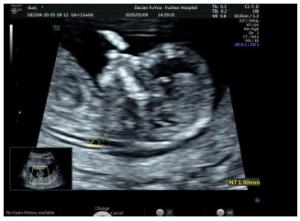
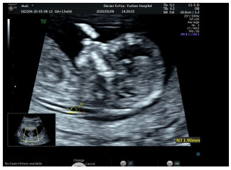
图 2 孕13周行NT超声,测量NT值为1.9 mm
Figure 2. NT ultrasound was performed at 13 weeks of gestation, and the NT value was 1.9 mm.

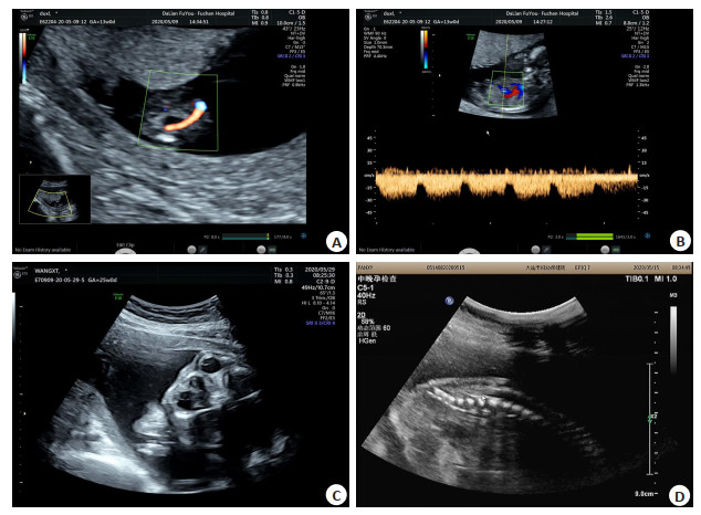
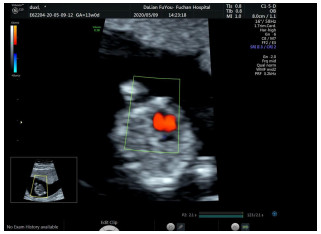
图 3 患者孕13周、24周超声影像学表现
A:孕13周行NT超声检出单脐动脉; B:孕13周行NT超声检出静脉导管a波正向; C:孕24周行二维、四维超声,显示眼眶及晶状体结构; D:孕24周行二维、四维超声检出脊柱圆锥位于L4-L5水平.
Figure 3. Ultrasound image of the patient at 13 and 24 weeks of gestation.
表 1 畸形胎儿和正常胎儿的NT检查水平比较
Table 1. Comparison of NT levels between malformed fetuses and normal fetuses
组别 NT值(mm, Mean±SD) NT异常[n(%)] 畸形胎儿(n=88) 4.02±0.59 82 (93.18) 正常胎儿(n=2329) 1.81±0.31 31 (1.33) t/χ2 62.7505 1605.2162 P 0.0000 0.0000 NT: Nuchal translucency.  下载: 导出CSV
表 2 NT超声检查、彩色多普勒超声检查结果比较[n(%)]
Table 2. Comparison of the results of NT ultrasound and color Doppler ultrasound
畸形类型 引产或分娩结果 NT超声检查 彩色多普勒超声检查 胎死宫内 38 (43.18) 36 (40.91) 37 (42.05) 全前脑 5 (5.68) 5 (5.68) 5 (5.68) 脑积水 3 (3.41) 3 (3.41) 3 (3.41) 胼胝体缺失 4 (4.55) 3 (3.41) 4 (4.55) 唇腭裂 2 (2.27) 2 (2.27) 2 (2.27) 双手内翻 6 (6.82) 5 (5.68) 5 (5.68) 足内翻 3(3.41) 3(3.41) 3(3.41) 四肢短小 3 (3.41) 3 (3.41) 3 (3.41) 隔离肺或肺囊腺瘤 1 (1.14) 1 (1.14) 1 (1.14) 腹部囊性肿块 2 (2.27) 2 (2.27) 2 (2.27) 膈疝 2 (2.27) 2 (2.27) 2 (2.27) 消化道发育异常 2 (2.27) 2 (2.27) 2 (2.27) 心血管发育异常 5 (5.68) 4 (4.55) 5 (5.68) 泌尿系统发育异常 8 (9.09) 7 (7.95) 7 (7.95) 水肿综合征 4(4.55) 4(4.55) 4(4.55) 合计 88 (100.00) 82 (93.18) 85 (96.59)
下载: 导出CSV
表 3 NT超声、彩色多普勒超声以及二者联合在诊断胎儿畸形中的诊断结果(n)
Table 3. Diagnostic results of NT ultrasound, color and their combination in the diagnosis of fetal malformations
检查方式及类型 引产或分娩结果 合计 畸形 正常 NT超声 畸形 82 31 113 正常 6 2298 2304 合计 88 2329 2417 彩色多普勒超声 畸形 85 22 107 正常 3 2307 2310 合计 88 2329 2417 二者联合 畸形 88 10 98 正常 0 2319 2319 合计 88 2329 2417
下载: 导出CSV
表 4 NT超声、彩色多普勒超声以及二者联合在诊断胎儿畸形中的诊断率比较(%)
Table 4. Comparison of diagnostic rates of NT ultrasound, color Doppler ultrasound and their combination in the diagnosis of fetal malformations
检查方式 敏感度 特异度 准确度 NT超声 93.18 (82/88) 98.67 (2298/2329) 98.47 (2380/2417) 彩色多普勒超声 96.59 (85/88) 99.06 (2307/2329) 98.97 (2392/2417) 二者联合 100.00 (88/88) 99.57 (2319/2329) 99.59 (2407/2417) χ2 6.2118 10.6676 15.4029 P 0.0448 0.0048 0.0005
下载: 导出CSV
-
[1] 刘凤林, 许占英, 李建荣.胎儿颈项透明层厚度超声与孕中期四维彩色多普勒超声联合诊断胎儿畸形的价值分析[J].中国药物与临床, 2019, 19(10): 1617-8. http://d.old.wanfangdata.com.cn/Periodical/zgywylc201910012 [2] Hellmuth SG, Pedersen LH, Miltoft CB, et al. Increased nuchal translucency thickness and risk of neurodevelopmental disorders[J]. Ultrasound Obstet Gynecol, 2017, 49(5): 592-8. doi: 10.1002/uog.15961/pdf [3] Ali MM, Chasen ST, Norton ME. Testing for Noonan syndrome after increased nuchal translucency[J]. Prenat Diagn, 2017, 37(8): 750-3. [4] Zhang DF. Application value of prenatal two-dimensional and fourdimensional ultrasound in fetal malformation screening[J]. J Prevent Med Chin PLA, 2019, 37 (7): 20-1, 23. http://en.cnki.com.cn/Article_en/CJFDTotal-YLYS201932008.htm [5] 王桂民, 刘晓英, 张辉.早期超声检测胎儿颈项透明层厚度在胎儿心脏畸形及染色体异常诊断中的应用及相关性[J].中国优生与遗传杂志, 2019, 27(7): 870-2. http://www.cqvip.com/qk/98444x/20197/7002655572.html [6] Holzer I, Husslein PW, Bettelheim D, et al. Value of increased nuchal translucency in the era of noninvasive prenatal testing with cell-free DNA[J]. Int J Gynaecol Obstet, 2019, 145(3): 319-23. http://www.researchgate.net/publication/331968874_Value_of_increased_nuchal_translucency_in_the_era_of_noninvasive_prenatal_testing_with_cell-free_DNA [7] 廖宏伟, 武小丁. NT超声检查技术联合孕中期四维彩超对胎儿结构畸形的诊断价值分析[J].中国实验诊断学, 2019, 23(11): 1888-91. http://www.zhangqiaokeyan.com/academic-journal-cn_health-horizon_thesis/0201270795397.html [8] 赵爱欣, 张效民, 邹爱霞, 等. MRI联合四维超声在胎儿心脏畸形筛查中的应用[J].中国CT和MRI杂志, 2019, 17(9): 73-5. [9] Vigneswaran TV, Homfray T, Allan LD, et al. Persistently elevated nuchal translucency and the fetal heart[J]. J Matern Fetal Neonatal Med, 2018, 31(18): 2376-80. doi: 10.1080/14767058.2017.1342804 [10] 王莉, 谢香梅.胎儿颈项透明层厚度超声检测联合孕中期四维彩超诊断胎儿畸形的应用价值[J].影像研究与医学应用, 2019, 3(11): 112-4. http://www.cnki.com.cn/Article/CJFDTotal-YXYY201911077.htm [11] Alanen J, Leskinen M, Sairanen M, et al. Fetal nuchal translucency in severe congenital heart defects: experiences in Northern Finland[J]. J Matern Fetal Neonatal Med, 2019, 32(9): 1454-60. doi: 10.1080/14767058.2017.1408067 [12] 孙园园, 时雅儒, 宋盼盼, 等.孕早期颈项透明层增厚与胎儿结构畸形的关系[J].中华实用诊断与治疗杂志, 2020, 34(3): 304-7. http://www.wanfangdata.com.cn/details/detail.do?_type=perio&id=syzdyzlzz202003023 [13] 王红军, 夏泽英, 钱土丽, 等.颈项透明层厚度检测联合孕中期的四维彩超检测在胎儿畸形诊断中的参考价值分析[J].中国优生与遗传杂志, 2019, 27(6): 752-4. http://d.wanfangdata.com.cn/periodical/zgysyyczz201906042 [14] 李春梅, 卢利羚.早孕期NT超声筛查联合孕母血清生化指标检测对胎儿结构畸形和染色体异常的诊断价值研究[J].湖南师范大学学报:医学版, 2020, 17(1): 178-82. http://www.cqvip.com/QK/87695A/20201/00002GOIK7807JP0MPDO8JPX6HR.html [15] Kenkhuis MJA, Bakker M, Bardi F, et al. Effectiveness of 12-13- week scan for early diagnosis of fetal congenital anomalies in the cell-free DNA era[J]. Ultrasound Obstet Gynecol, 2018, 51(4): 463-9. doi: 10.1002/uog.17487 [16] Nisbet D, Robertson A, Mannil B, et al. Quality management of nuchal translucency ultrasound measurement in Australia[J]. Aust N Z J Obstet Gynaecol, 2019, 59(1): 54-8. doi: 10.1111/ajo.12792/full [17] 刘吉庆, 苏静, 孟秋霞.四维超声心动图联合二维彩超在胎儿先天性心脏畸形筛查中的应用[J].中国妇幼保健, 2019, 34(19): 4574-6. [18] Barišić LS, Stanojević M, Kurjak A, et al. Diagnosis of fetal syndromes by three- and four-dimensional ultrasound: is there any improvement?[J]. J Perinat Med, 2017, 45(6): 651-65. http://europepmc.org/abstract/MED/28493822 [19] Merz E, Pashaj S. Advantages of 3D ultrasound in the assessment of fetal abnormalities[J]. J Perinat Med, 2017, 45(6): 643-50. http://www.degruyter.com/view/j/jpme.2017.45.issue-6/jpm-2016-0379/jpm-2016-0379.xml?format=INT [20] Syngelaki A, Hammami A, Bower S, et al. Diagnosis of fetal nonchromosomal abnormalities on routine ultrasound examination at 11-13 weeks' gestation[J]. Ultrasound Obstet Gynecol, 2019, 54(4): 468-76. doi: 10.1002/uog.20844 -

 点击查看大图
计量
- 文章访问数: 547
- HTML全文浏览量: 300
- PDF下载量: 2
- 被引次数: 0