


Investigation on the three-dimensional anatomy of supernumerary teeth in the upper anterior region based on CBCT
-
摘要:
目的探讨上颌前牙区多生牙解剖形态特点及其与周围组织的位置关系,为口腔多学科治疗与临床教学提供参考。 方法选取2011年11月~2016年4月在兰州大学口腔医院就诊经锥形束CT(CBCT)确诊为上颌前部多生牙的237例患者的309颗多生牙为研究对象,其中男性187例,女性50例,年龄6~47岁(平均20岁)。通过CBCT三维重建影像并使用扫描软件自带长度测量工具,分析测量上颌前部埋伏多生牙的数目、形态、大小、三维空间位置、唇腭侧骨壁厚度,以及对邻牙及其与周围组织毗邻关系的影响,并进行统计学分析。 结果多生牙多埋伏于颌骨腭侧及牙弓内,其中1颗占71.4%,2颗占27.5%,3颗占1.1%。11与21牙位间占89.5%,其他部位占11.5%。位于腭侧占87.5%,唇侧占4.5%(n=14),唇腭弓内占8.0%(n=25)。圆锥形占79.3%,结节形占9.8%,侧切牙形占10.9%,差异有统计学意义(P<0.05)。倒置占77.4 %,正位占17.0%,其他横向埋伏占5.6%,差异有统计学意义(P<0.05)。按Ⅰ类、Ⅱ类、Ⅲ类骨壁描述分别占83.1%、5.6%、11.3%,差异有统计学意义(P<0.05)。与邻近恒牙或恒牙胚接触占86.7%,与邻近恒牙或恒牙胚距离≥1 mm的占13.3%,差异有统计学意义(P<0.05)。牙齿长度为11.92±2.84 mm,最大冠周径为6.06±2.15 mm。就诊年龄与上颌前部埋伏多生牙临床分型的相关(P<0.05),性别方面以男性较为多见(P<0.05)。 结论上前牙区多生牙解剖形态以及空间位置相对恒定,CBCT能清晰显示多生牙与周围组织结构的三维空间位置关系,为早诊断、开展早期口腔临床干预提供实验测量的数据。 Abstract:ObjectiveDemonstrates the anatomical characteristics of the supernumerary teeth with the surrounding tissue in the middle maxilla to provide a reference for clinical practice. Methods309 supernumerary teeth in the middle maxilla from 237 patients were selected and diagnosed by CBCT at the Stomatological Hospital of Lanzhou University from November 2011 to April 2016, including 187 males and 50 females, with the age from 6 to 47 years old (average 20 years old). Identified the number, evaluated the size, the shape, 3D space, surrounding bone thickness, the neighboring teeth location, and the adjacency relationship of supernumerary teeth by using CBCT three- dimensional reconstruction of the image and the length measurement tool in the CBCT scanning. ResultsSupernumerary teeth are mostly ambushed in the jaw palatal side of the dental arch, one as 71.4% (n=221), two as 27.5% (n=85), and three as 1.1% (n=3). Located on the 11-21 occupy 89.5% (n=277), near the palatal side 87.5% (n=270), near the lip side 4.5% (n=15), internal arch 8.0% (n=25) and other locations occupy 11.5%. The appearance of the teeth covered comicalness (79.3%, n=245), no deform (9.8%, n=30), and lateral incisor (10.9%, n=34). Status of inverted impacted occupied 77.4% (n=239), vertical 17.0% (n=53), and other angulation 5.6% (n=17). According to the bone wall of type Ⅰ, type Ⅱ and type Ⅲ respectively occupied by 83.1% (n=257), 5.6% (n=17), 11.3% (n=35). Around 86.7% (n= 268) contact with permanent or germ teeth, among 13.3% (n=41) distance greater than 1 mm. Teeth average length is 11.92 mm and the largest diameter of the crown is 6.06 mm. ConclusionAnatomic morphology and spatial position of supernumerary teeth in the upper anterior region are relatively stable. CBCT can display the neighboring tissue in a three-dimensional way to provide an experimental basis for early diagnosis and early intervention into a dental clinic. -
Key words:
- supernumerary teeth /
- cone-beam CT /
- anatomy
-

图 1 CBCT示上颌正中倒置多生牙在上颌骨中矢状(A)、冠状(B)及横断(C)方向的位置
Figure 1. CBCT scan exhibiting a maxillary inverted supernumerary tooth in the anterior maxilla in the sagittal(A), coronal(B) and transverse(C) directions of the maxilla.
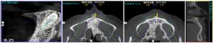
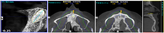
图 3 上颌正中倒置多生牙牙长测量(A)、最大冠周径(B)及多生牙到切牙孔最短距离(C)的测量及其上颌正中多生牙到鼻腔的最短距离(D)
Figure 3. Measurement of tooth length (A) and maximum crown circumference (B) of the supernumerary tooth; shortest distance from the maxillary inverted supernumerary tooth in the anterior maxilla to the incisive foramen (C) and nasal cavity (D) respectively
表 1 多生牙的分布特点(n=309)
Table 1. Distribution characteristics of supernumerary teeth
因素 牙数 埋伏牙位置分型 Pearson χ2 P 唇侧 腭侧 牙弓内 唇腭弓内 性别 8.597 0.035 男 221 9 200 2 10 女 88 5 70 1 12 年龄(岁) 8.338 0.040 6~20 292 11 256 3 22 ≥20 17 3 14 0 0 与牙的位置关系 12.067 0.007 11与21牙间 277 9 246 2 20 其他 32 5 24 1 2 形态 15.467 0.017 圆锥形 245 8 221 2 14 结节形 30 1 24 1 4 侧切牙形 34 5 25 0 4 牙齿数目(颗) 15.268 0.018 1 221 10 235 3 13 2 85 4 34 0 9 3 3 0 1 0 0 与邻近恒牙或恒牙胚的距离 11.976 0.007 接触(<1 mm) 268 10 241 2 15 不接触(≥1 mm) 41 4 29 1 7 唇腭侧骨壁厚度 15.077 0.020 Ⅰ类 257 10 232 2 13 Ⅱ类 17 1 12 0 4 Ⅲ类 35 3 26 1 5  下载: 导出CSV
-
[1] Ata-Ali F, Ata-Ali J, Peñarrocha-Oltra D, et al. Prevalence, etiology, diagnosis, treatment and complications of supernumerary teeth[J]. J Clin Exp Dent, 2014, 6(4): e414-8. [2] Mallineni SK. Supernumerary teeth: review of the literature with recent updates[J]. Conf Pap Sci, 2014, 2014: 1-6. [3] Rajab LD, Hamdan M. Supernumerary teeth: review of the literature and a survey of 152 cases[J]. Int J Paediatr Dent, 2010, 12(4): 244- 54. [4] Wang XP, Fan JB. Molecular genetics of supernumerary tooth formation[J]. Genesis, 2011, 49(4): 261-77. doi: 10.1002/dvg.20715 [5] Hattab FN. Double talon cusps on supernumerary tooth fused to maxillary central incisor: Review of literature and report of case[J]. J Clin Exp Dent, 2014, 6(4): e400-7. [6] Ferrazzano GF, Cantile T, Roberto L, et al. An impacted central incisor due to supernumerary teeth: a multidisciplinary approach[J]. Eur J Paediatr Dent, 2014, 15(2 Suppl): 187-90. [7] 石四箴.儿童口腔医学(第3版)[M].北京:人民卫生出版社, 2011. [8] Ferrés-Amat E, Ferrés-Amat E, Prats-Armengol J, et al. A survey of 183 paediatric patients from Barcelona including 239 supernumerary unerupted teeth[J]. Int J Oral Maxillofac Surg, 2013, 42 (10): 1315. [9] Singh VP, Sharma A, Sharma S. Supernumerary teeth in nepalese children[J]. Sci World J, 2014, 2014: 215396. [10] Fernández Montenegro P, Valmaseda Castellón E, Berini Aytés L, et al. Retrospective study of 145 supernumerary teeth[J]. Med Oral Patol Oral Cir Bucal, 2006, 11(4): E339-44. [11] Mossaz J, Kloukos D, Pandis N, et al. Morphologic characteristics, location, and associated complications of maxillary and mandibular supernumerary teeth as evaluated using cone beam computed tomography[J]. Eur J Orthod, 2014, 36(6): 708-18. doi: 10.1093/ejo/cjt101 [12] De Oliveira Gomes C, Drummond SN, Jham BC, et al. A survey of 460 supernumerary teeth in Brazilian children and adolescents[J]. Int J Paediatr Dent, 2008, 18(2): 98-106. doi: 10.1111/j.1365-263X.2007.00862.x [13] Khalaf K, Robinson DL, Elcock C, et al. Tooth size in patients with supernumerary teeth and a control group measured by image analysis system[J].Arch Oral Biol, 2005, 50(2): 243-8. doi: 10.1016/j.archoralbio.2004.09.013 [14] Finkelstein T, Shapira Y, Bechor N, et al. Surgical and orthodontic treatment of a fused maxillary central incisor and supernumerary tooth[J]. J Clin Orthod, 2014, 48(10): 654-8. [15] Gao YB, Lin ZY, Rodella LF, et al. Piezoelectric ultrasonic bone surgery system in the extraction surgery of supernumerary teeth[J]. J Craniomaxillofac Surg, 2014, 42(8): 1577-82. doi: 10.1016/j.jcms.2014.04.007 [16] 许竞, 张治勇, 邝拮喆, 等.以上下颌牙切线位X线片定位埋伏多生牙的效果探讨[J].实用口腔医学杂志, 2008, 24(2): 223-7. doi: 10.3969/j.issn.1001-3733.2008.02.017 [17] Sonick M, Abrahams J, Faiella RA. A comparison of the accuracy of periapical, panoramic, and computerized tomographic radiographs in locating the mandibular canal[J]. Int J Oral Maxillofac Implants, 1994, 9(4): 455-60. [18] Matherne RP, Angelopoulos C, Kulild JC, et al. Use of cone-beam computed tomography to identify root canal systems in vitro[J]. J Endod, 2008, 34(1): 87-9. doi: 10.1016/j.joen.2007.10.016 [19] 李晓敏, 陈蕾, 张治勇, 等. 3种不同口腔慢性炎症引起牙源性上颌窦炎的锥形束CT比较[J].分子影像学杂志, 2018, 41(2): 137-41. [20] Larheim TA, Abrahamsson AK, Kristensen M, et al. Temporomandibular joint diagnostics using CBCT[J]. Dentomaxillofac Radiol, 2015, 44(1): 20140235. doi: 10.1259/dmfr.20140235 [21] Terry DA, Triolo PT, Swift EJ. Fabrication of direct fiber-reinforced posts: a structural design concept[J]. J Esthet Restor Dent, 2010, 13 (4): 228-40. [22] Patel S, Dawood A, Ford TP, et al. The potential applications of cone beam computed tomography in the management of endodontic problems[J]. Int Endod J, 2007, 40(10): 818-30. doi: 10.1111/j.1365-2591.2007.01299.x [23] Tsuji M, Suzuki H, Suzuki S, et al. Three-dimensional evaluation of morphology and position of impacted supernumerary teeth in cases of cleidocranial dysplasia[J]. Congenit Anom (Kyoto), 2020, 60(4): 106-14. doi: 10.1111/cga.12358 [24] Jiang Y, Ma XW, Wu YP, et al. Epidemiological, clinical, and 3- dimentional CBCT radiographic characterizations of supernumerary teeth in a non- syndromic adult population: a single- institutional study from 60, 104 Chinese subjects[J]. Clin Oral Investig, 2020. DOI: 10.1007/s00784-020-03288-3. [25] Fardi A, Kondylidou- Sidira A, Bachour Z, et al. Incidence of impacted and supernumerary teeth- a radiographic study in a North Greek population[J]. Med Oral Patol Oral Cir Bucal, 2011, 16(1): 56- 61. [26] Cassetta M, Altieri F, Giansanti M, et al. Morphological and topographical characteristics of posterior supernumerary molar teeth: an epidemiological study on 25, 186 subjects[J]. Med Oral Patol Oral Cir Bucal, 2014, 19(6): e545-9. [27] Bereket C, Çakır- Özkan N, Şener İ, et al. Analyses of 1100 supernumerary teeth in a nonsyndromic Turkish population: a retrospective multicenter study[J]. Niger J Clin Pract, 2015, 18(6): 731-8. doi: 10.4103/1119-3077.154213 [28] McBeain M, Miloro M. Characteristics of supernumerary teeth in nonsyndromic population in an urban dental school setting[J]. J Oral Maxillofac Surg, 2018, 76(5): 933-8. doi: 10.1016/j.joms.2017.10.013 [29] 文陈妮, 李果, 任家银, 等.锥形束CT诊断上颌前牙区多生牙价值研究[J].华西口腔医学2012, 30(4): 399-401. doi: 10.3969/j.issn.1000-1182.2012.04.017 [30] Anthonappa RP, Omer RS, King NM. Characteristics of 283 supernumerary teeth in southern Chinese children[J]. Oral Surg Oral Med Oral Pathol Oral Radiol Endod, 2008, 105(6): e48-e54. doi: 10.1016/j.tripleo.2008.01.035 [31] 古向生, 李汝瑶, 王大为, 等.正中多生牙与中切牙间隙的关系[J].华西口腔医学杂志, 2002, 20(2): 128-30. doi: 10.3321/j.issn:1000-1182.2002.02.014 [32] 王桂红, 戴印和, 胡培, 等.早期拔除上颌正中多生牙与中切牙间隙之间关系的临床研究[J].口腔医学, 2013, 33(1): 33-6. doi: 10.3969/j.issn.1006-673X.2013.01.009 [33] 葛立宏, 王旭.多生牙发生的分子生物学研究进展[J].北京大学学报:医学版, 2013, 45(4): 661-5. doi: 10.3969/j.issn.1671-167X.2013.04.034 [34] 韩方凯, 王铁梅, 廖波, 等.应用多个因素分析方法曲面体层与CBCT对上颌前部埋伏牙定位的对照研究[J].口腔医学研究, 2012, 28(9): 903-6. [35] Dalili Z, Mahjoub P, Sigaroudi AK. Comparison between cone beam computed tomography and panoramic radiography in the assessment of the relationship between the mandibular canal and impacted class C mandibular third molars[J]. Dent Res J (Isfahan), 2011, 8(4): 203- 10. doi: 10.4103/1735-3327.86041 [36] Scarfe WC, Farman AG, Sukovic P. Clinical applications of conebeam computed tomography in dental practice[J]. J Can Dent Assoc, 2006, 72(1): 75-80. [37] Quereshy FA, Savell TA, Palomo JM. Applications of cone beam computed tomography in the practice of oral and maxillofacial surgery[J]. J Oral Maxillofac Surg, 2008, 66(4): 791-6. doi: 10.1016/j.joms.2007.11.018 [38] Garvey MT, Barry HJ, Blake M. Supernumerary teeth: an overview of classification, diagnosis and management[J]. J Can Dent Assoc, 1999, 65(11): 612-6. [39] Lai CS, Suter VG, Katsaros C, et al. Localization of impacted maxillary canines and root resorption of neighbouring teeth: a study assessing the diagnostic value of panoramic radiographs in two groups of observers[J]. Eur J Orthod, 2014, 36(4): 450-6. doi: 10.1093/ejo/cjt074 [40] Ferreira-Junior O, de Avila LD, Sampieri MB, et al. Impacted lower third molar fused with a supernumerary tooth: diagnosis and treatment planning using cone-beam computed tomography[J]. Int J Oral Sci, 2009, 1(4): 224-8. doi: 10.4248/IJOS09056 [41] Sano T, Tachibana T, Koide A, et al. Clinical Investigation of impacted maxillary anterior supernumerary teeth using cone beam computed tomography and panoramic radiography[J]. Jap J Ped Dent, 2015, (53): 22-6. [42] Ozan F, Kara I, Ay S. Impacted mandibular permanent incisors associated with a supernumerary tooth: a case report[J]. Eur J Dent, 2009, 3(4): 324-8. doi: 10.1055/s-0039-1697452 [43] Demiriz L, Hazar Bodrumlu E, İçen M, et al. Evaluation of the accuracy of cone beam computed tomography on measuring impacted supernumerary teeth[J]. Scanning, 2016, 38(6): 579-84. doi: 10.1002/sca.21303 [44] Liu DG, Zhang WL, Zhang ZY, et al. Three-dimensional evaluations of supernumerary teeth using cone-beam computed tomography for 487 cases[J]. Oral Surg Oral Med Oral Pathol Oral Radiol Endod, 2007, 103(3): 403-11. doi: 10.1016/j.tripleo.2006.03.026 [45] Silva MA, Wolf U, Heinicke F, et al. Cone-beam computed tomography for routine orthodontic treatment planning: a radiation dose evaluation[J]. Am J Orthod Dentofacial Orthop, 2008, 133(5): 640. e1-5. [46] Tuna EB, Kurklu E, Gencay K, et al. Clinical and radiological evaluation of inverse impaction of supernumerary teeth[J]. Med Oral Patol Oral Cir Bucal, 2013, 18(4): e613-8. [47] Lai CS, Bornstein MM, Mock L, et al. Impacted maxillary canines and root resorptions of neighbouring teeth: a radiographic analysis using cone-beam computed tomography[J]. Eur J Orthod, 2013, 35 (4): 529-38. doi: 10.1093/ejo/cjs037 -

 点击查看大图
计量
- 文章访问数: 638
- HTML全文浏览量: 369
- PDF下载量: 3
- 被引次数: 0