


Diagnostic value of MRI, MRV and DWI in cerebral venous sinus thrombosis at different times
-
摘要:
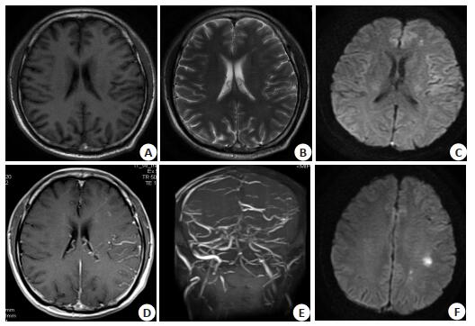
目的探讨MRI、磁共振静脉造影(MRV)及弥散加权成像(DWI)在不同时期脑静脉窦血栓中的诊断价值。 方法回顾性分析本院2015年11月~2019年8月经数字减影血管造影或临床随访确诊的27例脑静脉窦血栓患者的MRI资料,其中18例患者行DWI检查,9例患者接受MRI增强检查。 结果27例静脉窦血栓患者MRI平扫表现流空信号消失,静脉窦出现异常信号,3例脑静脉窦血栓患者MRI增强后表现为空三角征,MRV则显示静脉窦血流高信号不同程度缺失,局限性或阶段性血栓以血流高信号变细、中断为主。DWI对急性和亚急性脑静脉窦血栓及继发静脉性脑梗塞敏感。 结论MRI、MRV联合DWI有助于静脉窦血栓的早期诊断并可以反映血栓的演变过程,为临床及时治疗静脉窦血栓具有重要意义。 Abstract:ObjectiveTo evaluate the diagnostic value of MRI, MRV and DWI in cerebral venous sinus thrombosis at different times. MethodsMRI data of 27 patients with cerebral venous sinus thrombosis diagnosed by DSA or clinical follow-up from 2015 to 2019 were analyzed retrospectively. Eighteen patients underwent DWI examination and 9 patients underwent MRI enhancement. ResultsThe MRI showed that the emplacement signal disappeared and the venous sinus appeared abnormal signal in 27 patients with venous sinus thrombosis. 3 cases of CVST showed an empty triangular sign after MRI enhancement. MRV showed that the venous sinus flow signal was different in varying degrees, Or stage thrombosis with high blood flow signal thinning, interruption-based. DWI is sensitive to acute and subacute cerebral venous sinus thrombosis and secondary venous cerebral infarction. ConclusionMRI, MRV combined with DWI can help the early diagnosis of venous sinus thrombosis and reflect the evolution of thrombus.It has great significance for the clinical treatment of venous sinus thrombosis. -
[1] 李新瑜, 杨静, 鲁果果, 等.扩散加权成像静脉窦高信号在脑静脉窦血栓诊断及预测再通的价值[J].实用放射学杂志, 2019, 38(7): 1038-41. http://d.old.wanfangdata.com.cn/Periodical/syfsxzz201907003 [2] Dhadke VN, Dhadke SV, Kulkarni A. Clinical profile of cerebral venous sinus thrombosis[J]. J Assoc Physicians India, 2020, 68(3): 33-5. http://d.old.wanfangdata.com.cn/OAPaper/oai_doaj-articles_5cfbec050427f2e992d6f113e7ac013b [3] Kunz WG, Schuler F, Sommer WH, et al. Wavelet-based angiographic reconstruction of computed tomography perfusion data: diagnostic value in cerebral venous sinus thrombosis[J]. Invest Radiol, 2017, 52(5): 302-9. https://insights.ovid.com/crossref?an=00004424-201705000-00007 [4] 宋娟.颅内静脉窦血栓的CT表现[J].影像研究与医学应用, 2018, 2 (8): 189-90. http://d.old.wanfangdata.com.cn/Periodical/zgzxyjhyxxzz201605021 [5] 于磊.核磁诊断在脑静脉窦血栓患者的应用价值[J].血栓与止血学, 2020, 26(1): 46-7. http://d.old.wanfangdata.com.cn/Periodical/xsyzxx202001017 [6] Tang PH, Chai J, Chan YH, et al. Superior sagittal sinus thrombosis: subtle signs on neuroimaging[J]. Ann Acad Med Singap, 2008, 37 (5): 397-401. https://core.ac.uk/display/48790174 [7] 杜彦瑶, 许若梅, 王效春, 等.脑静脉窦血栓形成的影像学诊断及预后评估[J].中华放射学杂志, 2020, 54(4): 380-4. http://d.old.wanfangdata.com.cn/Periodical/zhfsx202004026 [8] 邢万平.磁共振成像以及磁共振静脉系成像平扫对颅内静脉窦血栓的诊断价值[J].血栓与止血学, 2019, 25(5): 798-9. http://d.old.wanfangdata.com.cn/Periodical/xsyzxx201905029 [9] 朱坤, 段建航. MRI检查对脑静脉窦血栓患者诊断准确率的影响[J].医学理论与实践, 2020, 33(8): 1325-6. http://d.old.wanfangdata.com.cn/Periodical/yxllysj202008063 [10] 庞华军, 赵峰, 卓兵芝, 等. CTV及MRV在颅内静脉窦血栓中的临床应用价值分析[J].农垦医学, 2019, 41(2): 144-6. http://d.old.wanfangdata.com.cn/Periodical/nkyx201902014 [11] 郝绍江.磁共振检查对脑静脉窦血栓行诊断试验相关性的评价[J].延安大学学报:医学科学版, 2019, 17(1): 72-4. http://d.old.wanfangdata.com.cn/Periodical/yadxxb-yxkxb201901020 [12] 朱丽明.脑静脉窦血栓形成MRI表现[J].中外医疗, 2019, 38(33): 172-4. http://d.old.wanfangdata.com.cn/Periodical/zgyxjsjcx200201002 [13] Kalita J, Singh VK, Jain N, et al. Cerebral venous sinus thrombosis score and its correlation with clinical and MRI findings[J]. J Stroke Cerebrovasc Dis, 2019, 28(11): 104324. https://www.sciencedirect.com/science/article/pii/S1052305719303775 [14] 李海云, 乔珊, 崔才三. 127例颅内静脉窦血栓形成的回顾性分析[J].山东大学学报(医学版), 2020, 58(5): 77-81. http://d.old.wanfangdata.com.cn/Periodical/shandykdxxb202005012 [15] 梁其洲, 李香营, 田宇, 等.多层螺旋CT在脑静脉窦血栓诊断中的价值[J].中国地方病防治杂志, 2017, 32(5): 554, 556. http://www.wanfangdata.com.cn/details/detail.do?_type=perio&id=zgdfbfzzz201705037 [16] 魏亚军. MRI对脑静脉窦血栓的诊断价值[J].河南医学研究, 2019, 28(22): 4149-50. http://d.old.wanfangdata.com.cn/Periodical/zhsjwkjbyjzz201205005 [17] Hiwatashi A, Kinoshita T, Moritani T, et al. Hypointensity on diffusion-weighted MRI of the brain related to T2 shortening and susceptibility effects[J]. AJR Am J Roentgenol, 2003, 181(6): 1705-9. doi: 10.2214/ajr.181.6.1811705 [18] Yildiz ME, Ozcan UA, Turk A, et al. Diffusion-weighted MR imaging findings of cortical vein thrombosis at 3 T[J]. Clin Neuroradiol, 2015, 25(3): 249-56. http://www.wanfangdata.com.cn/details/detail.do?_type=perio&id=98ad5a1ae9b56f30636dfc38037da997 [19] 张秋实, 曾小松, 赵耀. MRI及MRV在脑静脉和静脉窦血栓形成中的诊断价值[J].安徽医学, 2014, 35(11): 1512-4. http://d.old.wanfangdata.com.cn/Periodical/ahyx201411012 -

 下载:
下载:
 点击查看大图
图(1)
计量
- 文章访问数: 706
- HTML全文浏览量: 284
- PDF下载量: 4
- 被引次数: 0