


Clinical application of high frequency ultrasound in the diagnosis of superficial softtissue and small organ diseases
-
摘要:
目的探究高频超声在浅表软组织及小器官疾病诊断中的临床应用价值。 方法回顾性分析2014年5月~2017年5月我院收治的78例浅表软组织及小器官疾病患者临床资料,所有患者均采用高频超声进行诊断,将诊断结果与最终病理诊断结果对比,计算高频超声诊断的准确率及不同疾病的诊断的准确率是否具有差异,并对图像进行评价分析,计算图像优良率。 结果患者入院后均行组织病理学检查确诊。其中甲状腺疾病15例,乳腺疾病29例,男性外生殖器疾病18例,浅表软组织病变16例。甲状腺疾病高频超声检出率为93.33%,乳腺疾病高频超声检出率为93.10%,男性外生殖器疾病高频超声检出率为94.44%,浅表软组织疾病变高频超声检出率为87.50%,4种浅表软组织及小器官疾病高频超声检出率差异无统计学意义(P > 0.05)。对高频超声图像质量进行评价,其中优图像71例,良图像5例,差图像2例,图像优良率为97.44%。 结论高频超声对各类浅表软组织及小器官疾病的诊断具有较高的准确性,图像优良率高,且操作简单、无创、安全性高,值得临床推广。 Abstract:ObjectiveTo explore the clinical value of high frequency ultrasound in the diagnosis of superficial soft tissue and small organ diseases. MethodsWe retrospectively analyzed the clinical data of 78 patients with superficial soft tissue and small organ diseases admitted to the hospital from May 2014 to May 2017. All patients were diagnosed by high-frequency ultrasound and the diagnostic results were compared with the final pathological diagnostic results. The diagnostic accuracy of high-frequency ultrasound and the diagnostic accuracy rates for different diseases were calculated. The images were analyzed, and the excellent rate of images were calculated. ResultsAll patients were diagnosed by histopathological examination after admission. Among them, there were 15 cases of thyroid diseases, 29 cases of breast diseases, 18 cases of male external genital diseases and 16 cases of superficial soft tissue lesions. The detection rates of thyroid diseases, breast diseases, male external genital diseases and superficial soft tissue lesions by high-frequency ultrasound were 93.33%, 93.10%, 94.44% and 87.50%, respectively, without significant difference in the detection rates of the four superficial soft tissue or small organ diseases by high-frequency ultrasound (P < 0.05). The image quality of high-frequency ultrasound was evaluated excellent in 71 cases, good in 5 cases and poor in 2 cases. The excellent and good rate of images was 97.44%. ConclusionHigh-frequency ultrasound has high accuracy in the diagnosis of various superficial soft tissue and small organ diseases. The excellent and good rate of images is high. It is easy to operate and non-invasive, with high safety. -
Key words:
- high frequency ultrasound /
- superficial soft tissue /
- small organ /
- diagnosis
-

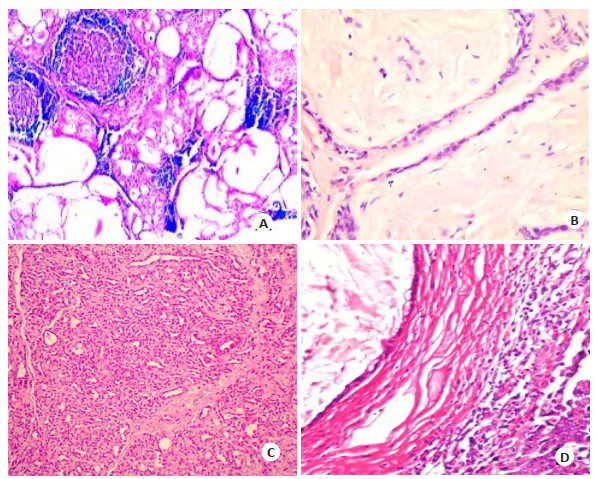
图 1 不同疾病典型病理学图像
A:弥漫中结节型甲状腺炎; B:局部结节增生型乳腺增生; C:血管瘤; D:前列腺囊肿.
Figure 1. Typical pathological images of different diseases.

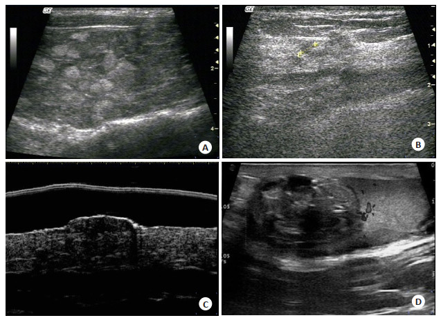
图 2 不同疾病典型高频超声图像
A:弥漫中结节型甲状腺炎; B:局部结节增生型乳腺增生; C:血管瘤; D:前列腺囊肿.
Figure 2. Typical high-frequency ultrasound images of different diseases.
表 1 高频超声检出率
Table 1. Detection rates by high-frequency ultrasound
疾病类型 病理结果(n) 高频超声结果(n) 检出率(%) 甲状腺疾病 甲状腺腺瘤 9 8 88.89 甲状腺炎 4 4 100.00 甲状腺癌 2 2 100.00 乳腺疾病 乳腺增生 13 13 100.00 乳腺纤维瘤 9 7 77.78 乳腺癌 7 7 100.00 男性外生殖器疾病 睾丸肿瘤 2 2 100.00 前列腺囊肿 12 11 91.67 前列腺结核 4 4 100.00 浅表软组织病变 脂肪瘤 8 8 100.00 皮肤纤维瘤 2 1 50.00 皮脂腺囊肿 3 3 100.00 血管瘤 1 1 100.00 神经纤维肉瘤 1 0 0.00 神经鞘瘤 1 1 100.00 总计 78 72 92.31  下载: 导出CSV
表 2 不同疾病高频超声检出率对比
Table 2. Comparison of detection rates of different diseases by high-frequency ultrasound[n(%)]
检查方式 甲状腺疾病 乳腺疾病 男性外生殖器疾病 浅表软组织疾病 总计 组织病理学 15 (100.00) 29 (100.00) 18 (100.00) 16 (100.00) 78 (100.00) 高频超声 14 (93.33) 27 (93.10) 17 (94.44) 14 (87.50) 72 (92.31) χ2 1.003 P 0.801
下载: 导出CSV
-
[1] 雷祖国.超声诊断对浅表器官疾病的价值[J].深圳中西医结合杂志, 2016, 26(17): 55-6. http://www.wanfangdata.com.cn/details/detail.do?_type=perio&id=szzxyjhzz201617028 [2] Iida Y, Kamijo T, Kusafuka K, et al. Depth of invasion in superficial oral tongue carcinoma quantified using intraoral ultrasonography[J]. Laryngoscope, 2018, 128(12): 2778-82. http://www.wanfangdata.com.cn/details/detail.do?_type=perio&id=10.1002/lary.27305 [3] 罗秀勤.高频超声在浅表器官疾病中的临床应用[J].临床合理用药杂志, 2015, (16): 9-13. http://d.old.wanfangdata.com.cn/Periodical/lchlyyzz201516006 [4] 张翠明, 聂宏娟, 王泽, 等.高频超声在浅表淋巴结疾病鉴别诊断中的应用[J].山西医药杂志, 2013, 35(20): 1118-9. http://d.old.wanfangdata.com.cn/Periodical/sxyyzz201320016 [5] 陈璐, 陈莉, 王婧玲, 等.外阴浅表性血管黏液瘤高频超声表现1例[J].中国医学影像技术, 2019, 35(4): 525-7. http://d.old.wanfangdata.com.cn/Periodical/zgyxyxjs201904013 [6] Zhang JL, Wang JH, Bai AF, et al. The significance of high-frequency ultrasound-guided breast mass biopsy in the diagnosis of breast cancer[J]. Eur J Gynaecologi Oncolo, 2017, 38(5): 741-4. doi: 10.12892/ejgo3814.2017 [7] He Y, Liao HM, Xiang X, et al. High-frequency ultrasonography and contrast-enhanced ultrasound for the evaluation of testicular capillary hemangioma: a case report[J]. Medicine (Baltimore), 2019, 98(11): e14779-87. http://cn.bing.com/academic/profile?id=2f400e21af15d86456b9c551518dcb6b&encoded=0&v=paper_preview&mkt=zh-cn [8] 刘文伟.研究高频超声在小器官疾病诊断中的应用价值[J].世界最新医学信息文摘:电子版, 2018, 18(31): 124-6. http://www.wanfangdata.com.cn/details/detail.do?_type=perio&id=sjzxyy-e201831097 [9] Schwartz TL, Margenthaler JA. Axillary ultrasound before neoadjuvant chemotherapy for breast cancer: don't discount the benefits yet[J]. Ann Surg Oncol, 2017, 24(3): 618-20. http://cn.bing.com/academic/profile?id=4ba7aab58b4be56d58955d2f216ca208&encoded=0&v=paper_preview&mkt=zh-cn [10] Qing T, Zhu HH, Hui L. Significance of contrast-enhanced ultrasonography in differential diagnosis of thyroid nodules[J]. Medicine, 2018, 97(40): e12688-95. http://cn.bing.com/academic/profile?id=b3b47d9671df6ad9d35246c06c19333f&encoded=0&v=paper_preview&mkt=zh-cn [11] Katayama H. Ultrasonic assessment of vascular function in Kawasaki disease[J]. J Med Ultrason (2001), 2019, 46(1): 1-2. http://www.wanfangdata.com.cn/details/detail.do?_type=perio&id=58ecbcab99f506a3a7db6dc04d2b754c [12] 刘桂梅, 葛辉玉, 冉维强, 等.高频超声对非性功能障碍男性不育患者阴囊疾病的诊断价值分析[J].中国全科医学, 2017, 20(30): 3791-5. http://d.old.wanfangdata.com.cn/Periodical/zgqkyx201730018 [13] Zhao H, Zou LW, Geng XP, et al. Limitations of mammography in the diagnosis of breast diseases compared with ultrasonography: a single-center retrospective analysis of 274 cases[J]. Eur J Med Res, 2015, 20(1): 49-56. http://cn.bing.com/academic/profile?id=1c0b44a53407eb53dd002e97464175e3&encoded=0&v=paper_preview&mkt=zh-cn [14] 莫泽来, 卢海兰, 吕立威, 等.联合应用高频浅表小器官及腔内探头对肛周脓肿的超声诊断[J].中国地方病防治杂志, 2017, 32(7): 834-7. http://d.old.wanfangdata.com.cn/Periodical/syyxzz200902081 [15] 袁峰.浅表器官疾病超声诊断的价值分析[J].中国社区医师, 2017, 33(8): 103-4. http://d.old.wanfangdata.com.cn/Periodical/zgsqys201708064 [16] Huang M, Tang XW, Cheng XX, et al. Clinical effect of high frequency ultrasound on the screening of dense breast cancer in women[J]. Clin Med, 2016, 15(2): 1275-8. http://www.en.cnki.com.cn/Article_en/CJFDTotal-EBED201604008.htm [17] van Mulder TJ, de Koeijer M, Theeten H, et al. High frequency ultrasound to assess skin thickness in healthy adults[J]. Vaccine, 2017, 35(14): 1810-5. http://www.wanfangdata.com.cn/details/detail.do?_type=perio&id=8d3102446078ceb394055005c5667b23 [18] 邓美玲, 尹剑, 张志龙, 等.婴幼儿化脓性肛瘘超声诊断52例分析[J].中国临床医生, 2014, 42(9): 43-4. http://d.old.wanfangdata.com.cn/Periodical/zglcys201409022 [19] 陈菲, 黄雪, 陈艳.高频超声检查对浅表软组织肿物的诊断价值研究[J].中国临床医生杂志, 2018, 46(2): 202-3. http://d.old.wanfangdata.com.cn/Periodical/zglcys201802026 [20] Piłat P, Borzęcki A, Jazienicki M, et al. High-frequency ultrasound in the diagnosis of selected non-melanoma skin nodular lesions[J]. Postepy Dermatol Alergol, 2019, 36(5): 572-80. http://cn.bing.com/academic/profile?id=4712a72f117e828054c331f4b9a704ac&encoded=0&v=paper_preview&mkt=zh-cn -

 点击查看大图
计量
- 文章访问数: 782
- HTML全文浏览量: 274
- PDF下载量: 10
- 被引次数: 0