


Diagnostic value of bedside color doppler ultrasonography in renal pseudoaneurysm hemorrhage after percutaneous nephrolithotripsy
-
摘要:
目的探讨床旁彩色多普勒超声对经皮肾镜碎石取石术(PCNL)术后并发肾假性动脉瘤出血的临床应用价值。 方法对2017年11月~2019年11月在南方医院采用介入栓塞治疗的9例PCNL术后并发肾出血患者的行床旁彩色多普勒超声检查、临床资料、数字减影血管造影检查及治疗效果进行回顾性分析。 结果9例行肾动脉栓塞术治愈的患者(造影结果详细列出:单纯动脉损伤5例,假性动脉瘤4例,动静脉瘘0例),动脉栓塞前均行床边彩超检查,其中床旁彩超诊断出4例肾假性动脉瘤,最终经肾动脉造影确诊。 结论床旁彩色多普勒超声,对PCNL术后继发肾假性动脉瘤的诊断有较大的优越性和临床价值。 Abstract:ObjectiveTo investigate the clinical value of bedside color doppler ultrasound in the diagnosis of renal pseudoaneurysm hemorrhage after percutaneous nephrolithotripsy (PCNL). MethodsNine patients with renal hemorrhage after PCNL treated by interventional embolization in Southern Hospital from November 2017 to November 2019 were enrolled. The bedside color doppler ultrasonography, clinical data and digital subtraction angiography were analyzed retrospectively. ResultsAmong 9 patients, 5 cases were finally diagnosed with arterial injury, 4 cases with pseudoaneurysm and no cases of arteriovenous fistula. All participants were examined by bedside color doppler ultrasound before arterial embolization. Four cases of renal pseudoaneurysm were initially diagnosed by bedside color Doppler Ultrasound and finally confirmed by renal arteriography. ConclusionBedside color doppler ultrasound. It has great superiority and clinical value in the diagnosis of renal pseudoaneurysm. -

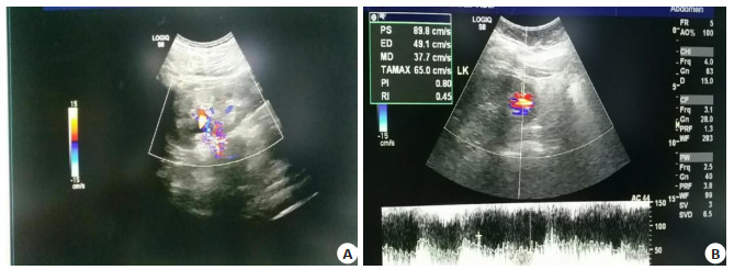
图 1 患者肾脏无回声囊性包块的二维超声及彩色多普超声表现
A:二维超声示左肾中上级无回声囊性包块, 无明显囊壁结构, 囊内透声差, 未见附壁血栓; B:彩色多普勒超声示无回声囊性包块内为红蓝相间的旋转血流信号.
Figure 1. Imaging of anechic cystic mass of kidney by two-dimensional ultrasound and color Doppler ultrasound.

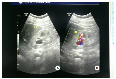
图 2 患者肾脏无回声囊性包块的二维超声及彩色多普勒超声表现
A:二维超声示左肾中上级无回声囊性包块, 无明显囊壁结构, 囊内透声差, 未见附壁血栓; B:彩色多普勒超声示无回声囊性包块内为红蓝相间的旋转血流信号.
Figure 2. Imaging of anechic cystic mass of kidney by two-dimensional ultrasound and color Doppler ultrasound.

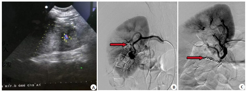
图 3 患者确诊为肾假性动脉瘤
A:床旁彩色多普勒超声发现肾假性动脉瘤; B: RAP栓塞前,肾血管造影显示肾动脉分支假性动脉瘤(红色箭头); C:弹簧圈栓塞后血管造影显示RAP消失(红色箭头).
Figure 3. The patient was diagnosed as renal pseudoaneurysm.
表 1 患者一般资料
Table 1. General information of patients
病例编号 年龄(岁) 性别 BMI (kg/m2) 出血时间(d) 动脉瘤大小(cm×cm) 出血类型 栓塞位置 1 54 女 23.53 7 1.1×1.2 假性动脉瘤 中级 2 57 女 20.28 9 单纯性肾动脉损伤 下级 3 45 男 24.09 10 1.9×1.2 假性动脉瘤 中级 4 57 男 21.71 0.25 单纯性肾动脉损伤 下级 5 68 男 23.94 15 2.2×1.8 假性动脉瘤 上级 6 62 女 25.34 8 1.1×1.6 假性动脉瘤 下级 7 55 女 20.90 4 单纯性肾动脉损伤 中级 8 60 男 27.68 7 单纯性肾动脉损伤 下级 9 59 女 23.52 11 单纯性肾动脉损伤 中级  下载: 导出CSV
-
[1] Türk C, Petřík A, Sarica K, et al. EAU guidelines on interventional treatment for urolithiasis[J]. Eur Urol, 2016, 69(3): 475-82. http://cn.bing.com/academic/profile?id=d02ee83591cf1bdc782237ae4c19e0a3&encoded=0&v=paper_preview&mkt=zh-cn [2] Wu WJ, Okeke Z. Current clinical scoring systems of percutaneous nephrolithotomy outcomes[J]. Nat Rev Urol, 2017, 14(8): 459-69. http://cn.bing.com/academic/profile?id=e4b1880a27a19906466f76942c78c2ba&encoded=0&v=paper_preview&mkt=zh-cn [3] Borofsky MS, Lingeman JE. The role of open and laparoscopic stone surgery in the modern era of endourology[J]. Nat Rev Urol, 2015, 12(7): 392-400. http://d.old.wanfangdata.com.cn/OAPaper/oai_pubmedcentral.nih.gov_3126085 [4] Sommer CM, Stampfl U, Bellemann N, et al. Patients with life-threatening arterial renal hemorrhage: CT angiography and catheter angiography with subsequent superselective embolization[J]. Cardiovasc Intervent Radiol, 2010, 33(3): 498-508. http://cn.bing.com/academic/profile?id=b7f17e5eb51658e4648a63f2552794b2&encoded=0&v=paper_preview&mkt=zh-cn [5] Lubas A, Wojtecka A, Smoszna J, et al. Hemodynamic characteristics and the occurrence of renal biopsy-related arteriovenous fistulas in native kidneys[J]. Int Urol Nephrol, 2016, 48(10): 1667-73. http://cn.bing.com/academic/profile?id=9314d3bd1cdb4c460630b5822f8cbf1b&encoded=0&v=paper_preview&mkt=zh-cn [6] Chavali JSS, Bertolo R, Kara O, et al. Renal arterial pseudoaneurysm after partial nephrectomy: literature review and single-center analysis of predictive factors and renal functional outcomes[J]. J Laparoendosc Adv Surg Tech A, 2019, 29(1): 45-50. http://cn.bing.com/academic/profile?id=a7f314b62d691ac2532baa9e6f7092f0&encoded=0&v=paper_preview&mkt=zh-cn [7] 胡孝贞.彩超诊断移植肾术后假性动脉瘤1例[J].医学影像学杂志, 2011, 21(7): 950, 1029. http://d.old.wanfangdata.com.cn/Periodical/yxyxxzz201107006 [8] Ferrara D, Esposito F, Blasio R, et al. Role of color Doppler ultrasound in the early diagnosis of a major complication after percutaneous renal biopsy: two case reports[J]. J Ultrasound, 2018, 21(4): 343-9. http://cn.bing.com/academic/profile?id=b068031ff71eef98c11760443362dd6e&encoded=0&v=paper_preview&mkt=zh-cn [9] 张余, 张辉, 牟玮.选择性肾动脉栓塞治疗经皮肾镜取石术后肾出血的临床应用分析[J].当代医学, 2019, 25(28): 4-8. http://d.old.wanfangdata.com.cn/Periodical/ddyx201928002 [10] Srivastava A, Singh KJ, Suri A, et al. Vascular complications after percutaneous nephrolithotomy: are there any predictive factors?[J]. Urology, 2005, 66(1): 38-40. http://d.old.wanfangdata.com.cn/NSTLQK/NSTL_QKJJ021963273/ [11] 王海岩, 张庆, 温晓飞, 等.经皮肾镜取石术后出血与肾动脉造影征象的相关性分析及介入治疗[J].中国临床医学影像杂志, 2013, 24(3): 207-9. http://d.old.wanfangdata.com.cn/Periodical/zglcyxyxzz201303018 [12] 梁卓寅, 曾国华, 吴文起, 等.经皮肾穿刺术后出血原因和介入栓塞治疗[J].中华腔镜泌尿外科杂志:电子版, 2007, 1(2): 81-3. http://d.old.wanfangdata.com.cn/Periodical/zhqjmnwkzz200702005 [13] 廖林楚, 银河, 张然昆, 等.经皮肾穿刺术中通道建立致大出血的原因分析[J].微创医学, 2015, 10(3): 394-5. http://d.old.wanfangdata.com.cn/Periodical/yxwx201503045 [14] 蒲小勇, 刘久敏, 毕学成, 等.腹腔镜肾盂切开取石术与经皮肾镜碎石取石术在大于2.5 cm肾盂结石处理中的临床效果比较[J].南方医科大学学报, 2017, 37(2): 251-5. doi: 10.3969/j.issn.1673-4254.2017.02.18 [15] Du N, Ma JQ, Luo JJ, et al. The efficacy and safety of transcatheter arterial embolization to treat renal hemorrhage after percutaneous nephrolithotomy[J]. Biomed Res Int, 2019, 2019: 6265183-9. http://cn.bing.com/academic/profile?id=94f0684ba18ae81472b58b707a68872c&encoded=0&v=paper_preview&mkt=zh-cn [16] El Tayeb MM, Knoedler JJ, Krambeck AE, et al. Vascular complications after percutaneous nephrolithotomy: 10 years of experience[J]. Urology, 2015, 85(4): 777-81. http://cn.bing.com/academic/profile?id=da2d7af6592a42b55bdea27d30c726ee&encoded=0&v=paper_preview&mkt=zh-cn [17] Kim HY, Lee KW, Lee DS. Critical causes in severe bleeding requiring angioembolization after percutaneous nephrolithotomy[J]. BMC Urol, 2020, 20(1): 22-9. http://cn.bing.com/academic/profile?id=5ae498f1e6a73f331191677006f906f9&encoded=0&v=paper_preview&mkt=zh-cn [18] 段正凡, 黄进帮, 罗自金, 等.经皮肾镜取石术后肉眼血尿与肾动脉造影表现的相关性分析及介入治疗时机[J].临床放射学杂志, 2015, 34 (10): 1668-72. http://d.old.wanfangdata.com.cn/Periodical/lcfsxzz201510034 [19] 郭朝森.肾部分切除术后肾动脉假性动脉瘤形成的危险因素分析[J].中国现代药物应用, 2016, 10(22): 45-6. http://d.old.wanfangdata.com.cn/Periodical/zhmnwk201109012 [20] 牛永华, 李凤华, 杜晶, 等.超声引导下瘤腔内注射凝血酶治疗IFAP合并动静脉瘘[J].上海交通大学学报:医学版, 2010, 30(9): 1079-81. http://d.old.wanfangdata.com.cn/Periodical/shdeykdxxb201009015 [21] 王贤明, 张文君, 贺祎, 等.彩色多普勒超声诊断内脏动脉瘤的临床价值[J].临床误诊误治, 2013, 26(3): 58-60. http://d.old.wanfangdata.com.cn/Periodical/lcwzwz201303026 [22] Ferro C, Rossi UG, Seitun S, et al. Aortic branch artery pseudoaneurysms associated with intramural hematoma: when and how to do endovascular embolization[J]. Cardiovasc Intervent Radiol, 2013, 36(2): 422-32. http://cn.bing.com/academic/profile?id=81db4a0f662f577923460fceaa9c30d5&encoded=0&v=paper_preview&mkt=zh-cn [23] 陈君耀, 易惠明.彩色多普勒血流显像对假性动脉瘤的诊断价值探讨[J].中国医学装备, 2015, 12(11): 99-102. doi: 10.3969/J.ISSN.1672-8270.2015.11.031 [24] 吴学惠.彩超在医源性假性动脉瘤中的诊断价值[J].河北联合大学学报:医学版, 2013, 15(2): 218-9. http://d.old.wanfangdata.com.cn/Periodical/hbmtyxyxb201302053 -

 点击查看大图
计量
- 文章访问数: 673
- HTML全文浏览量: 237
- PDF下载量: 3
- 被引次数: 0