


An animal imaging study of a novel magnetic resonance contrast agent
-
摘要:
目的 研究一种新型含钆磁共振对比剂(Gd-PPF-S-CAs)在小鼠体内正常器官的成像研究。 方法 首先将Gd-PPF-S-CAs与小分子磁共振对比剂(DTPA-Gd)在体外弛豫效能进行对比;其次选取正常Balb/C小鼠10只,随机分为两组,一组注射Gd-PPF-S-CAs,一组注射临床用DTPA-Gd,分别进行小鼠的肝、肾、膀胱扫描,将Gd-PPF-S-CAs组在体内增强效果、增强幅度以及强化持续时间3个方面与临床DTPA-Gd进行对比。 结果 体外弛豫效能结果显示,实验组Gd-PPF-S-CAs弛豫率为15.43 mmol-1s-1,是临床DTPA-Gd弛豫率(3.53 mmol-1s-1)的5倍;通过对小鼠肝、肾、膀胱的增强效果、增强幅度以及强化持续时间进行分析,Gd-PPF-S-CAs较临床用DTPA-Gd在小鼠肝、肾、膀胱有着更为明显的增强效果、较高的增强幅度以及长时间的强化持续窗口。 结论 Gd-PPF-S-CAs在体外有着较高的弛豫效能;在小鼠体内正常器官有着明显的增强效果和长效的强化持续时间,能够有效的解决小分子临床DTPA-Gd的增强幅度较低、组织对比度不高和成像窗口时间较短的不足;同时,Gd-PPF-S-CAs具有酶降解特性,能够在体内快速代谢,有效地解决了Gd3+对比剂的潜在毒性问题,具有良好的临床应用前景。 -
关键词:
- Gd-PPF-S-CAs /
- 磁共振对比剂 /
- 小分子磁共振对比剂
Abstract:Objective To explore the imaging of a novel gadolinium-containing magnetic resonance contrast agent (Gd-PPF-S-CAS) in normal organs in mice. Methods Firstly, the relaxation performance of GD-PPF-S-CAS and DTPA-GD in vitro was compared. Secondly, 10 normal Balb/C mice were randomly divided into two groups. One group was injected with Gd-PPF-S-CAs, and the other was injected with clinical DTPA-Gd. The liver, kidney and bladder of the mice were scanned respectively. The Gd-PPF-S-CAs group was compared with clinical DTPA-Gd in terms of in vivo enhancement effect, enhancement amplitude and duration of enhancement. Results The relaxation rate of Gd-PPF-S-CAs in the experimental group was 15.43 mmol-1s-1, which was five times that of the clinical DTPA-Gd relaxation rate of 3.53 mmol-1s-1. By analyzing the enhancement effect, amplitude and duration of liver, kidney and bladder in mice, GD-PPF-S-CAS had more obvious enhancement effect, higher enhancement amplitude and longer duration of enhancement than DTPA-Gd in mice liver, kidney and bladder. Conclusions Gd-PPF-S-CAs have a high relaxation effect in vitro. Normal organs in mice have obvious enhancement effects and long-lasting enhancement duration, which can effectively solve the shortcomings of small molecular DTPA-Gd enhancement, low tissue contrast and short imaging window time. At the same time, Gd-PPF-S-CAs have the characteristics of enzymatic degradation and can be metabolized in the body quickly, which effectively solves the potential toxicity of Gd3++ contrast agent and has good clinical application prospects. -
Key words:
- Gd-PPF-S-CAs /
- magnetic resonance imaging contrast agents /
- DTPA-Gd
-

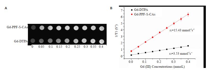
图 1 3.0 T磁共振测定纵向弛豫率
A: T1加权图像; B: r1值.
Figure 1. Measurement of longitudinal relaxivity by 3.0 T MR.

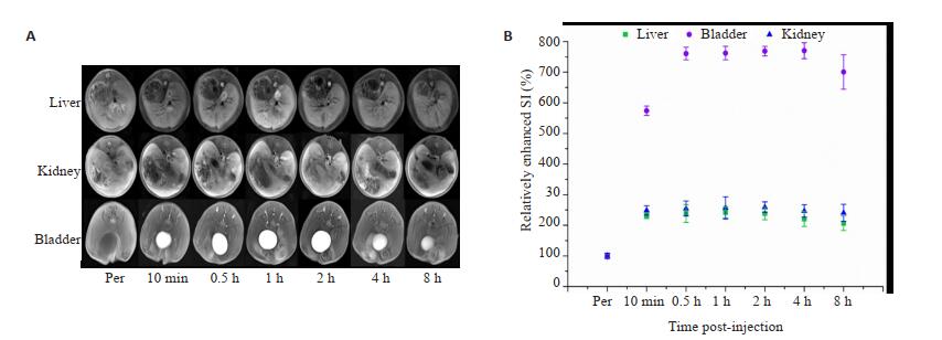
图 2 注射Gd-PPF-S-CAs后主要器官(肝、肾和膀胱)的MRI
A: T1加权图像; B:相对信号增强比值.
Figure 2. MRI of major organs (liver, kidney and bladder) after Gd-PPF-S-CAs injection.

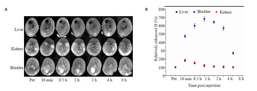
图 3 MRI of major organs (liver, kidney and bladder) after DTPA-Gd injection.
A: T1加权图像; B:相对信号增强比值.
-
[1] Lin SP, Brown JJ. MR contrast agents: physical and pharmacologic basics[J]. J Magn Reson Imaging, 2007, 25(5): 884-99. http://d.old.wanfangdata.com.cn/NSTLQK/NSTL_QKJJ025696577/ [2] Corr SA, Byrne SJ, Tekoriute R, et al. Linear assemblies of magnetic nanoparticles as MRI contrast agents[J]. J Am Chem Soc, 2008, 130(13): 4214-5. doi: 10.1021/ja710172z [3] Debroye E, Parac-Vogt TN. Towards polymetallic lanthanide complexes as dual contrast agents for magnetic resonance and optical imaging[J]. Chem Soc Rev, 2014, 43(23): 8178-92. doi: 10.1039/C4CS00201F [4] Tang JB, Sheng YQ, Hu HJ, et al. Macromolecular MRI contrast agents: Structures, properties and applications[J]. Prog Polym Sci, 2013, 38(3/4): 462-502. http://d.old.wanfangdata.com.cn/NSTLQK/NSTL_QKJJ0229009308/ [5] Zheng XY, Li LD, Sun LD, et al. Lanthanide nanoparticles: promising candidates for magnetic resonance imaging contrast enhancement[J]. Handb Phys Chem Rare Earths, 2016, 50: 301-35. doi: 10.1016/bs.hpcre.2016.05.001 [6] Lu KD, Aung T, Guo NN, et al. Nanoscale metal-organic frameworks for therapeutic, imaging, and sensing applications[J]. Adv Mater Weinheim, 2018, 30(37): e1707634-8. doi: 10.1002/adma.201707634 [7] Tang ZH, He CL, Tian HY, et al. Polymeric nanostructured materials for biomedical applications[J]. Prog Polym Sci, 2016. DOI: 10.1016/j.progpolymsci.2016.05.005. [8] Li YW, Huang YR, Wang Z, et al. Polycatechol nanoparticle MRI contrast agents[J]. Small, 2016, 12(5): 668-77. doi: 10.1002/smll.201502754 [9] Caravan P. Strategies for increasing the sensitivity of gadolinium based MRI contrast agents[J]. Chem Soc Rev, 2006, 35(6): 512-23. doi: 10.1039/b510982p [10] Duncan R. The dawning era of polymer therapeutics[J]. Nat Rev Drug Discov, 2003, 2(5): 347-60. doi: 10.1038-nrd1088/ [11] Hoshyar N, Gray S, Han HB, et al. The effect of nanoparticle size on in vivo pharmacokinetics and cellular interaction[J]. Nanomedicine (Lond), 2016, 11(6): 673-92. http://europepmc.org/abstract/MED/27003448 [12] Duncan R, Izzo L. Dendrimer biocompatibility and toxicity[J]. Adv Drug Deliv Rev, 2005, 57(15): 2215-37. doi: 10.1016/j.addr.2005.09.019 [13] Hu QY, Katti PS, Gu Z. Enzyme-responsive nanomaterials for controlled drug delivery[J]. Nanoscale, 2014, 6(21): 12273-86. doi: 10.1039/C4NR04249B [14] Wang YL, Ye FR, Jeong EK, et al. Noninvasive visualization of pharmacokinetics, biodistribution and tumor targeting of poly[N-(2-hydroxypropyl)methacrylamide]in mice using contrast enhanced MRI[J]. Pharm Res, 2007, 24(6): 1208-16. doi: 10.1007/s11095-007-9252-1 [15] Xu RZ, Wang YL, Wang XL, et al. In vivo evaluation of a PAMAM-cystamine-(Gd-DO3A) conjugate as a biodegradable macromolecular MRI contrast agent[J]. Exp Biol Med (Maywood), 2007, 232(8): 1081-9. doi: 10.3181/0702-RM-33 [16] Xu RZ, Kaneshiro TL, Jeong EK, et al. Synthesis and evaluation of nanoglobule-cystamine-(Gd-DO3A), a biodegradable nanosized magnetic resonance contrast agent for dynamic contrast-enhanced magnetic resonance urography[J]. Int J Nanomedicine, 2010, 5: 707-13. doi: 10.2147/IJN.S12224 [17] Soppimath KS, Aminabhavi TM, Kulkarni AR, et al. Biodegradable polymeric nanoparticles as drug delivery devices[J]. J Control Release, 2001, 70(1/2):1-20. http://d.old.wanfangdata.com.cn/OAPaper/oai_doaj-articles_f60daba3aa54b41252e2de1d775d15fe [18] Kumari A, Yadav SK, Yadav SC. Biodegradable polymeric nanoparticles based drug delivery systems[J]. Colloids Surfaces B: Biointerfaces, 2010, 75(1):1-18. doi: 10.1016/j.colsurfb.2009.09.001 [19] Ye F, Barrefelt A, Asem H, et al. Biodegradable polymeric vesicles containing magnetic nanoparticles, quantum dots and anticancer drugs for drug delivery and imaging[J]. Biomaterials, 2014, 35(12): 3885-94. doi: 10.1016/j.biomaterials.2014.01.041 [20] Dong JJ, Liu M, Zhang KC, et al. Biocleavable oligolysine-grafted poly(disulfide amine)s as magnetic resonance imaging probes[J]. Bioconjugate Chem, 2016, 27(1):151-8. doi: 10.1021/acs.bioconjchem.5b00569 [21] Ishiguchi DT, Takahashi S. Safety of gadoterate meglumine (Gd-DOTA) as a contrast agent for magnetic resonance imaging[J]. Drugs R & D, 2010, 10(3): 133-45. http://d.old.wanfangdata.com.cn/OAPaper/oai_pubmedcentral.nih.gov_3586093 [22] Thomsen HS, Morcos SK, Torsten A, et al. Nephrogenic systemic fibrosis and gadolinium-based contrast media: updated ESUR Contrast Medium Safety Committee guidelines[J]. Eur Radiol, 2013, 23(2): 307-18. http://d.old.wanfangdata.com.cn/NSTLQK/NSTL_QKJJ0228836058/ [23] de Campos FF, Enzweiler J. Anthropogenic gadolinium anomalies and rare earth elements in the water of Atibaia River and Anhumas Creek, Southeast Brazil[J]. Environ Monit Assess, 2016, 188(5): 281-90. doi: 10.1007/s10661-016-5282-7 [24] Nardone B, Saddleton E, Laumann AE, et al. Pediatric nephrogenic systemic fibrosis is rarely reported: a RADAR report[J]. Pediatr Radiol, 2014, 44(2): 173-180. doi: 10.1007/s00247-013-2795-x [25] Nguyen HV, Chen QX, Paletta JT, et al. Nitroxide-based macromolecular contrast agents with unprecedented transverse relaxivity and stability for magnetic resonance imaging of tumors[J]. ACS Cent Sci, 2017, 3(7): 800-11. doi: 10.1021/acscentsci.7b00253 -

 下载:
下载:
 点击查看大图
图(4)
计量
- 文章访问数: 835
- HTML全文浏览量: 304
- PDF下载量: 14
- 被引次数: 0