


Value of MRI diagnosis of degenerative knee joint lesions and its imaging features
-
摘要:
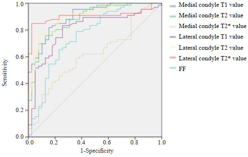
目的 探讨膝关节退行性病变MRI影像学特征,并分析MRI定量指标对膝关节退行性病变的诊断价值。 方法 选择2019年2月~2023年4月湖北民族大学附属民大医院接诊的疑似膝关节退行性病变患者162例。患者检查前均先休息30 min后行MRI扫描,并对对内侧髁、外侧髁T1、T2、T2*值及髌骨脂肪含量(FF)值进行测量。在MRI扫描后1周内行膝关节镜检查,根据镜检结果是否符合膝关节退行性病变将患者分为观察组(n=93)与对照组(n=69)。比较2组各MRI指标,并以ROC曲线分析各指标单独检测与联合检测对膝关节退行性病变的诊断价值。 结果 MRI常规扫描对关节软骨变性检出率仅为66.67%,半月板退变检出率为61.53%。关节边缘骨质增生、关节腔隙狭窄、关节积液检出率较高,分别为89.47%、92.86%、93.33%。观察组患者内侧髁T1值、内侧髁T2值、内侧髁T2*值、外侧髁T1值、外侧髁T2值、外侧髁T2*值及FF(%)均高于对照组,差异有统计学意义(P < 0.05)。ROC分析结果显示内侧髁T1值、内侧髁T2值、内侧髁T2*值、外侧髁T1值、外侧髁T2值、外侧髁T2*值及FF(%)对膝关节退行性该病诊断截断值分别为1018.92、39.27、22.34、994.71、39.91、22.74、79.18%,曲线下面积分别为0.911、0.892、0.619、0.813、0.904、0.917、0.791(P < 0.05)。 结论 MRI常规扫描对关节软骨变性、半月板退变检出率较低,内侧髁T1值、内侧髁T2值、内侧髁T2*值、外侧髁T1值、外侧髁T2值、外侧髁T2*值及FF(%)均可用于膝关节退行性病变的诊断。 Abstract:Objective To explore the changes in MRI imaging features of degenerative lesions of the knee joint and analyze the diagnostic value of MRI quantitative indicators for it. Methods A total of 162 patients with suspected degenerative lesions of the knee joint who were admitted to Minda Hospital, Hubei University for Nationalities from February 2019 to April 2023 were selected. Before examination, patients rested for 30 min before undergoing MRI scans, and measured the T1, T2, T2* values of the medial and lateral condyles, as well as the patellar fat fraction (FF) values. Knee arthroscopy was performed within 1 week after MRI scanning, and the patients were divided into observation group (n=93) and control group (n=69) according to whether the microscopic findings were consistent with degenerative lesions of the knee joint. The MRI indexes of the two groups were compared, and the diagnostic value of each index for degenerative lesions of the knee joint was analyzed by ROC curve. Results The detection rate of articular cartilage degeneration by routine MRI scanning was only 66.67%, and the detection rate of meniscus degeneration was 61.53%. The detection rates of hyperostosis at the joint margin, joint cavity stenosis, and joint effusion were 89.47%, 92.86%, and 93.33%, respectively. The medial condylar T1 value, medial condylar T2 value, medial condylar T2* value, lateral condylar T1 value, lateral condylar T2 value, lateral condylar T2* value and FF (%) of the patients in the observation group were higher than those in the control group, and the difference was statistically significant (P < 0.05). ROC analysis results showed that the cutoff values of medial condylar T1, medial condylar T2, medial condylar T2*, lateral condylar T1, lateral condylar T2, lateral condylar T2* and FF for the diagnosis of degenerative lesions of the knee joint were 1018.92, 39.27, 22.34, 994.71, 39.91, 22.74, 79.18%, respectively, and the AUCs were 0.911, 0.892, 0.619, 0.813, 0.904, 0.917, 0.791 (P < 0.05). Conclusion Routine MRI scanning has a low detection rate for articular cartilage degeneration and meniscus degeneration. The values of medial condyle T1, medial condyle T2, medial condyle T2*, lateral condyle T1, lateral condyle T2, lateral condyle T2* and FF (%) can be used for the diagnosis of degenerative lesions of the knee joint. -
Key words:
- degenerative lesions of the knee joint /
- MRI /
- diagnostic value /
- ROC curve analysis
-
表 1 2组患者一般资料比较
Table 1. Comparison of general information of patients in 2 groups
Group Gender (n) Age (year, Mean±SD) BMI(kg/m2, Mean±SD) Male Female Observation group(n=93) 35 58 53.89±10.94 20.42±4.82 Control group(n=69) 31 38 54.61±12.07 20.91±5.03 χ2/t 0.873 -0.396 -0.628 P 0.350 0.692 0.265  下载: 导出CSV
表 2 膝关节退行性病变患者病理特征及MRI检出情况
Table 2. Pathological features and MRI detection in patients with degenerative knee disease
Pathological characteristics n Number of cases detected by MRI [n(%)] Articular cartilage degeneration 18 12(66.67) Meniscal degeneration 13 8(61.53) Joint margin osteophytes 19 17(89.47) Articular stenosis 28 26(92.86) Joint effusion 15 14(93.33)
下载: 导出CSV
表 3 2组MRI定量指标比较
Table 3. Comparison of quantitative MRI indices between the 2 groups(Mean±SD)
Group Medial condyle T1 value Medial condyle T2 value Medial condyle T2* value Lateral condyle T1 value Lateral condyle T2 value Lateral condyle T2* value FF(%) Observation group(n=93) 1073.28±73.19 42.33±4.71 24.31±3.19 1093.29±78.18 42.29±3.88 24.91±2.37 81.34±3.93 Control group(n=69) 924.02±81.39 36.97±5.03 20.18±2.97 941.62±53.11 37.85±4.03 21.06±3.11 77.52±4.01 t 12.235 6.985 8.389 13.904 7.084 8.944 6.065 P <0.001 <0.001 <0.001 <0.001 <0.001 <0.001 <0.001 FF: Fat fraction.
下载: 导出CSV
表 4 ROC曲线分析
Table 4. ROC curve analysis
Targets Cut-off AUC 95% CI P Medial condyle T1 values 1018.92 0.911 0.841-0.966 0.013 Medial condyle T2 values 39.27 0.892 0.819-0.944 0.001 Medial condyle T2* values 22.34 0.619 0.517-0.732 0.041 Lateral condyle T1 values 994.71 0.813 0.721-0.897 0.045 Lateral condyle T2 values 39.91 0.904 0.848-0.951 0.032 Lateral condyle T2* values 22.74 0.917 0.849-0.961 0.007 FF(%) 79.18 0.791 0.648-0.839 0.044
下载: 导出CSV
-
[1] Khurshedovna AS, Khazratkulovich RZ, Servetovna AA, et al. Radiation visualization of chronic joint diseases[J]. Cent Asian J Med Nat Sci, 2021, 2(2): 12-17. [2] Shan S, Rehman A, Ehsan J. Diagnostic value of dynamic ultrasonography in detection of meniscal injury in correlation with MRI knee findings keeping it as reference standard[J]. Pakistan J Med Res, 2023, 62(2): 49-52. [3] Li L, Yang XF, Yang LF, et al. Biomechanical analysis of the effect of medial meniscus degenerative and traumatic lesions on the knee joint[J]. Am J Transl Res, 2019, 11(2): 542-56. [4] Lee LS, Chan PK, Fung WC, et al. Imaging of knee osteoarthritis: a review of current evidence and clinical guidelines[J]. Musculoskeletal Care, 2021, 19(3): 363-74. doi: 10.1002/msc.1536 [5] 朱莉, 邹文远, 敖锋. 三种MRI生理学定量技术对早期膝关节软骨损伤的诊断价值[J]. 陕西医学杂志, 2021, 50(3): 293-6. doi: 10.3969/j.issn.1000-7377.2021.03.009 [6] Bodden J, Ok AH, Joseph GB, et al. Joint-adjacent adipose tissue by MRI is associated with prevalence and progression of knee degenerative changes: data from the osteoarthritis initiative[J]. J Magn Reson Imaging, 2021, 54(1): 155-65. doi: 10.1002/jmri.27574 [7] Sieroń D, Jabłońska I, Lukoszek D, et al. Knee diameter and cross-section area measurements in MRI as new promising methods of chondromalacia diagnosis-pilot study[J]. Medicina, 2022, 58(9): 1142. doi: 10.3390/medicina58091142 [8] Berton A, Longo UG, Candela V, et al. Quantitative evaluation of meniscal healing process of degenerative Meniscus lesions treated with hyaluronic acid: a clinical and MRI study[J]. J Clin Med, 2020, 9(7): 2280. doi: 10.3390/jcm9072280 [9] Nebelung S, Dötsch L, Shah D, et al. Functional MRI mapping of human Meniscus functionality and its relation to degeneration[J]. Sci Rep, 2020, 10(1): 2499. doi: 10.1038/s41598-020-59573-4 [10] Huang GS, Peng YJ, Hwang DW, et al. Assessment of the efficacy of intra‑articular platelet rich plasma treatment in an ACLT experimental model by dynamic contrast enhancement MRI of knee subchondral bone marrow and MRI T2 measurement of articular cartilage[J]. Osteoarthritis Cartilage, 2021, 29(5): 718-27. doi: 10.1016/j.joca.2021.02.001 [11] 李建安, 何卫, 汪青山, 等. MRI检查与X线平片对膝关节退行性骨关节病变中的诊断价值对比[J]. 吉林医学, 2019, 40(4): 739-41. doi: 10.3969/j.issn.1004-0412.2019.04.024 [12] Gao KT, Pedoia V, Young KA, et al. Multiparametric MRI characterization of knee articular cartilage and subchondral bone shape in collegiate basketball players[J]. J Orthop Res, 2021, 39(7): 1512-22. doi: 10.1002/jor.24851 [13] Linka K, Thüring J, Rieppo L, et al. Machine learning-augmented and microspectroscopy-informed multiparametric MRI for the non-invasive prediction of articular cartilage composition[J]. Osteoarthr Cartil, 2021, 29(4): 592-602. doi: 10.1016/j.joca.2020.12.022 [14] 王珊珊, 陈祖钦, 贺思健, 等. 磁共振化学位移成像联合体质指数评估青年骨量减少的价值[J]. 武警医学, 2020, 31(3): 233-6. doi: 10.3969/j.issn.1004-3594.2020.03.012 [15] Mohammed HT, Yoon S, Hupel T, et al. Unnecessary ordering of magnetic resonance imaging of the knee: a retrospective chart review of referrals to orthopedic surgeons[J]. PLoS One, 2020, 15(11): e0241645. doi: 10.1371/journal.pone.0241645 [16] Zhao Y, Zhu ZH, Chang J, et al. Predictive value of the morphology of proximal tibiofibular joint for total knee replacement in patients with knee osteoarthritis[J]. J Orthop Res, 2021, 39(6): 1289-96. doi: 10.1002/jor.24862 [17] Ciliberti FK, Guerrini L, Gunnarsson AE, et al. CT- and MRI-based 3D reconstruction of knee joint to assess cartilage and bone[J]. Diagnostics, 2022, 12(2): 279. doi: 10.3390/diagnostics12020279 [18] Farag Abd El-Ati Mohamed AK, Farok Aggag M, Mohammed Zayed E, et al. Mri versus ultrasound in diagnosis of meniscal injury in the knee joint[J]. Al Azhar Med J, 2023, 52(2): 649-60. doi: 10.21608/amj.2023.291655 [19] Kapustina I, Morcos G, Wieland M, et al. Worrisome and incidental signs on knee radiographs in clinical practice: traumatic and degenerative lesions[J]. Appl Radiol, 2023: 8-16. [20] Ozeki N, Koga H, Sekiya I. Degenerative Meniscus in knee osteoarthritis: from pathology to treatment[J]. Life, 2022, 12(4): 603. doi: 10.3390/life12040603 [21] Ramezanpour S, Kanthawang T, Lynch J, et al. Impact of sustained synovitis on knee joint structural degeneration: 4-year MRI data from the osteoarthritis initiative[J]. J Magn Reson Imaging, 2023, 57(1): 153-64. doi: 10.1002/jmri.28223 [22] Uritani D, Koda H, Yasuura Y, et al. Factors associated with subjective knee joint stiffness in people with knee osteoarthritis: a systematic review[J]. Int J Rheum Dis, 2023, 26(3): 425-36. doi: 10.1111/1756-185X.14536 [23] Małecki K, Niedzielski K, Korczyc-Stępnicka A, et al. A clinical, radiological and isokinetic evaluation in patients with recurrent patellar dislocation undergoing MPFL reconstruction according to Avikainen: a prospective study evaluating early degenerative changes after a minimum 10‑year follow‑up period[J]. BMC Musculoskelet Disord, 2023, 24(1): 147. doi: 10.1186/s12891-023-06249-5 [24] Wang B, Wang L, Wang YY, et al. Clinical diagnostic value of magnetic resonance imaging in knee joint sports injury[J]. J Med Imaging Health Inform, 2021, 11(2): 453-61. doi: 10.1166/jmihi.2021.3301 [25] Hung TNK, Vy VPT, Tri NM, et al. Automatic detection of Meniscus tears using backbone convolutional neural networks on knee MRI[J]. J Magn Reson Imaging, 2023, 57(3): 740-9. doi: 10.1002/jmri.28284 [26] Rana S, Hossen M, Islamn A, et al. Interpretation of the common MRI findings in patients with painful knee joint[J]. Eur J Med Health Sci, 2021, 3(1): 19-26. [27] Elshimy A, Osman AM, Awad MES, et al. Diagnostic accuracy of point-of-care knee ultrasound for evaluation of meniscus and collateral ligaments pathology in comparison with MRI[J]. Acta Radiol, 2023, 64(7): 2283-92. doi: 10.1177/02841851211058280 [28] Ivanov S, Trizlov D. Painful anterior knee schwannoma: a case report[J]. JIMAB, 2023, 29(2): 4990-3. -

 点击查看大图
计量
- 文章访问数: 26
- HTML全文浏览量: 13
- PDF下载量: 3
- 被引次数: 0