


Intermediate value of grayscale ultrasound image-based radiomics in discriminating the pathological grade of bladder urothelial carcinoma
-
摘要:
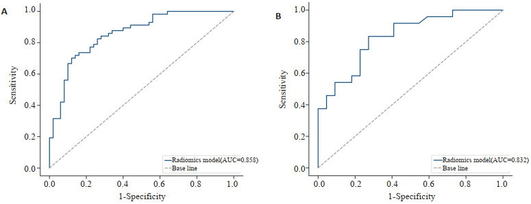
目的 通过灰阶超声影像组学特征鉴别膀胱尿路上皮癌病理分级。 方法 回顾性分析2016年4月~2023年5月山西白求恩医院153例经病理证实的膀胱尿路上皮癌患者。灰阶超声图像手工勾画肿瘤感兴趣区并提取组学特征,LASSO特征降维后采用3种机器学习方法建模并选出最优影像组学模型。采用ROC曲线对模型性能评估,采用Hosmer-Lemeshow适合度检验评价模型的拟合度,并绘制校正曲线,采用决策曲线分析进一步探讨模型的临床应用价值。 结果 3种机器学习模型中的支持向量机算法模型性能表现最优,此模型在训练集和测试集的曲线下面积分别为0.858(95% CI:0.787~0.928)和0.832(95% CI:0.708~0.936),校准曲线显示出良好的一致性。决策曲线分析结果显示具有较高的净收益。 结论 基于灰阶超声影像组学在鉴别膀胱尿路上皮癌病理分级具有术前诊断价值, 有助于临床精准诊疗。 Abstract:Objective To discriminate the pathological grade of bladder urothelial carcinoma through grayscale ultrasound image- based radiomics analysis. Methods A retrospective analysis was conducted on 153 patients with bladder urothelial carcinoma confirmed by pathology in our hospital from April 2016 to May 2023. The grayscale ultrasound images were manually delineated to outline the tumor region of interest and extract radiomics features. LASSO feature selection was utilized for dimensionality reduction, followed by modeling with three machine learning methods to identify the best model. The performance of the models was evaluated using ROC curves, and the goodness-of-fit was assessed using the Hosmer-Lemeshow test and calibration curve. Furthermore, decision curve analysis was conducted to explore the clinical utility of the model. Results Among the three machine learning models, the Support vector machine model exhibited the best performance, with an AUC of 0.858 (95% CI: 0.787-0.928) on the training set and 0.832 (95% CI: 0.708-0.936) on the test set. The calibration curve demonstrated good consistency. The decision curve analysis also showed a high net benefit. Conclusion Grayscale ultrasound image- based radiomics has preoperative diagnostic value in distinguishing the pathological grading of bladder urothelial carcinoma, which contributes to precise clinical diagnosis and treatment. -
Key words:
- bladder tumor /
- radiomics /
- pathological grading
-

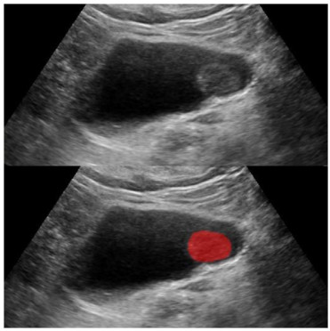
图 1 超声灰阶图像膀胱肿瘤ROI勾画
Figure 1. Ultrasound grayscale image delineation of bladder tumor ROI.

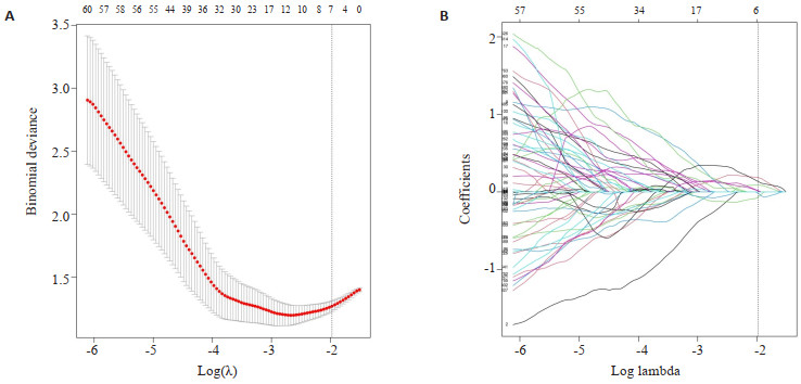
图 2 影像组学特征运用LASSO方法进行降维结果
Figure 2. Application of LASSO method for dimensionality reduction in radiomics features analysis. A: The selection of the optimal penalty coefficient λ, based on the criterion of minimum standard deviation, λ =0.139; B: Penalty coefficient graph for radiomics features, where with the variation of the penalty coefficient λ, the coefficients of most features are compressed to zero. At λ=0.139, 7 non-zero coefficient radiomics features are selected.

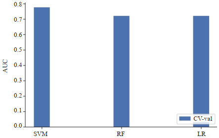
图 3 3种机器学习算法10折交叉验证AUC值,其中SVM算法模型最高
Figure 3. AUC values from 10- fold cross- validation of three machine learning algorithms, with the SVM model achieving the highest.

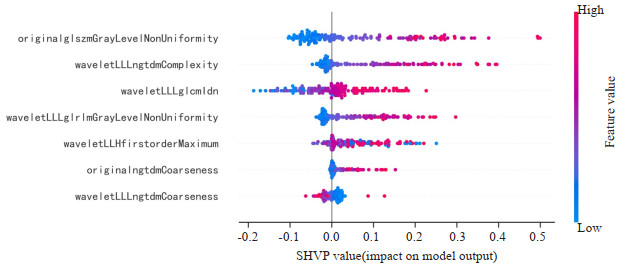
图 4 7个影像组学特征的SHAP值解释了对模型的贡献度
Figure 4. The SHAP values of the 7 radiomics features explained their contribution to the model.

图 5 影像组学模型的ROC曲线
Figure 5. ROC curve of the radiomics model. A: ROC curve of the radiomics model in the training set; B: ROC curve of the radiomics model in the test set.

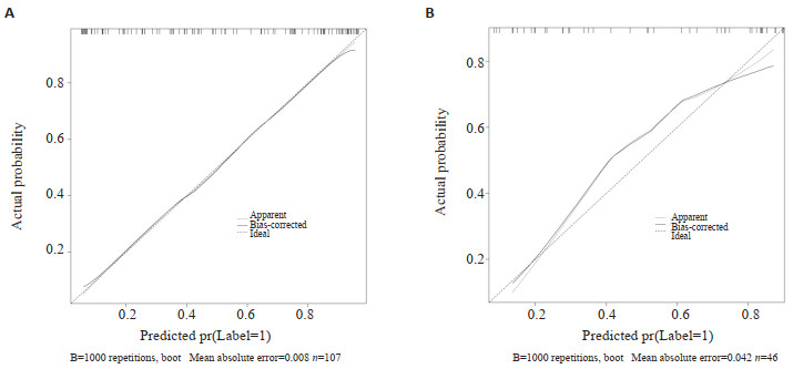
图 6 影像组学模型的校准曲线具有良好的一致性
Figure 6. Calibration curve of the radiomics model demonstrated good consistency. A: Calibration curve for the training set with a Brier score of 0.153; B: Calibration curve for the test set with a Brier score of 0.171.

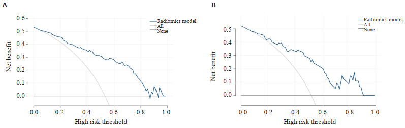
图 7 影像组学模型在大多数阈值都能获得更高的净收益
Figure 7. Radiomics model achieved higher net benefits at most thresholds. 'None' represents the assumption that all patients have LGUC, and 'All' represents the assumption that all patients have HGUC. A: DCA curve of the radiomics model in the training set; B: DCA curve of the radiomics model in the test set.
表 1 临床特征分析
Table 1. Analysis of clinical features.
Index Training set Test set LGUC HGUC P LGUC HGUC P Age (year, Mean±SD) 64.22±11.52 67.73±12.41 0.10 65.64±11.78 64.76±14.17 0.71 Gender (Male/Female, n) 39/11 47/10 0.74 19/3 22/2 0.92 Tumor length(cm, Mean±SD) 2.32±0.45 2.44±0.32 0.13 2.28±0.41 2.35±0.53 0.65 Smoking (Yes/No, n) 41/9 50/7 0.58 20/2 21/3 0.92 LGUC: Low grade urothelial carcinoma; HGUC: High grade urothelial carcinoma.  下载: 导出CSV
-
[1] Sung H, Ferlay J, Siegel RL, et al. Global cancer statistics 2020: GLOBOCAN estimates of incidence and mortality worldwide for 36 cancers in 185 countries[J]. CA Cancer J Clin, 2021, 71(3): 209-49. doi: 10.3322/caac.21660 [2] 吴素华, 叶璟威, 张意军, 等. 膀胱癌新辅助化疗后肿瘤消退分级的评估与应用[J]. 中华泌尿外科杂志, 2023, 44(11): 823-9. doi: 10.3760/cma.j.cn112330-20221105-00588 [3] 荣誉, 张锦英. 根治性经尿道膀胱肿瘤电切术治疗老年肌层浸润性膀胱癌的疗效及对生活质量的影响[J]. 中国老年学杂志, 2014, 34(12): 3351-3. doi: 10.3969/j.issn.1005-9202.2014.12.065 [4] Babjuk M, Burger M, Compérat EM, et al. European association of urology guidelines on non-muscle-invasive bladder cancer (TaT1 and carcinoma in situ)‑2019 update[J]. Eur Urol, 2019, 76(5): 639-57. doi: 10.1016/j.eururo.2019.08.016 [5] 孙林, 宋黎明, 魏后忆, 等. 加速康复外科方案在腹腔镜根治性膀胱切除+体腔内尿流改道术围手术期应用的效果分析[J]. 中华泌尿外科杂志, 2023, 44(5): 363-8. doi: 10.3760/cma.j.cn112330-20221021-00557 [6] Daoud D, Chacon Alberty L, Wei Q, et al. Incidence of primary graft dysfunction is higher according to the new ISHLT 2016 guidelines and correlates with clinical and molecular risk factors[J]. J Thorac Dis, 2021, 13(6): 3426-42. doi: 10.21037/jtd-20-3564 [7] 吴启开, 杨潇, 袁宝瑞, 等. Ⅵ-RADS评分在膀胱癌患者术后预后评估中的应用价值[J]. 中华泌尿外科杂志, 2023, 44(8): 611-5. doi: 10.3760/cma.j.cn112330-20220222-00061 [8] 杨贝贝, 徐金芝, 郑小菊. SWE联合尿脱落细胞学检测对膀胱尿路上皮癌级别鉴定的价值[J]. 影像科学与光化学, 2023, 41(1): 53-8. https://www.cnki.com.cn/Article/CJFDTOTAL-GKGH202301011.htm [9] 何卫阳, 许荧杰, 童行, 等. 无线智能胶囊膀胱镜动态检测猪膀胱黏膜状态的研究[J]. 中华泌尿外科杂志, 2023, 44(1): 52-5. [10] 王雪莲, 赵灿灿, 周牧野, 等. 基于磁共振影像组学列线图预测中晚期鼻咽癌放化疗疗效[J]. 分子影像学杂志, 2023, 46(4): 654-60. doi: 10.12122/j.issn.1674-4500.2023.04.14 [11] Xv Y, Lv FJ, Guo HM, et al. Machine learning-based CT radiomics approach for predicting WHO/ISUP nuclear grade of clear cell renal cell carcinoma: an exploratory and comparative study[J]. Insights Imaging, 2021, 12(1): 170. doi: 10.1186/s13244-021-01107-1 [12] 陈松丽. 基于超声影像组学预测膀胱肿瘤病理分级的临床研究[D]. 南昌: 南昌大学, 2022. [13] 朱怡鸣, 宋彬, 雷岩. 膀胱尿路上皮癌的CT表现及在术前病理分级中的价值[J]. 医学影像学杂志, 2021, 31(10): 1731-4. https://www.cnki.com.cn/Article/CJFDTOTAL-XYXZ202110032.htm [14] 韩引萍, 薛彩强, 刘显旺, 等. 能谱CT参数在膀胱尿路上皮癌术前病理分级预测中的价值[J]. 放射学实践, 2023, 38(1): 65-9. https://www.cnki.com.cn/Article/CJFDTOTAL-FSXS202301013.htm [15] 王健阁, 朱照伟, 范雅峰, 等. 肌层浸润性膀胱癌青年患者行机器人膀胱根治性切除术的疗效分析[J]. 中华医学杂志, 2021, 101(12): 861-5. doi: 10.3760/cma.j.cn112137-20200629-01987 [16] Hansel DE, Amin MB, Comperat E, et al. A contemporary update on pathology standards for bladder cancer: transurethral resection and radical cystectomy specimens[J]. Eur Urol, 2013, 63(2): 321-32. doi: 10.1016/j.eururo.2012.10.008 [17] Huang XZ, Zhou AY, Liu MW, et al. Shear wave elasticity differentiation between low‑and high‑grade bladder urothelial carcinoma and correlation with collagen fiber content[J]. J Ultrasound Med, 2021, 40(1): 113-22. doi: 10.1002/jum.15381 [18] Guo SP, Xu P, Zhou AY, et al. Contrast‑enhanced ultrasound differentiation between low‑and high‑grade bladder urothelial carcinoma and correlation with tumor microvessel density[J]. J Ultrasound Med, 2017, 36(11): 2287-97. doi: 10.1002/jum.14262 [19] Wei W, Liu ZY, Rong Y, et al. A computed tomography-based radiomic prognostic marker of advanced high-grade serous ovarian cancer recurrence: a multicenter study[J]. Front Oncol, 2019, 9: 255. doi: 10.3389/fonc.2019.00255 [20] 冯源, 兰晓莉. 影像组学介绍[J]. 中华核医学与分子影像杂志, 2023, 43(1): 55-60. https://www.cnki.com.cn/Article/CJFDTOTAL-XFCR201904008.htm [21] 周志鹏, 赵春雷. PET影像组学临床应用进展[J]. 中国医学影像学杂志, 2023, 31(4): 424-8. https://www.cnki.com.cn/Article/CJFDTOTAL-ZYYZ202304022.htm [22] 王梓华, 吴红珍, 梁莹莹, 等. 基于增强CT影像组学模型预测膀胱尿路上皮癌组织学分级[J]. 中国医学影像学杂志, 2022, 30(11): 1166-70. https://www.cnki.com.cn/Article/CJFDTOTAL-ZYYZ202211013.htm [23] 张添辉, 龙曦, 王丽琼, 等. 基于MRI影像组学特征构建膀胱尿路上皮癌病理分级预测模型的价值[J]. 临床放射学杂志, 2022, 41(1): 116-20. https://www.cnki.com.cn/Article/CJFDTOTAL-LCFS202201022.htm [24] 李正良, 彭思思, 王涛. 基于k-fold交叉验证的代理模型序列采样方法[J]. 计算力学学报, 2022, 39(2): 244-9. https://www.cnki.com.cn/Article/CJFDTOTAL-JSJG202202015.htm [25] Chen H, Lundberg SM, Lee SI. Explaining a series of models by propagating Shapley values[J]. Nat Commun, 2022, 13: 4512. [26] Ji GW, Zhu FP, Xu Q, et al. Radiomic features at contrast-enhanced CT predict recurrence in early stage hepatocellular carcinoma: a multi-institutional study[J]. Radiology, 2020, 294(3): 568-79. [27] Liang WJ, Yang PF, Huang R, et al. A combined nomogram model to preoperatively predict histologic grade in pancreatic neuroendocrine tumors[J]. Clin Cancer Res, 2019, 25(2): 584-94. [28] Zheng YL, Zhou D, Liu H, et al. CT-based radiomics analysis of different machine learning models for differentiating benign and malignant parotid tumors[J]. Eur Radiol, 2022, 32(10): 6953-64. [29] 梁洁, 杜昕, 王现亮, 等. 基于灰度共生矩阵的MRI纹理分析在椎管内脊膜瘤和神经鞘瘤鉴别诊断中的应用价值[J]. 磁共振成像, 2022, 13(8): 84-7. https://www.cnki.com.cn/Article/CJFDTOTAL-CGZC202208016.htm [30] 刘士远. 加强医学影像数据库建设推动行业快速发展[J]. 中华放射学杂志, 2022, 56(9): 931-4. -

 点击查看大图
计量
- 文章访问数: 26
- HTML全文浏览量: 14
- PDF下载量: 4
- 被引次数: 0