


A quantitative study of magnetic resonance ESWAN sequence on the establishment of a hierarchy of cerebral microbleeds lesions
-
摘要:
目的 应用磁共振三维增强多梯度回波T2*加权血管成像(ESWAN)序列的相位值(PV)完善脑微出血(CMBs)影像诊断的分级标准。 方法 回顾性分析2021年5月~2022年5月在中南大学湘雅医院行ESWAN序列扫描的100例患有CMBs的脑小血管病(CSVD)患者、29例脑内无异常影像表现且无其他基础疾病的健康志愿者。根据病变诊断结果将CSVD组分为CMBs组、腔隙性脑梗死(LI)组(CMBs单独合并LI)、脑白质病损(WML)组(CMBs单独合并WML)、LI+WML组。将局限于两个脑区内且LI的病灶数小于3个的定义为散发LI,否则为多发LI;Fazekas Ⅰ级定义为轻度WML,否则为重度WML。采用医院后处理软件勾画并测量CSVD组各CMBs病灶以及健康组红核与黑质的PV值,分析CMBs病灶PV值与健康红核黑质的差异以及LI、WML病变发生状况与CMBs PV值的关系。 结果 CSVD组各脑区CMBs病灶与健康组红核、黑质的平均PV值差异均有统计学意义(F=65.599,P < 0.001);不同WML病变程度、LI病灶个数时CMBs平均PV值间的差异均有统计学意义(P < 0.05)且呈负相关关系(P=0.027、0.047)。根据影响程度初步将CMBs Ⅰ级定义为PV值>-0.74,CMBs Ⅱ级定义为PV值-0.81~-0.74,CMBs Ⅲ级定义为PV值-0.84~-0.81,CMBs Ⅳ级定义为PV值-0.89~-0.84,CMBs Ⅴ级定义为PV值< -0.89。 结论 CMBs病变与LI、WML病变程度相互影响;磁共振ESWAN序列的PV值能量化反映CMBs的病变程度,以此为依据建立CMBs的病变等级制度,有望取代目前相对主观的人工计数评估方式,辅助实现CMBs早期病灶的精准检出、CSVD疾病初期的快速诊断。 -
关键词:
- 脑微出血 /
- 三维增强多梯度回波T2*加权血管成像 /
- 磁共振 /
- 定量诊断
Abstract:Objective To enhance the grading criteria for cerebral microbleeds (CMBs) imaging diagnosis by applying the phase value (PV) of magnetic resonance enhanced gradient echo GRE T2-star weighted angiography (ESWAN) sequences. Methods A retrospective analysis was conducted on 100 patients with cerebral small vessel disease (CSVD) presenting CMBs and 29 healthy volunteers without any abnormal brain imaging findings or underlying diseases, who underwent ESWAN sequence scans at Xiangya Hospital of Central South University from May 2021 to May 2022. The CSVD group was divided into CMBs group, lacunar infarction (LI) group (CMBs with LI only), white matter lesions (WML) group (CMBs with WML only), and LI + WML group based on the lesion diagnosis results. LI confined to two brain regions and with fewer than three lesions was defined as scattered LI, otherwise as multiple LI; Fazekas grade Ⅰ was defined as mild WML, otherwise as severe WML. Hospital post-processing software was used to outline and measure the PV of each CMBs lesion in the CSVD group and the red nucleus and substantia nigra in the healthy group, analyzing the differences in PV values between CMBs lesions and the healthy red nucleus and substantia nigra, and the relationship between LI, WML lesions occurrence and PV values of CMBs. Results Statistical significance was found in the differences between the average PV values of CMBs lesions in various brain regions of the CSVD group and the red nucleus and substantia nigra of the healthy group (F=65.599, P < 0.001); Differences in average PV values of CMBs were statistically significant across different degrees of WML lesions and numbers of LI lesions (P < 0.05), showing a negative correlation (P=0.027, 0.047). Based on the degree of impact, preliminary definitions were established: Grade Ⅰ CMBs as PV values >-0.74, grade Ⅱ CMBs as PV values between -0.81 and -0.74, grade Ⅲ CMBs as PV values between -0.84 and -0.81, grade Ⅳ CMBs as PV values between -0.89 and -0.84, and grade Ⅴ CMBs as PV values < -0.89. Conclusion CMBs lesions and the severity of LI and WML lesions mutually influence each other; PV values from magnetic resonance ESWAN sequences can quantitatively reflect the severity of CMBs lesions. Based on this, a grading system for CMBs lesions is established, helping to replace the currently more subjective manual counting assessment method. This aids in the precise detection of early CMBs lesions and rapid diagnosis of CSVD at its initial stages. -

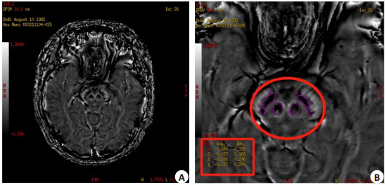
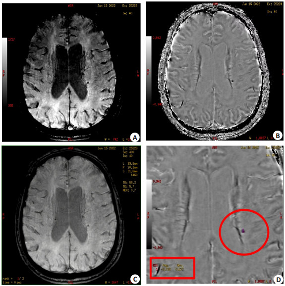
图 1 CMBs病灶的诊断与PV值的测量
Figure 1. The diagnosis of CMBs lesions and the measurement of PV value. A: Magnetic moment diagram of ESWAN sequence; B: Phase diagram; C: Amplitude diagram; D: Circle drawing of CMBs lesions on the phase map, the box showed the corresponding measurement results.

图 2 健康对照组红核与黑质PV值的测量
Figure 2. Measurement of PV value of red nucleus and substantia nigra in healthy control group. A: The phase diagram of ESWAN sequence; B: Circle frame was the circle drawing of the left and right red nuclei and substantia nigra on the phase diagram, the box showed the corresponding measurement results.

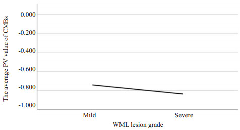
图 3 WML不同病变等级与CMBs平均PV值的变化趋势
Figure 3. The change trend of different grades of WML and the average PV value of CMBs.

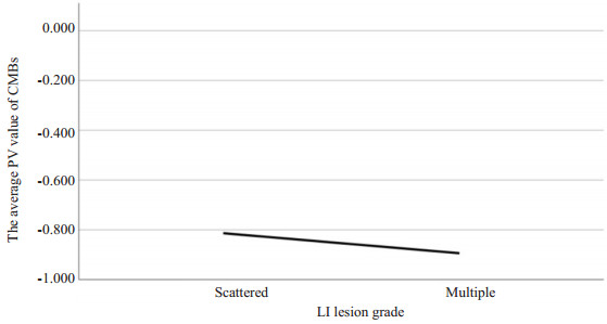
图 4 LI、WML病变共存时不同LI病灶个数与CMBs平均PV值的变化趋势
Figure 4. The change trend of the number of different LI lesions and the average PV value of CMBs in the coexistence of LI and WML lesions.
表 1 健康组红核、黑质与不同脑区CMBs平均PV值的比较
Table 1. Comparison of mean PV values of CMBs between red nucleus, substantia nigra and different brain regions in healthy group(Mean±SD)
Group n PV Red nucleus in healthy group 29 -0.16±0.07 Substantia nigra in healthy group 29 -0.20±0.07 CSVD group Cortex 37 -0.80±0.20*# Subcortical white matter 105 -0.83±0.23*# Internal capsule / external capsule 12 -0.83±0.20*# Thalamus 47 -0.86±0.22*# Basal ganglia gray matter 43 -0.92±0.21*# Brain stem 26 -0.82±0.22*# Cerebellum 32 -0.86±0.24*# *P < 0.001 vs red nucleus in healthy group; #P < 0.001 vs substantia nigra in healthy group.  下载: 导出CSV
-
[1] 于淼. 脑小血管病患者MRI总负荷评分与认知功能的相关性[D]. 大连: 大连医科大学, 2020. [2] 刘丽, 王丽华. 脑微出血相关危险因素研究[J]. 中风与神经疾病杂志, 2022, 39(6): 568-70. https://www.cnki.com.cn/Article/CJFDTOTAL-ZFSJ202206025.htm [3] Charidimou A, Boulouis G, Haley K, et al. White matter hyperintensity patterns in cerebral amyloid angiopathy and hypertensive arteriopathy[J]. Neurology, 2016, 86(6): 505-11. doi: 10.1212/WNL.0000000000002362 [4] Low A, Mak E, Rowe JB, et al. Inflammation and cerebral small vessel disease: a systematic review[J]. Ageing Res Rev, 2019, 53: 100916. doi: 10.1016/j.arr.2019.100916 [5] 黄慧琴, 杨期明. 脑微出血的研究进展[J]. 中国实用神经疾病杂志, 2021, 24(16): 1458-64. https://www.cnki.com.cn/Article/CJFDTOTAL-HNSJ202116010.htm [6] 朱娇艳. 原发性高血压脑微出血磁敏感成像ESWAN序列的初步研究[D]. 长沙: 中南大学, 2014. [7] 白小曦, 张立欧, 黄丽萍. 磁共振ESWAN成像技术进展及其临床应用[J]. 现代肿瘤医学, 2019, 27(9): 1621-4. doi: 10.3969/j.issn.1672-4992.2019.09.037 [8] 彭旭红, 雷苑麟, 赖碧玉, 等. MR ESWAN序列对高血压患者脑微出血的定量诊断价值[J]. 临床放射学杂志, 2019, 38(6): 1143-6. https://www.cnki.com.cn/Article/CJFDTOTAL-LCFS201906051.htm [9] 中国研究型医院学会脑小血管病专业委员会《中国脑小血管病诊治专家共识》编写组. 中国脑小血管病诊治专家共识2021[J]. 中国卒中杂志, 2021, 16(7): 716-26. doi: 10.3969/j.issn.1673-5765.2021.07.013 [10] 闫蕾. 磁共振ESWAN序列对高血压患者脑微出血的研究[D]. 太原: 山西医科大学, 2016. [11] Yang JH, Yang ZX, Wu HZ, et al. Quantification of iron deposition in the brain of hypertensive patients using3D-enhanced susceptibility- weighted angiography (ESWAN)[J/OL]. Curr Med Imag Former Curr Med Imag Rev, 2023. doi: 10.2174/1573405620666230627112146 .[12] Pfefferbaum A, Adalsteinsson E, Rohlfing T, et al. MRI estimates of brain iron concentration in normal aging: comparison of field-dependent (FDRI) and phase (SWI) methods[J]. NeuroImage, 2009, 47(2): 493-500. doi: 10.1016/j.neuroimage.2009.05.006 [13] 付建辉, 赵辉. 脑小血管病研究进展[J]. 中华脑血管病杂志: 电子版, 2011, 5(5): 355-61. doi: 10.3969/j.issn.1672-9248.2011.05.002 [14] 高中宝, 赵杏丽, 王振福, 等. 脑微出血与脑白质病变及腔隙性梗死关系研究[J]. 中国卒中杂志, 2015, 10(10): 822-6. doi: 10.3969/j.issn.1673-5765.2015.10.003 [15] Gao ZB, Zhai YZ, Zhao XL, et al. Deep cerebral microbleeds are associated with the severity of lacunar infarcts and hypertension: a retrospective analysis[J]. Medicine, 2018, 97(23): e11031. doi: 10.1097/MD.0000000000011031 [16] Luo Q, Tang HD, Xu XX, et al. The prevalence and risk factors of cerebral microbleeds: a community-based study in China[J]. Ther Clin Risk Manag, 2021, 17: 165-71. doi: 10.2147/TCRM.S297708 [17] Yamada S, Saiki M, Satow T, et al. Periventricular and deep white matter leukoaraiosis have a closer association with cerebral microbleeds than age[J]. Eur J Neurol, 2012, 19(1): 98-104. doi: 10.1111/j.1468-1331.2011.03451.x [18] 舒俊龙, 黄一宁, 李凡, 等. 脑血管病患者脑微出血的危险因素分析[J]. 中国全科医学, 2019, 22(23): 2793-7. doi: 10.12114/j.issn.1007-9572.2019.00.099 [19] 吕晓培. 缺血性脑血管病微出血与脑白质病变的临床特征及相关性研究[D]. 石家庄: 河北医科大学, 2016. [20] Zhou YN, Gao HY, Zhao FF, et al. The study on analysis of risk factors for severity of white matter lesions and its correlation with cerebral microbleeds in the elderly with lacunar infarction[J]. Medicine, 2020, 99(4): e18865. doi: 10.1097/MD.0000000000018865 [21] Poels MMF, Ikram MA, van der Lugt A, et al. Incidence of cerebral microbleeds in the general population: the Rotterdam Scan Study [J]. Stroke, 2011, 42(3): 656-61. doi: 10.1161/STROKEAHA.110.607184 [22] Beaman C, Kozii K, Hilal S, et al. Cerebral microbleeds, cerebral amyloid angiopathy, and their relationships to quantitative markers of neurodegeneration[J]. Neurology, 2022, 98(16): e1605-e1616. [23] Wang PN, Chou KH, Peng LN, et al. Strictly lobar cerebral microbleeds are associated with increased white matter volume[J]. Transl Stroke Res, 2020, 11(1): 29-38. doi: 10.1007/s12975-019-00704-z [24] 韩建成, 高培毅, 林燕, 等. 缺血性脑卒中患者脑内微出血的磁共振成像研究[J]. 中华老年心脑血管病杂志, 2008, 10(3): 181-4. https://www.cnki.com.cn/Article/CJFDTOTAL-LNXG200803008.htm [25] Kato H, Izumiyama M, Izumiyama K, et al. Silent cerebral microbleeds on T2*-weighted MRI: correlation with stroke subtype, stroke recurrence, and leukoaraiosis[J]. Stroke, 2002, 33 (6): 1536-40. doi: 10.1161/01.STR.0000018012.65108.86 [26] Fan YH, Mok VCT, Lam WWM, et al. Cerebral microbleeds and white matter changes in patients hospitalized with lacunar infarcts [J]. J Neurol, 2004, 251(5): 537-41. doi: 10.1007/s00415-004-0359-6 -

 点击查看大图
计量
- 文章访问数: 20
- HTML全文浏览量: 8
- PDF下载量: 3
- 被引次数: 0