


Application value of amide proton transfer combined with quantitative susceptibility mapping in the diagnosis of Parkinson's disease
-
摘要:
目的 探讨酰胺质子转移成像(APT)和定量磁化率成像(QSM)对帕金森病(PD)诊断的临床应用价值。 方法 纳入2022年6月~2023年6月新疆医科大学第二附属医院明确诊断为PD的患者38例作为PD组,另招募同时期相匹配的健康志愿者22例作为对照(HC)组。对所有受试者进行QSM和APT序列扫描,利用后处理软件获取所有受试者黑质区域的磁化率值(MSV)及非对称性磁化转移率(MTRasym),利用Logistic回归建模两种参数联合诊断时的预测概率,采用ROC曲线比较分析单一成像技术及两种成像技术联合的诊断效能。 结果 对比HC组,PD组双侧黑质的平均MSV值升高,平均MTRasym值减低,差异有统计学意义(P < 0.001)。运动症状受影响较重侧黑质的MSV值高于受影响较轻侧(P < 0.001),运动症状受影响较重侧黑质的MTRasym值低于受影响较轻侧(P < 0.05)。使用双侧黑质MSV和MTRasym的平均值,APT、QSM以及QSM联合APT的ROC曲线下面积分别为0.812、0.873、0.897,使用运动症状受影响较重侧黑质的MSV和MTRasym值,QSM联合APT的ROC曲线下面积为0.928。 结论 QSM和APT对PD均具有较高的诊断效能,与单独使用QSM或APT相比,QSM联合APT对PD的诊断效能更高,能够为PD的准确诊断提供影像学依据。 Abstract:Objective To explore the clinical application value of amide proton transfer (APT) and quantitative susceptibility mapping (QSM) for the diagnosis of Parkinson's disease (PD). Methods A total of 38 patients who were diagnosed with PD in the Second Affiliated Hospital of Xinjiang Medical University from June 2022 to June 2023 were included as the PD group, and 22 matched healthy control volunteers were recruited during the same period. QSM and APT images of all subjects were collected. The magnetic susceptibility value (MSV) and magnetization transfer asymmetry (MTRasym) of the substantia nigra region were obtained. The prediction probability of the combined diagnosis of two kinds of parameters was modeled by Logistics regression, and the diagnostic efficiency of the single imaging technology and the combined diagnosis of two kinds of imaging technology was compared by the ROC curve. Results Compared with the healthy control group, the average MSV value of substantia nigra in the PD group was significantly higher, while the average MTRasym value of substantia nigra in the PD group was significantly lower (P < 0.001). MSV value of substantia nigra on the more heavily affected side was higher than that on the less lightly affected side(P < 0.001), and MTRasym value of substantia nigra on the more heavily affected side was lower than that on the less lightly affected side (P < 0.05). Using the average values of bilateral substantia nigra, the area under the ROC curve of APT, QSM and QSM combined with APT were 0.812, 0.873, 0.897. While using the values of the more affected side, the area under the ROC curve of QSM combined with APT was 0.928. Conclusion Both QSM and APT have high diagnostic efficacy for PD. QSM combined with APT has higher diagnostic efficacy for PD compared with QSM or APT alone and can provide an imaging basis for accurate diagnosis of PD. -

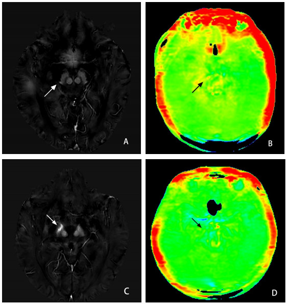
图 1 PD与HC典型病例影像
Figure 1. PD and HC typical case images. QSM(A) image and APT (B) image of a typical normal control (female, 66 years old). QSM (C) image and APT (D) image of a PD patient (female, 69 years old, this patient was diagnosed with Parkinson's disease, right upper limb tremor with motor delay for 5 years). The MSV in regions of the right substantia nigra (white arrow) were higher in PD patients than in normal controls. The MTRasym in regions of the right substantia nigra (black arrow) were lower in PD patients than in normal controls.

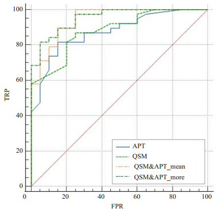
图 2 QSM、APT以及联合QSM和APT的ROC曲线比较
Figure 2. ROC curve of QSM, APT and QSM combined with APT.
表 1 PD与HC组间差异分析
Table 1. Analysis of differences between PD and HC groups (Mean±SD)
Index PD group (n=38) HC group (n=20) t P MSV_Mean 0.096±0.015 0.074±0.012 5.891 < 0.001 MSV_More 0.102±0.017 - 7.485 < 0.001 MSV_Less 0.091±0.017 - 4.032 < 0.001 MTRasym_Mean 1.196±0.481 1.723±0.488 4.067 < 0.001 MTRasym_More 1.029±0.658 - 4.300 < 0.001 MTRasym_Less 1.389±0.734 - 1.897 0.063 MSV: Magnetic susceptibility value; MTRasym: Magnetization transfer asymmetry; Mean: Mean values; More: Values in the more-affected side in PD; Less: Values in the less-affected side in PD.  下载: 导出CSV
表 2 QSM、APT及两者联合在PD的诊断价值
Table 2. The diagnostic value of QSM, APT and their combination in PD
Index AUC SE P 95% CI Sensitivity(%) Specificity (%) APT 0.812 0.062 <0.001 0.691-0.934 81.6 77.3 QSM 0.873 0.044 <0.001 0.865-0.992 86.8 72.7 QSM&APT_Mean 0.897 0.044 <0.001 0.812-0.983 84.2 81.8 QSM&APT_More 0.928 0.032 <0.001 0.787-0.959 94.7 77.3 QSM&APT_Mean: Combining QSM and APT values in mean substantia nigra; QSM&APT_More: Combining QSM and APT values in the more-affected side of substantia nigra.
下载: 导出CSV
-
[1] Zheng ZL, Zhu ZY, Zhou C, et al. Burden of parkinson disease in China, 1990-2019: findings from the 2019 global burden of disease study[J]. Neuroepidemiology, 2023, 57(1): 51-64. doi: 10.1159/000527372 [2] Qi SG, Yin P, Wang LH, et al. Prevalence of Parkinson's disease: a community- based study in China[J]. Mov Disord, 2021, 36(12): 2940-4. doi: 10.1002/mds.28762 [3] Armstrong MJ, Okun MS. Diagnosis and treatment of parkinson disease: a review[J]. JAMA, 2020, 323(6): 548-60. doi: 10.1001/jama.2019.22360 [4] Tolosa E, Garrido A, Scholz SW, et al. Challenges in the diagnosis of Parkinson's disease[J]. Lancet Neurol, 2021, 20(5): 385-97. doi: 10.1016/S1474-4422(21)00030-2 [5] Li G, Ma JF, Cui SS, et al. Parkinson's disease in China: a forty-year growing track of bedside work[J]. Transl Neurodegener, 2019, 8: 22. doi: 10.1186/s40035-019-0162-z [6] Harada T, Kudo K, Fujima N, et al. Quantitative susceptibility mapping: basic methods and clinical applications[J]. Radiographics, 2022, 42(4): 1161-76. doi: 10.1148/rg.210054 [7] Mahoney-Sánchez L, Bouchaoui H, Ayton S, et al. Ferroptosis and its potential role in the physiopathology of Parkinson's Disease[J]. Prog Neurobiol, 2021, 196: 101890. doi: 10.1016/j.pneurobio.2020.101890 [8] Shi LQ, Huang C, Luo QH, et al. The association of iron and the pathologies of Parkinson's diseases in MPTP/MPP +- induced neuronal degeneration in non- human Primates and in cell culture [J]. Front Aging Neurosci, 2019, 11: 215. doi: 10.3389/fnagi.2019.00215 [9] Ward RJ, Zucca FA, Duyn JH, et al. The role of iron in brain ageing and neurodegenerative disorders[J]. Lancet Neurol, 2014, 13(10): 1045-60. doi: 10.1016/S1474-4422(14)70117-6 [10] Dexter DT, Wells FR, Lees AJ, et al. Increased nigral iron content and alterations in other metal ions occurring in brain in Parkinson's disease[J]. J Neurochem, 1989, 52(6): 1830-6. doi: 10.1111/j.1471-4159.1989.tb07264.x [11] 米日班·买买提库尔班, 张树贤, 马景旭, 等. 酰胺质子转移磁共振成像在帕金森病中的研究进展[J]. 磁共振成像, 2023, 14(6): 94-8. https://www.cnki.com.cn/Article/CJFDTOTAL-CGZC202306016.htm [12] Reichenbach JR, Schweser F, Serres B, et al. Quantitative susceptibility mapping: concepts and applications[J]. Clin Neuroradiol, 2015, 25(2): 225-30. [13] 王钧豪, 苏一飞, 成睿, 等. 酰胺质子转移成像在脑胶质瘤诊断及分子分型预测中的研究进展[J]. 分子影像学杂志, 2023, 46(4): 765-8. doi: 10.12122/j.issn.1674-4500.2023.04.33 [14] Koike H, Morikawa M, Ishimaru H, et al. Amide proton transfer-chemical exchange saturation transfer imaging of intracranial brain tumors and tumor- like lesions: our experience and a review[J]. Diagnostics, 2023, 13(5): 914. doi: 10.3390/diagnostics13050914 [15] Zhou JY, Heo HY, Knutsson L, et al. APT- weighted MRI: techniques, current neuro applications, and challenging issues[J]. J Magn Reson Imaging, 2019, 50(2): 347-64. doi: 10.1002/jmri.26645 [16] Postuma RB, Berg D, Stern M, et al. MDS clinical diagnostic criteria for Parkinson's disease[J]. Mov Disord, 2015, 30(12): 1591-601. doi: 10.1002/mds.26424 [17] 陈永平, 商慧芳. 2016中国帕金森病诊断标准解读[J]. 中国实用内科杂志, 2017, 37(2): 124-6. https://www.cnki.com.cn/Article/CJFDTOTAL-SYNK201702010.htm [18] An HD, Zeng XY, Niu TF, et al. Quantifying iron deposition within the substantia nigra of Parkinson's disease by quantitative susceptibility mapping[J]. J Neurol Sci, 2018, 386: 46-52. doi: 10.1016/j.jns.2018.01.008 [19] Li KR, Avecillas-Chasin J, Nguyen TD, et al. Quantitative evaluation of brain iron accumulation in different stages of Parkinson's disease[J]. J Neuroimaging, 2022, 32(2): 363-71. doi: 10.1111/jon.12957 [20] Zhao QY, Tao YQ, Zhao K, et al. Structural insights of Fe3+ induced α-synuclein fibrillation in Parkinson's disease[J]. J Mol Biol, 2023, 435(1): 167680. doi: 10.1016/j.jmb.2022.167680 [21] Herman S, Djaldetti R, Mollenhauer B, et al. CSF- derived extracellular vesicles from patients with Parkinson's disease induce symptoms and pathology[J]. Brain, 2023, 146(1): 209-24. doi: 10.1093/brain/awac261 [22] Zhang JQ, Zhang YL, Wang J, et al. Characterizing iron deposition in Parkinson's disease using susceptibility-weighted imaging: an in vivo MR study[J]. Brain Res, 2010, 1330: 124-30. doi: 10.1016/j.brainres.2010.03.036 [23] Zhang Y, Wu IW, Buckley S, et al. Diffusion tensor imaging of the nigrostriatal fibers in Parkinson's disease[J]. Mov Disord, 2015, 30 (9): 1229-36. doi: 10.1002/mds.26251 [24] Baumann CR, Held U, Valko PO, et al. Body side and predominant motor features at the onset of Parkinson's disease are linked to motor and nonmotor progression[J]. Mov Disord, 2014, 29(2): 207-13. doi: 10.1002/mds.25650 [25] Li CM, Chen M, Zhao XN, et al. Chemical exchange saturation transfer MRI signal loss of the substantia nigra as an imaging biomarker to evaluate the diagnosis and severity of Parkinson's disease[J]. Front Neurosci, 2017, 11: 489. doi: 10.3389/fnins.2017.00489 [26] Li CM, Wang R, Chen HB, et al. Chemical exchange saturation transfer MR imaging is superior to diffusion-tensor imaging in the diagnosis and severity evaluation of Parkinson's disease: a study on substantia nigra and Striatum[J]. Front Aging Neurosci, 2015, 7: 198. [27] Zhao YJ, Wee HL, Chan YH, et al. Progression of Parkinson's disease as evaluated by Hoehn and Yahr stage transition times[J]. Mov Disord, 2010, 25(6): 710-6. doi: 10.1002/mds.22875 -

 点击查看大图
计量
- 文章访问数: 31
- HTML全文浏览量: 14
- PDF下载量: 10
- 被引次数: 0