


Ultrasonic features of breast masses in diagnosis of breastcarcinoma
-
摘要:
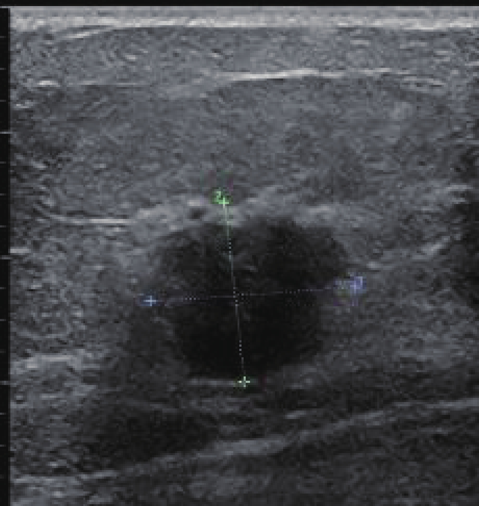
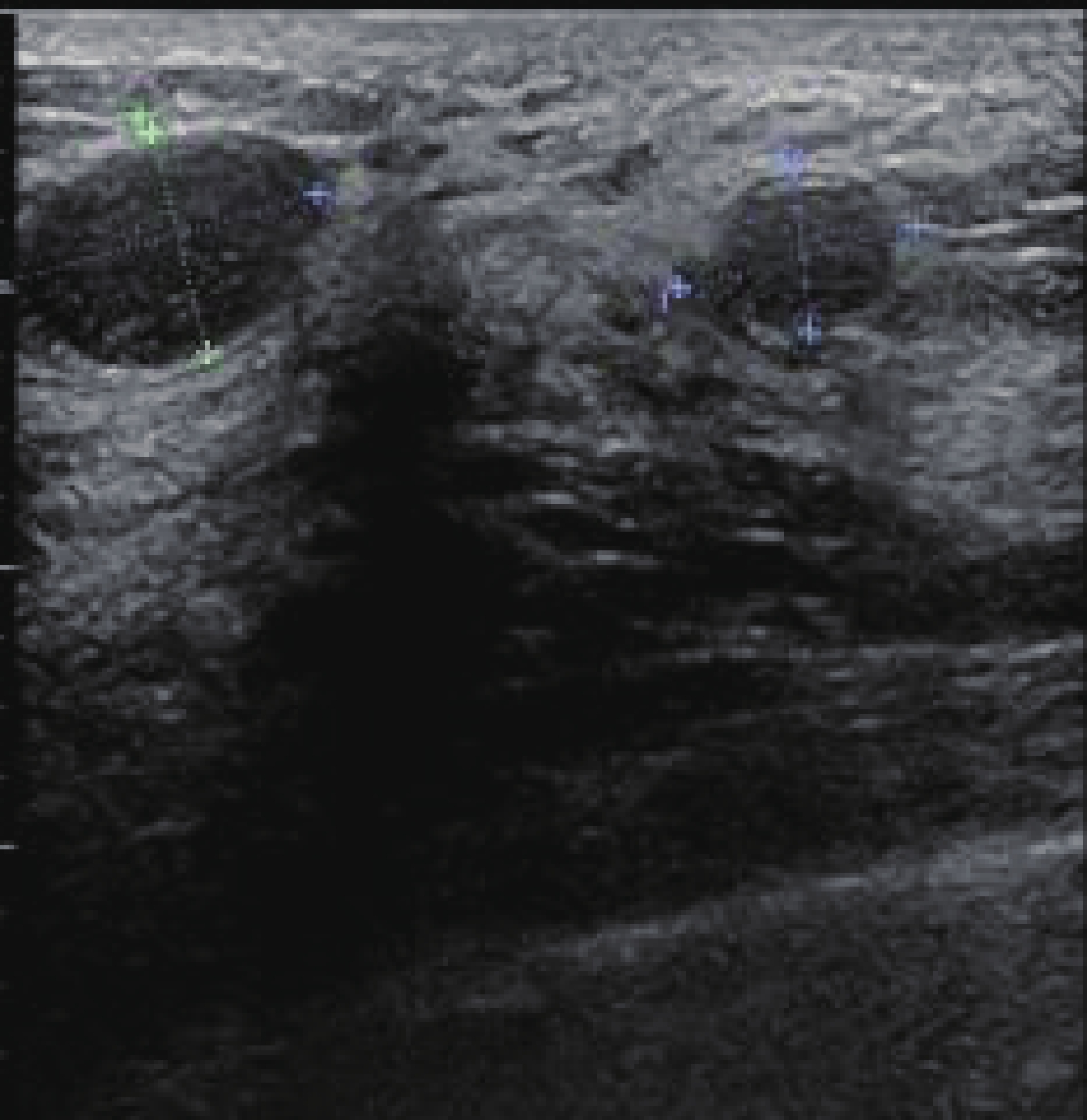
目的 评价乳腺肿块主要超声特征,提高乳腺癌诊断的准确性。 方法 对2014年1月~2016年12月在我院行乳腺肿块切除术的108例女性患者的超声特征作回顾性分析。分析肿块大小、部位、形态、边界、内部回声、钙化、后方回声、纵横比、动脉频谱及同侧腋下淋巴结情况等,并与术后病理结果比较,运用统计学方法计算肿块超声特征的灵敏度、特异度、阳性预测值、阴性预测值、诊断符合率。 结果 108例患者共发现乳腺肿块117个,有70例病理确诊为乳腺癌,其中有10例伴同侧腋下淋巴结肿大。肿块在大小、部位及各项超声特征方面比较差异均有统计学意义(P<0.05)。 结论 超声诊断乳腺癌的诊断符合率以肿块内钙化、肿块后方回声衰减及肿块纵横比≥1最高。 Abstract:Objective To evaluate the accuracy of breast masses ultrasonic features in diagnostic of breast carcinoma. Methods Theultrasonic features of suspicious breast masses in 108 female patients with breast resection in our hospital from January 2014 to December 2016 were retrospectively analyzed. We analyzed the mass size, location, form, border, internal echo, calcification, posterior echo, "taller-than-wide" shape, arterial spectrum and the axillary lymph node. The sensitivity, specificity, postive predictive values, negative predictive values, diagnostic coincidence rate of each ultrasonic feature were calculated by comparing with postoperatic pathological results . Results A total of 117 suspicious masses were found in 108 patients. Seventy cases were diagnosed as breast cancer and 10 cases with ipsilateral axillary lymph node enlargement.There were significant differences in the size of the tumor and ultrasound characteristics (P<0.05). Conclusion The highest diagnostic coincidence ratio of breast carcinoma is calcification within the tumor, acoustic attenuation behind the masses, "taller-than-wide" shape≥1. -
Key words:
- breast mass /
- breast cancer /
- ultrasonic feature /
- diagnostic accuracy
-
表 1 乳腺可疑肿块超声特征分析(n)
病理 钙化 边界 内部回声 形态 后方回声 纵横比 血管阻力指数 有 无 不清楚 清楚 不均 均匀 不规则 规则 衰减 不衰减 ≥1 <1 ≥0.7 <0.7 恶性 43 27 47 23 51 19 57 13 39 31 40 30 50 20 良性 7 40 17 30 20 27 11 36 5 42 7 40 19 28 P <0.05 <0.05 <0.05 <0.05 <0.05 <0.05 <0.05  下载: 导出CSV
表 2 乳腺可疑肿块超声特征各参数分析(%)
超声特征 灵敏度 特异度 阳性预测值 阴性预测值 漏诊率 误诊率 诊断符合率 钙化 61.4 85.1 86.0 59.7 38.5 14.9 70.9 边界不清 67.1 63.8 73.4 56.6 32.9 36.2 65.8 内部回声不均 72.8 57.4 71.8 58.6 27.2 42.6 66.6 形态不规则 81.4 76.5 83.8 73.4 18.6 23.5 29.4 后方回声衰减 55.7 89.3 88.6 57.5 44.3 10.7 69.0 纵横比≥1 57.1 85.1 85.1 57.1 42.9 14.9 68.3 血管RI≥0.7 71.4 59.5 72.4 58.3 28.6 40.3 66.6 同侧腋下淋巴结 11.2 100 100 43.3 88.8 0 100
下载: 导出CSV
-
[1] 张 斌, 周爱芬, 陈 忠, 等. 武汉市20余万农村妇女宫颈癌和乳腺癌筛查情况分析[J]. 中国妇幼保健, 2013, 28(9): 1398-402. doi: 10.7620/zgfybj.j.issn.1001-4411.2013.09.07 [2] 刘晓芳, 金小珠. 2012-2013年郑州市管城回族区宫颈癌和乳腺癌筛查结果分析[J]. 河南医学研究, 2014, 23(2): 135-7. http://d.wanfangdata.com.cn/Periodical/hnyxyj201402069 [3] 赵巧玲, 李 芬, 张 华, 等. 乳腺肿块术前超声诊断的评价[J]. 中国普通外科杂志, 2010, 19(5): 497-500. http://d.wanfangdata.com.cn/Periodical/zgptwkzz201005011 [4] 刘武岩, 郭 敏, 宋晓东, 等. 彩色多普勒超声对乳腺良恶性肿瘤的诊断价值分析[J]. 中国超声医学杂志, 2013, 5(S1): 25-6. [5] 王小健, 曹晓斌, 刘海峰, 等. 江苏省海安县2004-2011年女性乳腺癌流行趋势分析[J]. 中华肿瘤防治杂志, 2014, 28(12): 894-8. http://d.wanfangdata.com.cn/Periodical/qlzlzz201412003 [6] 石 薇, 郭玲玲, 雷 华, 等. 利用GoPubMed对乳腺癌研究的文献计量学分析[J]. 中华妇幼临床医学杂志, 2015, 79(7): 780-812. [7] 陈万青, 郑荣寿, 曾红梅, 等. 2011年中国恶性肿瘤发病和死亡分析[J]. 中国肿瘤, 2015, 24(1): 1-10. http://d.wanfangdata.com.cn/Periodical/zgzl201501001 [8] Shaniskar DM, Krishna PD, Newhouse V. Advantuges of subharmonic over second harmonic backscatter for contrast-to-tissue echo enhancement[J]. Ultrasond Med Biol, 2012, 24(3): 395-9. [9] 张沙沙, 易景娜. 女大学生对乳腺癌年轻化的认识几对策[J]. 河南医学高等专科学校学报, 2016, 29(5): 168-73. [10] 张永辉, 陈建国, 朱 健, 等. 启东市1972~2011年乳腺癌发病趋势分析[J]. 中国肿瘤, 2014, 23(8): 636-41. doi: 10.11735/j.issn.1004-0242.2014.08.A003 [11] Guyer PB, Dewbury KC, Rubin CM, et al. Ultrasonic attenuation in fibroadenoma of the breast[J]. Clin Radiol, 1992, 45(3): 175-8. doi: 10.1016/S0009-9260(05)80635-X [12] Omori LM, Hisa N, Ohkuma K, et al. Breast masses with mixed cystic-solid sonographic appearance[J]. J Clin Ultrasound, 1993, 21(8): 489-95. doi: 10.1002/(ISSN)1097-0096 [13] Kossoff G. Causes of shadowing in breast sonography[J]. Ultrasound Med Biol, 1988, 14(Suppl 1): 211-5. [14] Adler DD, Carson PL, Rubin JM, et al. Doppler ultrasound color flow imaging in the study of breast cancer: preliminary findings[J]. Ultrasound Med Biol, 1990, 16(6): 553-9. doi: 10.1016/0301-5629(90)90020-D [15] Cheung YC, Wan YL, Chen SC, et al. Sonographic evaluation of mammographically detected microcalcifications without a mass prior to stereotactic core needle biopsy[J]. J Clin Ultrasound, 2002, 30(6): 323-31. doi: 10.1002/(ISSN)1097-0096 [16] 陈灵焕. 乳腺肿瘤钙化的分类及意义[J]. 吉林医学, 2012, 33(20): 4356-7. doi: 10.3969/j.issn.1004-0412.2012.20.061 [17] 赵 锋, 邢 虎, 张 歆, 等. 高频超声对乳腺肿瘤内部微钙化的敏感性和特异度的研究[J]. 中国实验诊断学, 2009, 40(1): 52-68. [18] 胡静怡, 邓学东. 乳腺癌超声BI-RADS分级与病理分型及免疫组化的对照研究[J]. 苏州大学学报: 医学版, 2011, 31(6): 994-7. http://d.wanfangdata.com.cn/Periodical/szyxyxb201106035 [19] 蒋 田, 周如海, 袁 瑞, 等. 高频彩色多普勒超声在早期乳腺癌诊断中的临床应用价值[J]. 中国妇幼保健, 2011, 26(32): 5078-9. http://d.wanfangdata.com.cn/Periodical/zgfybj201132056 [20] Genzen JR, Tormey CA, Acad Clinical Lab Phys Scientists. Pathology consultation on reporting of critical values[J]. Am J Clin Pathol, 2011, 135(4): 505-13. doi: 10.1309/AJCP9IZT7BMBCJRS [21] 许 萍, 王文平, 王 怡, 等. 乳腺肿块超声血流显示的影响因素及良恶性诊断价值[J]. 中国医学影像技术, 2010, 16(2): 325-8. -

 点击查看大图
图(2) / 表(2)
计量
- 文章访问数: 684
- HTML全文浏览量: 263
- PDF下载量: 4
- 被引次数: 0