


Clinical value of magnetic resonance diffusion tensor imaging in the prognosis evaluation of acute lacunar infarction
-
摘要:
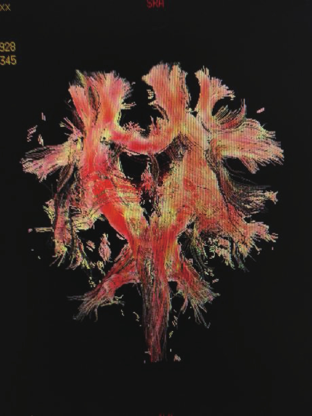
目的 探讨磁共振弥散张量成像(DTI)在急性腔隙性脑梗死患者预后评估中的临床价值及相关性。 方法 急性腔隙性脑梗死患者40例,在MRI常规检查的同时加做DTI检查,得到各向异性分数伪彩图并测量分析梗死灶各向异性分数值,重建双侧皮质脊髓束,以梗死灶在双侧皮质脊髓束的空间位置分为纤维束外、纤维束旁、纤维束内3组;每次磁共振检查之前进行Fugl-Meyer运动功能评分评定,最后分析梗死灶位置与运动功能评分之间的关系。 结果 康复治疗前后病灶与皮质脊髓束越空间关系越密切的,运动功能评分增加的百分数越低,呈负相关(P<0.01)。 结论 通过DTI在急性腔隙性脑梗死灶与双侧皮质脊髓束的空间位置关系的水分子扩散各向异性分值及扩散张量纤维束成像分级,可以较准确、直观地显示皮质脊髓束的损伤程度,为脑梗死患者临床治疗及预后提供明确的依据。 Abstract:Objective To explore spatial position of the acute lacunar infarction lesions in corticospinal tract, the relationship between limb motor function and acute lacunar infarction, and assess the clinical value of magnetic resonance diffusion tensor imaging (DTI) in prognosis of patients with acute lacunar infarction. Methods Forty cases of lacunar infarction in patients with acute were performed with the conventional MRI and DTI examination at the same time. Fractional anisotropy and the measurement of pseudocolor value FA infarction, reconstruction bilateral cortical spinal tract were analyzed.In the spatial position of CST, the infarct were divided into 3 groups: fiber bundle, fiber bundle and fiber bundle.Fugl-Meyer motor function score was assessed before each MR examination. Relationship between the location of the infarct and the Fugl-Meyer motor function score was analyzed. Results There was a negative correlation between the increase of Fugl-Meyer exercise score and the lesion before and after the rehabilitation treatment (P<0.01). Conclusion DTI diffusion of water molecules through the spatial relationship of acute lacunar infarction foci and the CST anisotropy and DTT classification can accurately display the degree of injury of the corticospinal tract, cerebral infarction patients. It would provide a clear basis for the clinical treatment and prognosis of patients with cerebral infarction. -
Key words:
- cerebral infarction /
- cortical spinal tract /
- magnetic resonance imaging /
- motor function
-
表 1 皮质脊髓束(CST)与梗死灶的空间关系的DTT分级结果及FA、FM评分之间的关系(
$\, \overline{{x}}{{±}}{{s}}$ )DTT分级 梗死灶与皮质脊髓束的关系(例) FA(分) FA下降的百分数(%) FM(分) FM增加的百分数(%) 纤维束外 纤维束旁 纤维束内 康复前 康复后 P 康复前 康复后 1级 7 1 0 0.6153±0.0448 0.5469±0.0468 <0.01 11.21±0.15 56.69±4.06 90.56±3.56 37.41±0.06 2级 1 7 8 0.6201±0.0421 0.3839±0.0956 <0.01 38.10±0.17 45.32±4.24 66.35±3.98 31.69±0.07 3级 0 6 10 0.6178±0.0335 0.1485±0.0569 <0.01 75.91±0.16 25.63±4.87 35.11±3.12 27.01±0.05  下载: 导出CSV
-
[1] 张 瑞, 陈增爱, 沈加琳, 等. 脑卒中后运动功能重组的功能影像学分析[J]. 中国康复医学杂志, 2010, 25(3): 281-5. http://d.wanfangdata.com.cn/Periodical/zgkfyxzz201003024 [2] Masutani Y, Aoki S, Abe O, et al. Mr diffusion tensor imaging: recent advance and new techniques for diffusion tensor visualization[J]. Eur J Radiol, 2003, 46(1): 53-66. doi: 10.1016/S0720-048X(02)00328-5 [3] Ito R, Mori S, Melhem E. Diffusion tensor brain imaging and tractography[J]. Neuroimag Clin N Am, 2002, 12(1): 1-19. doi: 10.1016/S1052-5149(03)00067-4 [4] 张晓丹, 陈正光. 扩散张量成像和纤维束示踪成像的原理及其在颅脑的临床应用进展[J]. 中国CT与MRI杂志, 2008, 6(4): 64-6. http://d.wanfangdata.com.cn/Periodical/zgcthmrizz200805019 [5] Melhem ER, Mori S, Mukundan G, et al. Diffusion tensor Mr imaging of the brain and white matter tractography[J]. AJR Am J Roentgenol, 2002, 178(1): 3-16. doi: 10.2214/ajr.178.1.1780003 [6] 张霞, 邢悦, 张芸, 等. 弥散张量成像技术在缺血性脑卒中的临床应用[J]. 四川大学学报: 医学版, 2009, 40(3): 551-4. http://d.wanfangdata.com.cn/Periodical/hxykdxxb200903045 [7] 陈前丽, 万智勇, 王海栋, 等. 16层螺旋CT灌注成像诊断超急性期脑梗死的价值[J]. 实用放射学杂志, 2008, 8(24): 1107-9. http://d.wanfangdata.com.cn/Periodical/syfsxzz200808003 [8] Carrera E, Jones PS, Iglesias S, et al. The vascular mean Transit time: a surrogate for the penumbra flow threshold[J]. J Cereb Blood Flow Metab, 2011, 31(4): 1027-35. doi: 10.1038/jcbfm.2010.197 [9] 林志超, 王秀河, 黄力, 等. 对比分析DW-FLAIR和FLAIR成像技术在脑梗死病变中的应用[J]. 中国医学影像技术, 2005, 21(12): 1818-20. doi: 10.3321/j.issn:1003-3289.2005.12.008 [10] 丁庆国, 陈振湖, 陆永明, 等. 急性脑缺血磁共振弥散加权成像及灌注加权成像[J]. 中国CT和MRI杂志, 2006, 2(4): 17-9. http://d.wanfangdata.com.cn/Periodical/zgcthmrizz200602007 [11] 符惠, 宏李, 曼黄勇. 三种不同磁共振成像方法检查脑梗死的差异性分析[J]. 中国基层医药, 2013, 20(10): 1506-8. doi: 10.3760/cma.j.issn.1008-6706.2013.10.030 [12] Cvoro V, Marshall I, Armitage PA, et al. Mr diffusion and perfusion parameters: relationship to metabolites in acute ischaemic stroke[J]. J Neurol Neurosurg& Psych, 2010, 81(2): 185-91. [13] 李正侠. 196例急性脑梗死DWI与MRA分析[J]. 中国实用神经疾病杂志, 2011, 14(16): 34-6. doi: 10.3969/j.issn.1673-5110.2011.16.017 [14] 张步环, 王 宏, 贾文霄, 等. DTI及DTT技术与NIHSS评分系统在急性脑梗死患者预后评价中的对比研究[J]. 临床放射学杂志, 2014, 33(6): 812-7. http://d.wanfangdata.com.cn/Periodical/lcfsxzz201406002 [15] 刘树学, 王本国, 莫雪玲, 等. 磁共振弥散张量成像(DTI)在脑梗死皮质脊髓束损伤与运动功能转归相关性中的应用研究[J]. 中国CT与MRI杂志, 2011, 9(5): 28-31. http://d.wanfangdata.com.cn/Periodical/zgcthmrizz201105009 [16] 王海滨, 陈文辉, 乔 松, 等. 磁共振DTI及DTT在脑梗死白质纤维束损伤中的应用[J]. 放射学实践, 2010, 25(3): 267-70. http://d.wanfangdata.com.cn/Periodical/fsxsj201003009 [17] 张升华, 秦东京, 姜兴岳, 等. 脑梗死DTI分型及其与临床分期相关性的研究[J]. 临床放射学杂志, 2011, 30(4): 465-8. http://d.wanfangdata.com.cn/Periodical/lcfsxzz201104004 [18] Wu DH, Liu XJ, Wu LX, et al. Effects of early rehabilitation training on hemiplegia patients with cerebral infarction[J]. 中国现代医学杂志, 2006, 16(8): 3783-8. [19] Zhang YM, Zhang N, Zhou Y, et al. Basic theory of diffusion tensor imaging in prognostic evaluation of patients with cerebral infarction[J]. Chin J Clin Rehabilitat, 2005, 34(9): 104-7. [20] Hsieh CT, Chen CY, Chiang YH, et al. Role of diffusion tensor imaging in a patient with spontaneous intracerebral hematoma treated by stereotactic evacuation[J]. Surg Neurol, 2008, 70((1): 75-8. doi: 10.1016/j.surneu.2007.04.004 -

 点击查看大图

图(2) / 表(1)
计量
- 文章访问数: 801
- HTML全文浏览量: 291
- PDF下载量: 3
- 被引次数: 0