


Clinical application of superb microvascular imaging in thyroid dysfunction
-
摘要:
目的 应用超微血管成像探讨甲状腺上动脉血流参数(收缩期最大流速Vmax、舒张期末流速Vmin、阻力指数RI、血流量)在甲状腺功能异常的超声表现。 方法 初次进行甲状腺检查的患者114例,根据血清检验结果,正常对照组84例,甲亢组34例,甲减组22例。使用方差分析比较3组甲状腺上动脉血流参数的差异,应用SNK比较法进行两两比较分析组间血流参数的差异。 结果 正常对照组与甲减组的Vmax(33.89±17.56 cm/s vs 68.23±15.41 cm/s)、Vmin(12.31±7.59 cm/s vs 25.62±9.41 cm/s)以及血流量(14.92±10.61 mL/min vs 46.67±10.17mL/min)之间的差异有统计学意义(P<0.01),同时,比较甲减组与甲亢组的Vmax(68.23±15.41 cm/s vs 38.36±16.61cm/s)、Vmin(25.62±9.41 cm/s vs 14.15±7.06 cm/s)、血流量(46.67±10.17mL/min vs 14.63±9.69 mL/min )差异有统计学意义(P<0.01),3组间RI值差异无统计学意义(P>0.05),正常对照组与甲亢组的Vmax、Vmin、血流量差异无统计学意义(P>0.05)。 结论 甲状腺上动脉血流参数作为定量参数,能反映甲状腺功能减低的血流情况。 Abstract:Objective To explore ultrasound findings of the thyroid hemodynamic parameters in thyroid disorders by micro vascular imaging. Methods Fifty-six patients with untreated thyroid dysfunction and 84 in control group were enrolled. According to the results of serum tests, 56 patients were divided into hyperthyroidism group (n=34) and hypothyroidism group (n=22). The differences of vascularity index among the three groups were analyzed. Results There were significant differences of Vmax(33.89±17.56 cm/s vs 68.23±15.41 cm/s)、Vmin(12.31±7.59 cm/s vs 25.62±9.41 cm/s) and vascularity flow (14.92±10.61 mL/min vs 46.67±10.17mL/min) in 3 groups (P<0.05). Resistive index in 3 groups had no significant differences. Conclusion The hemodynamic parameters of thyroid can reflect the blood flow of hypothyroidism. -
Key words:
- micro vascular /
- thyroid disorders /
- vascularity parameter
-
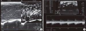
图 1 甲减患者甲状腺实质血流信号
甲状腺实质血供明显增多, Vmax是85.9 cm/s, Vmin是10.7 cm/s, RI是0.69, 甲状腺上动脉内径1.5 mm, 血流量是29.06 mL/min; A: 甲减患者甲状腺实质mSMi成像模式; B: 甲状腺上动脉频谱参数.
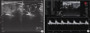
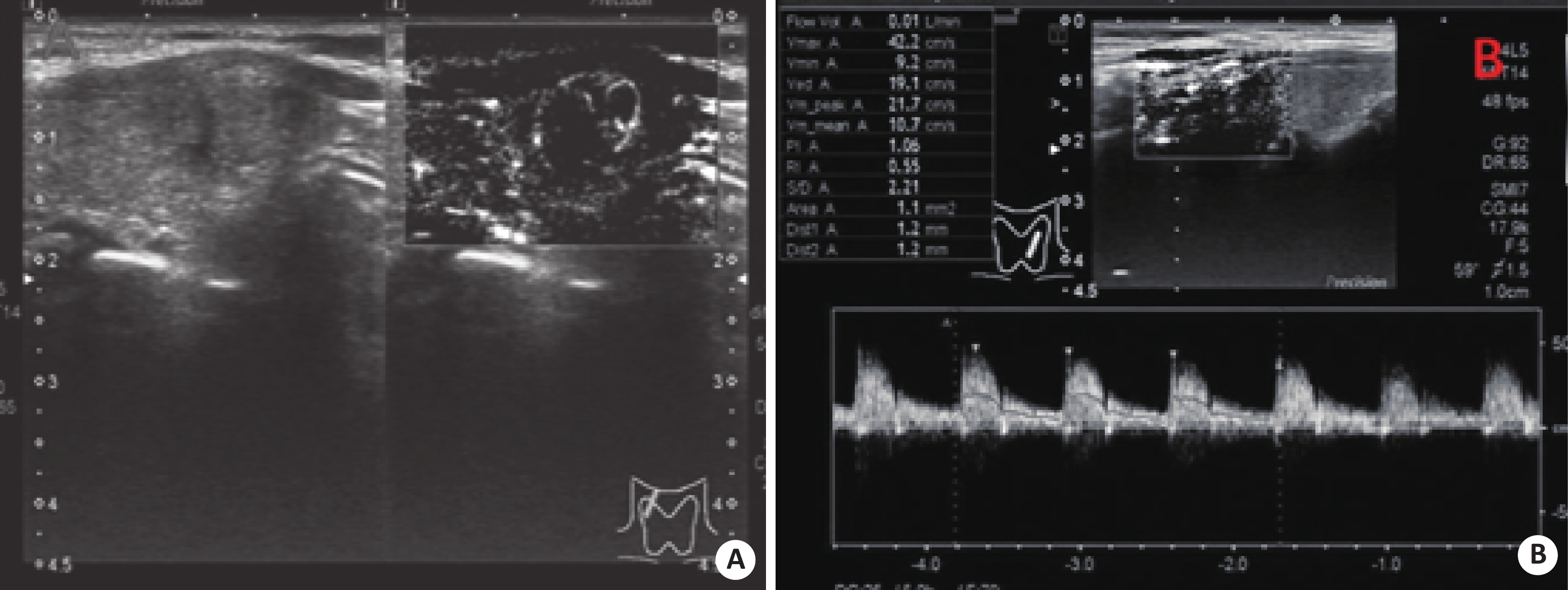
图 2 甲亢患者甲状腺实质血流信号
甲状腺实质血供增多, Vmax是42.2 cm/s, Vmin是9.2 cm/s, RI是0.55, 甲状腺上动脉内径1.2 mm, 血流量是7.1 mL/min; A: 甲亢患者的甲状腺实质mSMi成像模式; B: 甲状腺上动脉频谱参数.
表 1 3组组间Vmax、Vmin、RI、血流量的两两比较
变量 组别 P Vmax 正常组 甲亢组 0.555 甲减组 0.000 甲亢组 正常组 0.555 甲减组 0.008 Vmin 正常组 甲亢组 0.533 甲减组 0.000 甲亢组 正常组 0.533 甲减组 0.008 RI 正常组 甲亢组 0.993 甲减组 0.045 甲亢组 正常组 0.993 甲减组 0.090 血流量 正常组 甲亢组 0.972 甲减组 0.000 甲亢组 正常组 0.972 甲减组 0.001  下载: 导出CSV
-
[1] 左春洁. 桥本氏甲状腺炎不同功能状态下的超声指标对比分析[J]. 川北医学院学报, 2017, 32(2): 267-70. http://d.wanfangdata.com.cn/Periodical/cbyxyxb201702032 [2] 王珍珍, 田家玮, 任 敏. CDFI鉴别甲状腺功能亢进与亚临床甲状腺功能减退的应用价值[J]. 中国超声医学杂志, 2008, 24(6): 503-5. http://d.wanfangdata.com.cn/Periodical/zgcsyxzz200806007 [3] 李 响, 康 姝, 王学梅, 等. 超微血管成像与彩色多普勒血流成像在乳腺肿瘤诊断中的应用[J]. 中国医学影像技术, 2015, 31(5): 663-7. [4] Teng W, Shan Z, Teng X, et al. Effect of Iodine intake on thyroid diseases in China[J]. N Engl J Med, 2006, 354(26): 2783-93. doi: 10.1056/NEJMoa054022 [5] 连小兰, 白 耀, 孙梅励, 等. 血清抗甲状腺球蛋白抗体和抗甲状腺过氧化物酶抗体测定的临床诊断意义[J].中国医学科学院学报,2004, 26(6): 677-81. http://d.wanfangdata.com.cn/Periodical/zgyxkxyxb200406018 [6] 丛淑珍, 冯占武, 吴丽桑, 等. 超声在桥本氏甲状腺炎诊断中的应用价值[J]. 中国超声医学杂志, 2015, 31(9): 840-1. http://d.wanfangdata.com.cn/Periodical/jgzz201304051 [7] Vitti P, Rago T, Mazzeo S, et al. Thyroid blood flow evaluation by color-flow Doppler sonography distinguishes Graves' disease from Hashimoto's thyroiditis[J]. J Endocrinol Invest, 1995, 18(11): 857-61. doi: 10.1007/BF03349833 [8] Caruso G, Attard M, Caronia A, et al. Color doppler measurement of blood flow in the inferior thyroid artery in patients with autoimmune thyroid diseases[J]. Eur J Radiol, 2000, 36(1): 5-10. doi: 10.1016/S0720-048X(00)00147-9 [9] 何 静. 超声在评估桥本氏甲状腺炎甲状腺功能减退中的应用价值[D]. 郑州: 郑州大学, 2016. [10] Fukunari N, Nagahama M, Sugino K, et al. Clinical evaluation of color Doppler imaging for the differential diagnosis of thyroid follicular lesions[J]. World J Surg, 2004, 28(12): 1261-5. doi: 10.1007/s00268-004-7597-8 [11] 丛淑珍, 梁 婷, 甘科红, 等. 采用红细胞指数评价甲状腺实质内血流[J]. 中国医学影像技术, 2012, 28(3): 457-60. http://d.wanfangdata.com.cn/Periodical/zgyxyxjs201203021 [12] Sarikaya B, Demirbilek H, Akata D, et al. The role of the resistive index in Hashimoto's thyroiditis: a sonographic pilot study in children[J]. Clinics, 2012, 67(11): 1253-7. doi: 10.6061/clinics [13] Machado P, Segal S, Lyshchik A, et al. A novel microvascular flow technique: initial results in thyroids[J]. Ultrasound Q, 2016, 32(1): 67-74. doi: 10.1097/RUQ.0000000000000156 [14] Uchida T, Takeno K, Goto M, et al. Superior thyroid artery mean peak systolic velocity for the diagnosis of thyrotoxicosis in Japanese patients[J]. Endocr J, 2010, 57(5): 439-43. doi: 10.1507/endocrj.K09E-263 [15] Zhao X, Chen L, Li L, et al. Peak systolic velocity of superior thyroid artery for the differential diagnosis of thyrotoxicosis[J]. PLoS One, 2012, 7(11): e50051-4. doi: 10.1371/journal.pone.0050051 [16] 叶 华, 孙广宏, 赵丽娟. 彩色多普勒超声在监测人工动静脉内瘘中的应用价值[J]. 宁夏医学杂志, 2011, 33(5): 426-7. http://d.wanfangdata.com.cn/Periodical/nxyxzz201105017 [17] 裴月颖. 动态观察不同临床类型桥本甲状腺炎超声改变的价值探讨[D]. 石家庄: 河北医科大学, 2012. [18] 薛 萌, 石秋玲, 谭坤能, 等. 彩色多普勒超声、甲状腺功能及甲状腺自身抗体检查在鉴别妊娠甲状腺功能亢进和Graves病中的价值[J]. 中华内科杂志, 2016, 55(6): 470-3. http://d.wanfangdata.com.cn/Periodical/zhnk201606014 [19] 刘 娟. 超声引导射频消融甲状腺良性结节及两年随访研究[D]. 广州: 南方医科大学, 2014. [20] Yan M, Gang L, Jing L, et al. The diagnostic value of superb microvascular imaging(SMI) in detecting blood flow signals of breast lesions[J]. Medicine, 2015, 94(36): 1-6. -

 点击查看大图

图(2) / 表(1)
计量
- 文章访问数: 778
- HTML全文浏览量: 258
- PDF下载量: 4
- 被引次数: 0