


Cannulated compression screws assist with 3D printing technique in treatment of odontoid fracture of Andersonll A type
-
摘要:
目的 研究3D打印技术辅助空心加压螺钉治疗Andersonll A型齿状突骨折的临床疗效。 方法 选取我院2014年1月~2017年1月收治的AndersonⅡA型齿状突骨折患者40例,按随机数字表法分为观察组(n=21,行3D打印辅助空心加压螺钉治疗)和对照组(n=19,行单纯空心加压螺钉治疗)。比较两组手术相关指标(手术成功率、手术时间、术中出血量、透视次数、置钉次数),骨折临床愈合时间和螺钉松动断裂发生率,术前和术后1月日本骨科学会评分(JOA),颈椎活动度(CROM),美国脊髓损伤学会评分(ASIA)。 结果 两组手术成功率均为100%,观察组相比于对照组,手术时间更短、术中出血量更少、透视次数更少、置钉次数更少,差异具有统计学意义(P<0.05)。观察组骨折临床愈合时间短于对照组,螺钉松动断裂发生率低于对照组,差异具有统计学意义(P<0.05)。两组术前JOA、CROM、ASIA差异无统计学意义(P>0.05)。两组术后1月JOA、CROM、ASIA评分高于术前,差异具有统计学意义(P<0.05)。两组术后1月JOA、CROM、ASIA评分差异无统计学意义(P>0.05)。 结论 3D打印技术辅助空心加压螺钉能减少手术时间、术中出血量、透视次数和置钉次数,骨愈合快,螺钉松动断裂少。 Abstract:Objective To explore the clinical effects of cannulated compression screws assist with 3D printing technique in treatment of odontoid fracture of Andersonll A type. Methods Forty patients with odontoid fracture of Anderson ⅡA type in our hospital from January 2014 to January 2017 were selected. They were divided into 2 groups according to the random number table. The observation group (n=21)was treated by 3D printing with annulated compression screws and the control group (n=19) was treated by annulated compression screws. Success rate, operation time, amount of bleeding, X-ray frequency, screw placement frequency, bone healing time, rate of screw loose and JOA, CROM, ASIA score were compared. Results Two groups had the 100% success rate of operation. The operation time, amount of bleeding, X-ray frequency and screw placement frequency of observation group were lower than that of control group (P<0.05). The bone healing time and the rate of screw loose of observation group was less than that of control group (P<0.05). The Japanese Orthopaedic Association Scores (JOA), Cervical range of motion (CROM), American Spinal Injury Association (ASIA) score in preoperative of two groups had no significant difference (P>0.05). The JOA, CROM, ASIA score at a month after operation were higher than preoperative groups (P<0.05). The JOA, CROM, ASIA score at a month after the operation of two groups had no significant difference (P>0.05). Conclusion 3D printing with annulated compression screws can decrease the operation time, amount of bleeding, X-ray frequency and screw placement frequency. It can promte the bone healing, and decrease the rate of screw loose. -
Key words:
- 3D printing /
- annulated compression screws /
- odontoid fracture
-
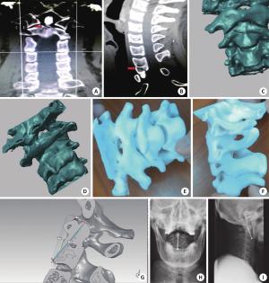
图 1 患者相关资料
A: 术前CT冠状位; B: 为术前CT矢状位; C、D: Mimics软件建立模型(C为正面观, D为后面观); E、F: 实物模型(E为正面观, F为侧面观); G: 在模型上手术, 确定进针角度深度和螺钉直径长度; H、I: 术后X线平片(H为正位片, I为侧位片).
表 1 两组一般资料比较
组别 n 年龄(岁) 性别(男/女) 受伤时间(d) 观察组 21 48.7±7.8 14/7 2.3±0.9 对照组 19 49.5±7.2 12/7 2.1±0.8 t/χ2 0.335 0.054 0.739 P 0.739 0.816 0.463  下载: 导出CSV
表 2 两组手术相关指标(
$\, \overline{{x}}{{±}}{{s}}$ )组别 手术时间(min) 术中出血量(mL) 透视次数(次) 置钉次数(次) 观察组(n=21) 114.5±27.9 164.5±22.7 9.5±1.8 3.1±0.5 对照组(n=19) 72.8±19.5 81.9±20.5 4.1±0.9 1.9±0.4 t 5.442 12.030 11.800 8.323 P <0.01 <0.01 <0.01 <0.01
下载: 导出CSV
表 3 两组骨折临床愈合时间和螺钉松动断裂发生率
组别 骨折临床愈合时间( $\, \overline{{x}}{{±}}{{s}}$ ,周)

螺钉松动断裂 [n(%)] 观察组(n=21) 12.1±1.7 0(0) 对照组(n=19) 13.8±2.0 4(10.5%) t/χ2 2.905 4.912 P 0.006 0.027
下载: 导出CSV
表 4 两组JOA、CROM、ASIA评分比较(
$\overline{{x}}{{±}}{{s}}$ ,分)组别 阶段 JOA CROM ASIA 观察组(n=21) 术前 8.1±1.6 23.8±8.9 70.5±9.8 术后1月 15.1±2.1# 46.6±10.4# 104.8±12.3# 对照组(n=19) 术前 8.2±1.4 21.3±9.1 68.3±10.1 术后1月 14.8±2.2# 50.1±11.5# 106.4±9.4# #P<0.05vs同组术前.
下载: 导出CSV
-
[1] 美国脊髓损伤协会, 国际脊髓损伤学会, 李建军, 等. 脊髓损伤神经学分类国际标准(2011年修订) [J]. 中国康复理论与实践, 2011, 17(10): 963-71. doi: 10.3969/j.issn.1006-9771.2011.10.009 [2] White AP, Hashimoto R, Norvell DC, et al. Morbidity and mortality related to odontoid fracture surgery in the elderly population[J]. Spine, 2010, 35(9 Suppl): S146-57. [3] 黄威, 蔡贤华, 李彦锦. Ⅱ型齿状突骨折手术治疗与并发症分析[J]. 中国矫形外科杂志, 2013, 21(4): 359-62. http://d.wanfangdata.com.cn/Periodical/zgjxwkzz201304010 [4] 杜浩, 赵致良, 干阜生, 等. 三维重建结合快速成形术制作模板定位髋臼假体的临床应用[J]. 中国矫形外科杂志, 2009, 17(10): 737-40. http://d.wanfangdata.com.cn/Periodical/zgjxwkzz200910006 [5] 丁焕文, 沈健坚, 涂强, 等. 计算机辅助技术在骨关节疾病中的应用[J]. 中国组织工程研究与临床康复, 2011, 15(17): 3113-8. doi: 10.3969/j.issn.1673-8225.2011.17.018 [6] 李新春, 康麟, 庞渊. 3D 打印技术在 Pilon 骨折手术治疗中的应用[J]. 新疆医科大学学报, 2015, 40(4): 471-3. http://d.wanfangdata.com.cn/Periodical/xjykdxxb201504021 [7] 李健伟, 佘黎平, 周世梅, 等. 3D打印技术在踝关节骨折手术中的应用[J]. 中国骨科临床与基础研究杂志, 2014, 16(6): 333-8. http://d.wanfangdata.com.cn/Periodical/zggklcyjcyjzz201406003 [8] 庞骄阳, 赵岩, 肖宇龙, 等. 3D打印技术在脊柱外科的应用[J]. 中国组织工程研究, 2016, 20(4): 577-82. doi: 10.3969/j.issn.2095-4344.2016.04.022 [9] 于乃春, 吉光荣. 快速成型技术在复杂脊柱畸形矫形手术中的应用[J]. 实用临床医药杂志, 2013, 17(15): 34-5, 41. http://d.wanfangdata.com.cn/Periodical/jslcyxzz201315012 [10] 梁亮科, 禤天航. 个体化3D打印技术辅助空心加压螺钉治疗AndersonⅡ型齿状突骨折的临床应用[J]. 中国医药导报, 2017, 14(9): 98-102. [11] Cheung WY, Arvinte D, Wong YW, et al. Neurological recovery after surgical decompression in patients with cervical spondylotic myelopathy- a prospective study[J]. Int Orthop, 2008, 32(2): 273-8. doi: 10.1007/s00264-006-0315-4 [12] Vernon H, Mior S. The neck disability index: a study of reliability and validity[J]. J Manipulative Physiol Ther, 1991, 14(7): 409-15. [13] 吴玉杰, 朱彤, 沈康平, 等. 颈椎前路单枚空心螺钉置入治疗Ⅱ型齿状突骨折[J]. 中国组织工程研究, 2013, 35(17): 3192-9. http://d.wanfangdata.com.cn/Periodical/xdkf201317023 [14] 史玉升, 张李超, 白宇, 等. 3D打印技术的发展及其软件实现[J]. 中国科学: 信息科学, 2015, 25(02): 197-203. [15] 鲍立杰, 张志平, 吴培斌. 3D打印技术在骨科的研究及应用进展[J]. 中国矫形外科杂志, 2015, 23(4): 325-7. http://d.wanfangdata.com.cn/Periodical/zgjxwkzz201504009 [16] 张云峰, 杨栋. 3D打印骨科模型临床应用的初步探索[J]. 中国临床研究, 2014, 27(10): 1260-1. http://d.wanfangdata.com.cn/Periodical/zgckyx201410039 [17] 王燎, 戴尅戎. 骨科个体化治疗与3D打印技术[J]. 医用生物力学, 2014, 29(3): 193-9. doi: 10.3871/j.1004-7220.2014.03.199. [18] 魏新旺, 杨志, 姚军, 等. 3D打印技术在骨盆骨折修复中的应用[J]. 中国组织工程研究, 2015, 44(4): 7163-6. http://d.wanfangdata.com.cn/Periodical/xdkf201544020 [19] 杨龙, 王建吉, 孙琦, 等. 胫骨平台骨折植入物内固定修复中3D打印技术的辅助应用[J]. 中国组织工程研究, 2016, 20(13): 1904-10. doi: 10.3969/j.issn.2095-4344.2016.13.011 [20] 邓爱文, 熊日波, 何伟明, 等. 髋臼骨折中应用3D打印技术术后的康复策略[J]. 南方医科大学学报, 2014, 34(4): 591-3. http://d.wanfangdata.com.cn/Periodical/dyjydxxb201404035 -

 点击查看大图

图(1) / 表(4)
计量
- 文章访问数: 566
- HTML全文浏览量: 234
- PDF下载量: 4
- 被引次数: 0