


CT features and evolution of lung abnormalities in patients suffered from acute pulmona.ry embolism
-
摘要:
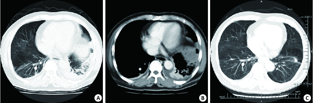
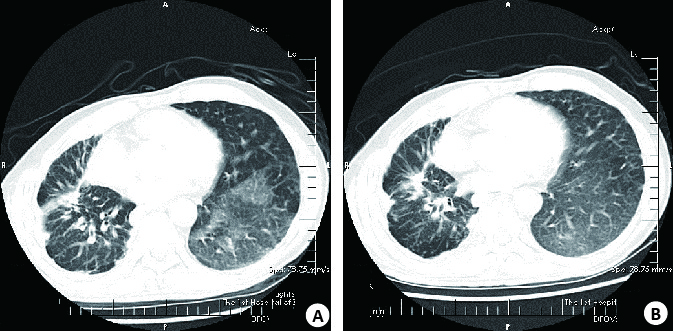
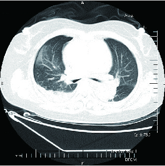
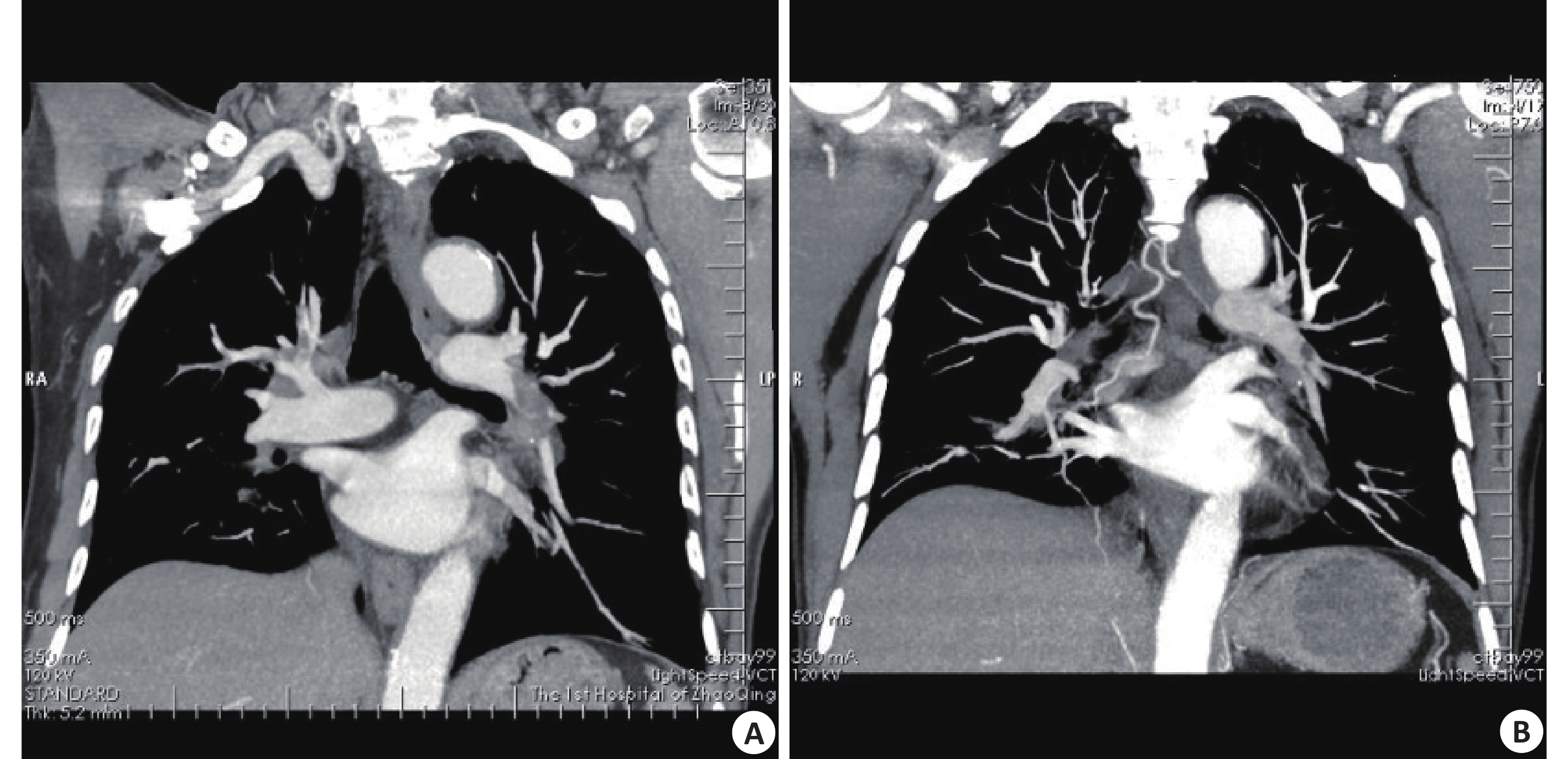
目的 探讨急性肺动脉栓塞患者肺部异常的CT征象及其演变特点。 方法 回顾性分析34例急性肺动脉栓塞患者的临床及CT影像资料,观察肺动脉栓塞的直接征象即肺动脉充盈缺损,及肺部异常的CT征象,包括肺梗死、“马赛克”征、磨玻璃影、局限性肺气肿、肺不张等,分析病灶的分布、影像特征等,及复查时病灶演变特点。 结果 初查CT可见肺梗死累及18名患者,共28个病灶;“马赛克”累及6名患者,10个病灶,磨玻璃影累及12名患者,28个病灶,局限性肺气肿累及5名患者,6个病灶,肺不张累及13名患者,19个病灶。13名患者CT复查,随着肺动脉栓子缩小,绝大部分肺部异常征象消失,仅肺梗死区遗留纤维条索灶。 结论 肺梗死、“马赛克”征、磨玻璃影、局限性肺气肿、肺不张等肺部异常是急性肺动脉栓塞常见CT征象,熟悉CT征象及其演变特点将有助于肺动脉栓塞的诊断及治疗效果的评估。 Abstract:Objective To investigate the CT features and evolution of lung abnormalities in patients suffered from acute pulmonary embolism. Methods The CT and clinical data of 34 patients suffered from acute pulmonay embolism were analyzed retrospectively. The direct sign, that is, thefilling defect within the pulmonary arteries, and the abnormal CT signs in the lungs, including pulmonaryinfarction, mosaic sign, ground-glass opacity, Westermark'ssign, and pulmonary atelectasis were assessed, and their location and radiologic features of lesions were described too. On the follow-up CT examinations, the evolution of lesions were carried out by comparing to their appearance shown on previous CT images. Results On initial CT exminations, pulmonaryinfarction involved 18 patients, resulting to 28 lesions, while mosaic sign involving 6 patients, resulting to 10 lesions, ground glass opacity involving 12 patients, resulting to 28 lesions, Westermark'ssign involving 5 patients, resulting to 6 lesions, and pulmonary atelectasis involving 13 patients, resulting to 19 lesions. Thirteen patients peformed at least one time follow-up CT exminations in various period. As the filling defectdecreased while followed up, almost of the lung abnormalities shrinked or disappeared on CT, but the pulmonaryinfarction turned to be focal fibrosis lesions. Conclusion Pulmonaryinfarction, mosaic sign, ground-glass opacity, Westermark'ssign, and pulmonary atelectasis were common signs on CT in patients suffered from acute pulmonary embolism, and acknowledging the signs and their evolution would contribute to the diagnosis and therapeutic effects evaluation of pulmonary embolism. -
Key words:
- pulmonary embolism /
- X -ray computed tomography /
- three-dimensional reconstruction /
- lung
-
[1] Giuntini C, Di Ricco G, Marini C, et al. Pulmonary embolism: epidemiology[J]. Chest, 1995, 107(1 Suppl): 35. [2] Zhang LJ, Lu GM, Meinel FG, et al. Computed tomography of acute pulmonary embolism: state-of-the-art[J]. Eur Radiol, 2015, 25(9): 2547-57. doi: 10.1007/s00330-015-3679-2 [3] Doğan H, de Roos A, Geleijins J, et al. The role of computed tomography in the diagnosis of acute and chronic pulmonary embolism[J]. Diagn Interv Radiol, 2015, 21(4): 307-16. doi: 10.5152/dir [4] 姜永宏, 刘正华, 张玉婷, 等. CT肺动脉栓塞指数与右心功能及动脉血气分析指标的相关性研究[J]. 实用放射学杂志, 2016, 32(12): 1864-6. doi: 10.3969/j.issn.1002-1671.2016.12.011 [5] Witkin AS, Channick RN. Chronic thromboembolic pulmonary hypertension: the end result of pulmonary embolism[J]. Curr Cardiol Rep, 2015, 17(8): 63. doi: 10.1007/s11886-015-0621-9 [6] Grosse C, Grosse A. CT findings in diseases associated with pulmonary hypertension: a current review[J]. Radiographics, 2010, 30(7): 1753-77. doi: 10.1148/rg.307105710 [7] 刘 康, 张耀森, 郝跃文. 肺动脉CTA在肺动脉栓塞的诊断及疗效观察的价值[J]. 中国CT和MRI杂志, 2016, 14(9): 50-1, 58. http://www.cnki.com.cn/Article/CJFDTOTAL-CTMR201609016.htm [8] Vitarelli A, Barillà F, Capotosto L, et al. Right ventricular function in acute pulmonary embolism: a combined assessment by three-dimensional and speckle-tracking echocardiography[J]. J Am Soc Echocardiogr, 2014, 27(3): 329-38. doi: 10.1016/j.echo.2013.11.013 [9] Kirchner J, Obermann A, Stückradt S, et al. Lung infarction following pulmonary embolism: a comparative study on clinical conditions and CT findings to identify predisposing factors[J]. Rofo, 2015, 187(6): 440-4. doi: 10.1055/s-00000066 [10] Kligerman SJ, Henry T, Lin CT, et al. Mosaic attenuation: etiology, methods of differentiation, and pitfalls[J]. Radiographics, 2015, 35(5): 1360-80. doi: 10.1148/rg.2015140308 [11] Ameli-Renani S, Rahman F, Nair A, et al. Dual-energy CT for imaging of pulmonary hypertension: challenges and opportunities[J]. Radiographics, 2014, 34(7): 1769-90. doi: 10.1148/rg.347130085 [12] Kim HY, Shim YM, Lee KS, et al. Persistent pulmonary nodular ground-glass opacity at thin-section CT: Histopathologic comparisons[J]. Radiology, 2007, 245(1): 267-75. doi: 10.1148/radiol.2451061682 [13] 袁焕初, 郑晓林, 邹玉坚, 等. 肺局限性磨玻璃影与支气管关系的多层螺旋CT表现[J]. 分子影像学杂志, 2017(1): 12-5. http://www.cnki.com.cn/Article/CJFDTOTAL-FZYX201701005.htm [14] Gao JW, Rizzo S, Ma LH, et al. Pulmonary ground-glass opacity: computed tomography features, histopathology and molecular pathology[J]. Transla Lung Cancer Res, 2017, 6(1): 68-75. doi: 10.21037/tlcr [15] Castañer E, Gallardo X, Ballesteros E, et al. CT diagnosis of chronic pulmonary thromboembolism[J]. Radiographics, 2009, 29(1): 31-50; discussion 50-3. doi: 10.1148/rg.291085061 [16] Pontana F, Remy-Jardin M, Duhamel AA, et al. Lung perfusion with dual-energy multi-detector row CT can it help recognize ground glass opacities of vascular origin[J]. Acad Radiol, 2010, 17(5): 587-94. doi: 10.1016/j.acra.2009.12.013 [17] 马建勇, 张 雷, 高 煜, 等. 肺动脉高压的多层螺旋CT表现[J]. 苏州大学学报:医学版, 2011, 31(4): 671-2. http://www.cnki.com.cn/Article/CJFDTOTAL-ZYKX200601012.htm [18] 蒋 明, 王昌明, 林 云, 等. 低氧性肺动脉高压和慢性支气管炎、肺气肿并肺动脉高压动物模型的异同[J]. 临床与实验病理学杂志, 2011, 27(4): 400-4. http://www.cnki.com.cn/Article/CJFDTOTAL-LSBL201104019.htm [19] Kazzaz F, Demla V, Cherian S. Unilateral pulmonary edema, westermark's sign and palla's sign in pulmonary embolism[J]. QJM, 2017, 25(8): 75. [20] Findik S. Pleural effusion in pulmonary embolism[J]. Curr Opin Pulm Med, 2012, 18(4): 347-54. doi: 10.1097/MCP.0b013e32835395d5 [21] Bray TJ, Mortensen KH, Gopalan D. Multimodality imaging of pulmonary infarction[J]. European J Radiol, 2014, 83(12): 2240-54. doi: 10.1016/j.ejrad.2014.07.016 -

 下载:
下载:

 点击查看大图
图(5)
计量
- 文章访问数: 845
- HTML全文浏览量: 349
- PDF下载量: 5
- 被引次数: 0