


Diagnostic value of pulmonary artery pressure value combined with right lower pulmonary artery diameter in pulmonary heart disease
-
摘要:
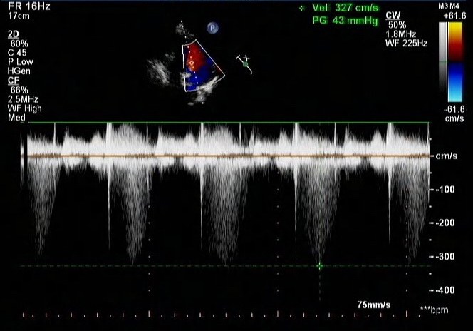
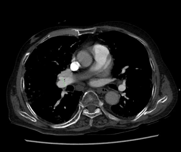
目的探讨右下肺动脉直径联合肺动脉压力值在肺心病诊断中的应用价值。 方法随机选取我院2014年6月~2016年5月49例确诊肺心病患者为肺心病组;以同期收治的诊断为慢性阻塞性肺疾病的31例患者为COPD组;以及无心肺疾病的30例就诊患者为对照组。分别使用CT测量上述各组的右下肺动脉直径,心脏彩超测量肺动脉压力,比较各组的指标变化,并分析各指标与肺心病病程的关系。 结果随着肺心病病程的进展,右下肺动脉直径增大;对照组和肺心病组的主肺动脉直径及肺动脉压力值差异有统计学意义;肺心病组与COPD组、对照组组间的右下肺动脉直径/肺动脉压力值比较,差异均有统计学意义。 结论右下肺动脉直径/肺动脉压力值变化与肺心病病程进展程度有关, 右下肺动脉直径/肺动脉压力值是诊断肺心病及其进程的CT监测指标。 Abstract:ObjectiveTo explore the diagnostic value of pulmonary artery pressure value combined with right lower pulmonary artery diameter in pulmonary heart disease. MethodsRandomly selected from June 2014 to May 2016, 49 cases with pulmonary heart disease diagnosed as pulmonary heart disease group; 31 cases with chronic obstructive pulmonary disease were treated with the same time as the COPD group; and 30 cases of normal adults as control group. CT was used to measure the diameter of the right lower pulmonary artery in each group, and the pulmonary artery pressure was measured by echocardiography. The indexes of each group were compared and the relationship between the indexes and the course of pulmonary heart disease were analyzed. ResultsWith the development of pulmonary heart disease, the diameter of the right pulmonary artery increased, there were significant differences in the diameter of the main pulmonary artery and pulmonary artery pressure between the control group and the pulmonary heart disease group. Pulmonary heart disease group and COPD group, the control group of the right lower pulmonary diameter/pulmonary artery pressure value comparison, the difference was statistically significant. ConclusionThe change of the right lower pulmonary diameter and pulmonary pressure value are related to the degree of the disease course of pulmonary heart disease. The value of right lower pulmonary diameter and pulmonary artery pressure value is a CT monitoring index in the diagnosis of pulmonary heart disease and its process. -
表 1 肺心病病程与RPA、肺动脉压力变化(PASP)的关系
指标 病程 n 直径(mm)或压力值(mmHg) RPA 对照组 30 10.87±2.08 ≤10 年 8 16.33±1.86* 11~15 年 12 17.66±3.88* 16~20 年 13 20.06±3.07*△ ≥21 年 16 22.22±4.06*△ PASP 对照组 30 12.37±4.48 ≤10 年 8 51.13±10.25* 11~15 年 12 57.17±8.86* 16~20 年 13 57.31±10.37* ≥21 年 16 60.94±11.20*△ *P<0.01 vs 对照组;△ P<0.05 vs ≤10 年.  下载: 导出CSV
表 2 各组间右下肺动脉直径、肺动脉压力值比较(x±s)
RPA(mm) PASP(mmHg) 对照组 10.87±2.08 12.37±4.48 COPD组 11.74±2.58 54.13±10.87* 肺心病组 19.57±4.06*# 57.45±10.49* *P<0.01 vs 对照组;#P<0.01 vs COPD组.
下载: 导出CSV
-
[1] 薛燕, 买明江·艾肯木. CT检查肺心病的临床意义[J]. 临床研究,2014, 2(8): 150-1. [2] 丁芳, 杨朝,耑冰,等. 慢性阻塞性肺病相关肺动脉高压的临床特点及压力相关因素分析[J]. 宁夏医学杂志, 2014, 36(3): 235-8. http://www.cnki.com.cn/Article/CJFDTOTAL-NXYX201403021.htm [3] 钟南山. 呼吸病学[M]. 北京: 人民卫生出版社, 2012: 45-59. [4] 中华医学会呼吸病学分会慢性阻塞性肺疾病学组. 慢性阻塞性肺疾病诊治指南(2013年修订版)[J]. 中国医学前沿杂志: 电子版, 2014, 36(2): 67-79, 80. http://www.cnki.com.cn/Article/CJFDTOTAL-YXQY201402028.htm [5] Tan RT, Kuzo R, Goodman LR, et al. Utility of CT scan evaluation for predicting pulmonary hypertension in patients with parenchymal lung disease[J]. Chest, 1998, 113(5): 1250-6. doi: 10.1378/chest.113.5.1250 [6] Ng S, Wells U, Padley P. A CT sign of chronic pulmonary arterial hypertension: the ratio of main pulmonary artery to aortic diameter [J]. J Thorac Imaging, 1999, 14(4): 270-8. doi: 10.1097/00005382-199910000-00007 [7] Wrobel P, Thompson R, Williams J. Mechanisms of pulmonary hypertension in chronic obstructive pulmonary disease: a pathophysiologic review[J]. J Heart Lung Transplant, 2012, 31(6):557-64. doi: 10.1016/j.healun.2012.02.029 [8] 高靳, 余建群, 白红利, 等. 慢性阻塞性肺部疾病病程与肺动脉直径变化关系的多层螺旋CT评价[J]. 实用放射学杂志, 2009, 25(3): 332-7. [9] 宁欣. CT检查应用于肺源性心脏病肺动脉高压的临床价值[J]. 中国医学创新, 2015, 12(6): 51-3. http://www.cnki.com.cn/Article/CJFDTOTAL-ZYCX201506019.htm [10] 谭卫丽. 慢性肺源性心脏病的临床诊断与治疗[J]. 中国伤残医学,2015,35(10): 116-7. [11] 郭璐, 刘跃建, 解郑良, 等. 不同病因的肺动脉高压患者肺动脉压力水平研究[J]. 中国全科医学, 2013, 16(13): 1487-9, 1492. http://www.cnki.com.cn/Article/CJFDTOTAL-QKYX201317013.htm [12] Xiao LT, Zhi HL, Xing GS, et al. Preliminary analysis of the characteristic changes of pulmonary hypertension and its clinical significance in patients with pulmonary function tests[J]. 中国实用内科杂志:临床前沿版, 2013, 33(1): 70-4. [13] 林延斌. 肺心病合并冠心病、心力衰竭的临床特点及治疗观察[J]. 哈尔滨医药, 2016, 36(4): 448-9. http://www.cnki.com.cn/Article/CJFDTOTAL-HBYY201604024.htm [14] 孙静. 慢性阻塞性肺疾病合并肺动脉高压研究进展[J]. 医学综述,2014, 20(10): 1829-32. http://www.cnki.com.cn/Article/CJFDTOTAL-YXZS201410033.htm [15] 李永强. 慢性阻塞性肺疾病合并肺源性心脏病的危险因素[J]. 中国老年学杂志, 2013, 33(23): 5872-3. http://www.cnki.com.cn/Article/CJFDTOTAL-ZLXZ201323046.htm [16] 潘茜. 心脏超声在诊断慢性肺心病方面的临床价值分析[J]. 当代医药论丛, 2014,29(13): 48-9. http://www.cnki.com.cn/Article/CJFDTOTAL-QYWA201413038.htm [17] Iyer AS, Wells JM, Vishin S, et al. CT scan-measured pulmonary artery to aorta ratio and echocardiography for detecting pulmonary hypertension in severe COPD[J]. Chest, 2014, 145(4): 824-32. doi: 10.1378/chest.13-1422 -

 点击查看大图
图(2) / 表(2)
计量
- 文章访问数: 732
- HTML全文浏览量: 250
- PDF下载量: 1
- 被引次数: 0