


MSCT findings of the relationship between pulmonary ground-glass opacity and bronchiale
-
摘要:
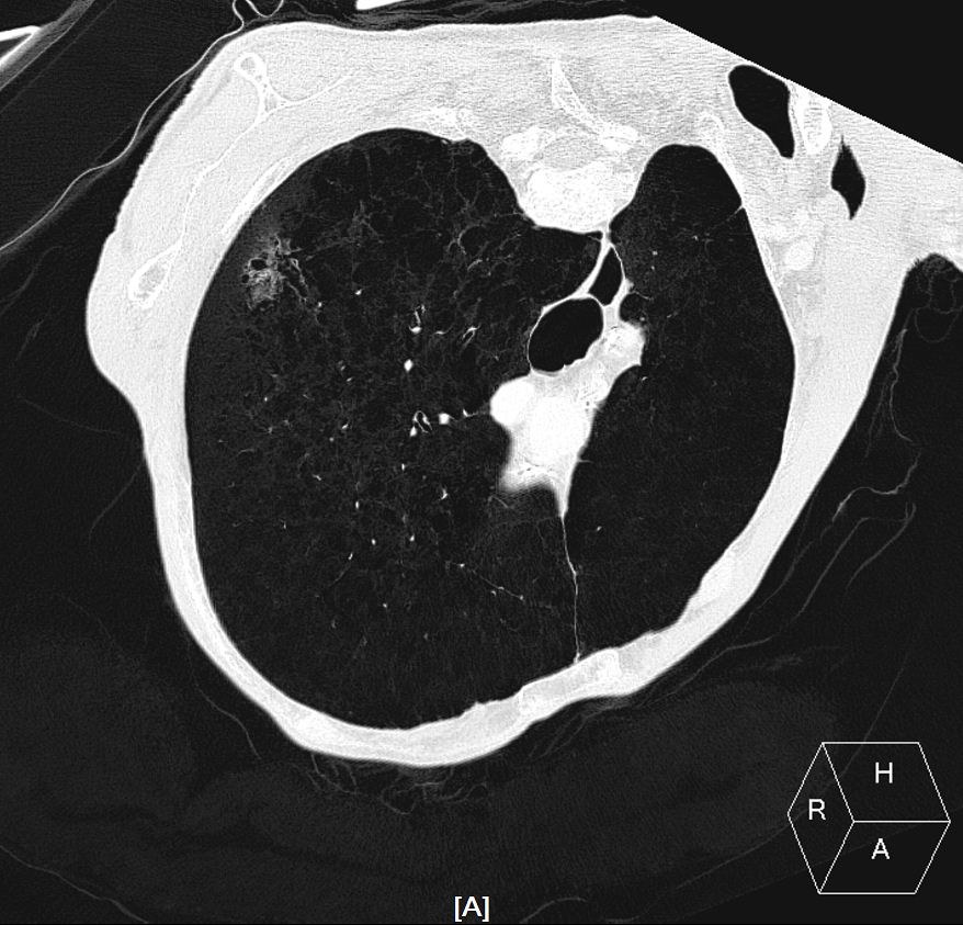
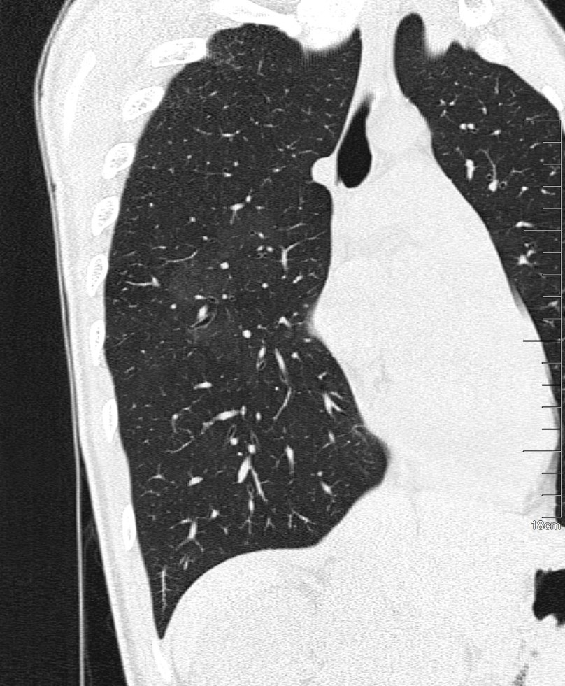
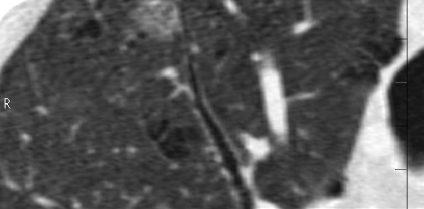
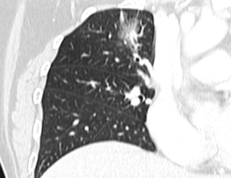
目的利用多层螺旋CT(MSCT)探讨肺局限性磨玻璃影(fGGO)与支气管的关系及与病理类型的相关性。 方法选择行256层iCT检查所检出的最大直径≤3.0 cm的fGGO患者86例,共89个fGGO作为观察对象,分为良性组29个,浸润前病变组11个,浸润性腺癌组49个,在横断位、薄层重组、曲面重建、最小密度投影(MinIP)图像上观察fGGO中磨玻璃部分的比例及其与支气管的关系。根据薄层CT图像上病灶内磨玻璃成分所占比例,将肺内磨玻璃密度影(fGGO)分为3类,A类为纯fGGO;B类为磨玻璃部分占51%~99%的混合fGGO;C类为磨玻璃部分≤50%的混合fGGO。将fGGO与支气管的关系分为5型:Ⅰ型:fGGO中的支气管被截断;Ⅱ型:fGGO实性成分内的支气管走行扭曲、扩张;Ⅲ型:fGGO磨玻璃区内的支气管扭曲、扩张;Ⅳ型:支气管在磨玻璃区走行正常;Ⅴ型:支气管在病灶旁边绕行,未进入病灶内部。 结果(1)3组GGO与支气管存在关系的比率分别为65.5%、36.4%及95.9%,3组之间的差异有统计学意义(χ2=26.758,P < 0.001);(2)GGO与支气管类型显示为Ⅰ型时,良性组、浸润前病变组及浸润性腺癌组分别为0、0、16个;Ⅱ型在3组中分别为2、0、15个;Ⅲ型在3组中分别为2、0、6;Ⅳ型在3组中分别为9、2、5个;Ⅴ型在3组中分别为6、2、5个,差异有统计学意义(P < 0.001)。良性病变组以Ⅳ、Ⅴ型多见,浸润前病变组多与支气管无关系,浸润型腺癌则以Ⅰ、Ⅱ型多见;(3)按CT图像上病灶内磨玻璃成分比例,A类13个,B类32个,C类44个,病灶内磨玻璃成分含量与支气管分型间存在相关性(r=0.442,P < 0.000)。不同的病理类型的fGGO及GGO含量不同的病变与支气管存在不同的关系,差距具有统计学意义。 结论多层螺旋CT扫描和多种重组方法,能清晰显示fGGO与支气管的关系及其形态特征,推断其组织学类型,对临床治疗具有重要意义。 Abstract:ObjectiveTo investigate the relationship between pulmonary focal ground-glass opacity (fGGO) and bronchial and the correlation with the pathological types by MSCT. MethodsA total of 86 patients and 89 lesions who had≤3.0 cm fGGO that detected on 256 iCT scan were enrolled in this study, including 29 benign lesions, 11 lesions were preinvasive lesions and 49 lesions of pulmonary adenocarcinoma.The solid component proportion of fGGO and its relationship with bronchiole were determined on the cross sectional plane、multiple plane reconstruction (MPR)、curve planar reformation (CPR)、MinIP images According to the proportion of solid component which was measured on CT images, fGGO lesions were divided into class A (pure GGO), class B (51%-99% ground glass) and class C (1%-50% ground glass).The correlation of fGGO with bronchiole were divided into 5 types: type Ⅰwith the bronchiole obstructed; type Ⅱ with the bronchiole of tortuous and dilated fGGO shadow; type Ⅲ within ground glass area the bronchiole was tortuous and dilated; type Ⅳ with normal bronchial course and ground glass area; type Ⅴ with the bronchiole running its way by the side of the lesion and did not enter fGGO. Results(1) The presence rate of certain relationship between GGO and bronchiole in the three groups were 65.5%(19/29)、36.4% (4/11) and 95.9% (47/49), respectively, with significant differences (χ2=26.758, P < 0.001). (2) In type Ⅰ, the lesion number of the benign lesion group, preinvasive group and invasive group were 0, 0 and 16 respectively, which was 2, 0 and 15 respectively in type Ⅱ, 2.0 and 6 respectively in type Ⅲ, 9, 2 and 5 respectively in type Ⅳ, and 6, 2 and 5 respectively in type Ⅴ.The differences were significant (P < 0.001).In benign lesion group type Ⅳ and Ⅴ were frequent seen, in pre-invasive lung cancer group, the lesions usually showed no certain relations with the bronchi, while in infiltrative adenocarcinomas type Ⅰ and Ⅱ were commonly found. (3) The lesion number of class A, B and C were 13, 32 and 44 respectively. A positive correlation existed between the proportion of ground glass and the bronchiole-related type (r=0.442, P < 0.000). The different nature of fGGO were different from the type of bronchus with significant differences. ConclusionMSCT scanning and multiple reconstruction Methods can clearly show the correlation of fGGO with bronchi and the morphological characteristics. It can infer the organization type with vital significance to the clinical treatment. -
Key words:
- focal ground-glass opacity /
- bronchus /
- pulmonary adenocarcinoma /
- MSCT
-
表 2 3类含不同密度GGO的病理类型(个)
GGO分类 病理类型 病灶总数 良性病变组 浸润前病变组 浸润性腺癌组 A类pGGO 13 7 6 0 B类mGGO 32 8 5 19 C类mGGO 44 14 0 30 pGGO: 纯磨玻璃影; mGGO: 混合性磨玻璃影.  下载: 导出CSV
表 3 3类含不同密度的GGO 与支气管的关系(个)
病变分类 病灶总数 GGO 与支气管的类型 Ⅰ Ⅱ Ⅲ Ⅳ Ⅴ A类pGGO 13 0 0 0 2 2 B类mGGO 32 2 2 3 8 7 C类mGGO 44 14 15 5 6 4 pGGO: 纯磨玻璃影; mGGO: 混合性磨玻璃影.
下载: 导出CSV
-
[1] Austin J, Muller NL, Friedman PJ, et al. Glossary of terms for CT of the lung:recommendations of the Nomenclature Committee of the Fleischner Society[J]. Radiology, 1996,200(2): 327-31. doi: 10.1148/radiology.200.2.8685321 [2] 范丽, 于红, 刘士远, 等. 3 cm以下肺恶性局灶性磨玻璃结节与实性结节螺旋CT征象对照[J]. 中华放射学杂志, 2010, 44(1): 16-9. [3] 何慧, 孙鹏飞, 曹向荣, 等. 肺局灶性磨玻璃密度结节的多层螺旋CT诊断[J]. 中国医学影像学杂志, 2014, 22(2): 121-3, 126. http://www.cnki.com.cn/Article/CJFDTOTAL-ZYYZ201402015.htm [4] Kim HY, Shim YM, Lee KS, et al. Persistent pulmonary nodular ground-glass opacity at thin-section CT: Histopathologic comparisons[J]. Radiology, 2007, 245(1): 267-75. doi: 10.1148/radiol.2451061682 [5] Aoki T, Tomoda Y, Watanabe H, et al. Peripheral lung adenocarcinoma:correlation of thin-section CT findings with histologic prognostic factors and survival[J]. Radiology, 2001,220(13): 803-7. http://cn.bing.com/academic/profile?id=09b7b888d09bc22ba9513e95fc3ac37d&encoded=0&v=paper_preview&mkt=zh-cn [6] 高丰, 葛虓俊, 李铭, 等. 经多层螺旋CT探讨肺磨玻璃结节与支气管的关系[J]. 中华放射学杂志, 2013, 47(2): 157-61. [7] Travis WD, Brambilla E, Noguchi M, et al. International association for the study of lung cancer/American thoracic society/European respiratory society international muhidisciplinary classification of lung adenocarcinoma[J]. J Thorac Oncol, 2011, 6(9): 244-8. http://cn.bing.com/academic/profile?id=421b2e68eefe52f9ca343055b565cf38&encoded=0&v=paper_preview&mkt=zh-cn [8] Collins J, Stern EJ. Ground-glass opacity at CT: the ABCs[J]. AJR Am J Roentgenol, 1997, 169(2): 355-67. doi: 10.2214/ajr.169.2.9242736 [9] Nakajima R, Yokose T, Kakinuma R, et al. Localized pure ground-glass opacity on high-resolution CT: Histologic characteristics [J]. J Comput Assist Tomogr, 2002, 26(3): 323-9. doi: 10.1097/00004728-200205000-00001 [10] Henschke CI, Yankelevitz DF, Mirtcheva R, et al. CT screening for lung cancer: Frequency and significance of part-solid and nonsolid nodules[J]. Am J Roentgen, 2002, 178(5): 1053-7. doi: 10.2214/ajr.178.5.1781053 [11] 强金伟, 周康荣, 蒋亚平, 等. 多层螺旋CT与病理对照研究孤立性肺结节与支气管的关系[J]. 中华放射学杂志, 2003, 37(11): 992-6. http://www.cnki.com.cn/Article/CJFDTOTAL-ZHGS200311009.htm [12] 强金伟, 周康荣, 蒋亚平, 等. 多排螺旋CT显示支气管与外周肺癌关系的价值[J]. 中华肿瘤杂志, 2004, 26(1): 45-8. http://www.cnki.com.cn/Article/CJFDTOTAL-ZHZL200401017.htm [13] Takaashima S, Maruyama Y, Hasegawa M, et al. CT findings and progression of smmallperipheral lung neoplasms having a replacement growth pattern[J]. AJR, 2003, 180(3): 817-26. doi: 10.2214/ajr.180.3.1800817 [14] 陈天忠, 韦乐心, 余绍立, 等. 多层螺旋CT对肺磨玻璃结节与支气管关系的初探[J]. 临床放射学杂志, 2014, 33(5): 711-5. http://www.cnki.com.cn/Article/CJFDTOTAL-LCFS201405018.htm [15] Gaeta M, Barone M, Russi EG, et al. Carcinomatous solitary pulmonary nodules:evaluation of the tumor-bronchi relationship with thin-section CT[J]. Radiology, 1993, 187(2): 535-9. doi: 10.1148/radiology.187.2.8475303 [16] Miser WF. Cancer screening in the primary care setting-The role of the primary care physician in screening for breast, cervical,colorectal, lung, ovarian, and prostate cancers[J]. Prim Care, 2007,34(1): 137-42. doi: 10.1016/j.pop.2007.02.002 -


 点击查看大图
图(5) / 表(3)
计量
- 文章访问数: 751
- HTML全文浏览量: 235
- PDF下载量: 3
- 被引次数: 0