


Value comparison of MSCT and MRI in brucella spondylitis
-
摘要:
目的 探讨多排螺旋CT和MRI在布氏杆菌脊柱炎中的应用价值。 方法 回顾性分析36例布氏杆菌脊柱炎患者64排128层螺旋CT及MRI图像。 结果 MR显示2个椎体受累者29例,3个椎体受累者5例,4个椎体受累者2例;腰1椎体受累3例,腰2椎体受累6例,腰3椎体受累15例,腰4椎体受累24例,腰5椎体受累18例,骶1椎体受累3例;30例椎间盘有不同程度累及,21例有椎间隙变窄,15例椎间隙未见明显变窄;27例有不同程度椎旁软组织肿胀。CT显示2个椎体受累27例,3个椎体受累4例,有骨质破坏椎体63个,有6例椎体骨质破坏不明显,9例椎体软组织肿胀不明显。MRI较CT更能清晰显示椎体骨髓水肿及椎旁软组织情况,但对细微骨质破坏不如CT。 结论 MRI和MSCT相结合,能提高布氏杆菌脊柱炎侵犯程度和累及范围。 Abstract:Objective To investigate the value of Multi-slice spiral CT and magnetic resonance (MR) imaging in Brucella spondylitis. Methods The imaging findings of 36 patients with brucellosis spondylitis retrospectively analyzed. The patients were performed 64 layer 128 slice spiral CT and MRI. Results In MRI, 29 had 2 lumbar, 5 had 3 lumbar and 2 had four lumbar vertebrae involvement. Three had the first lumbar, 6 had the second lumbar, 15 had the third lumbar, 24 had the fourth lumbar, 18 had the fifth lumbar and 3 had the first sacral vertebra involvement. Thirty patients had different degrees intervertebral disc involvement. There were 21 patients had the positive findings of intervertebral space narrowing, however, 15 patients had no obvious narrowing. Twenty-seven patients had different levels of paraspinal soft tissue swelling. In CT, 27 had two lumbar and 4 had three lumbar vertebrae involvement. Additionally vertebral destruction was detected in 63 vertebrae. There were 6 cases had no obvious vertebral destructions and 9 cases had no significant vertebral soft tissue swelling. Compared with CT, MRI is a highly sensitive imaging technique in showing the vertebral marrow edema and soft tissue involvement, but lack in the ability of showing tiny vertebral destruction. Conclusion The combination of MSCT and MRI can improve the diagnosis of Brucella spondylitis, especially in the involvement degrees and scope. -
Key words:
- brucellosis /
- spondylitis /
- multi-slice spiral CT /
- magnetic resonance imaging
-
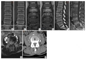
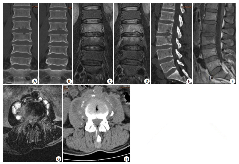
图 1 脊柱受累情况MRI、CT图对比
A、B、E: 为同一患者CT冠状及矢状位显示骨质破坏, 未能显示椎体骨髓水肿; C、D、F: 为该患者MRI 压脂冠状位及T1WI矢状位显示骨髓水肿但骨质破 坏显示不明; G、H: 分别用CT和MRI显示周围软组 织肿胀, MRI比CT更能清晰显示椎旁软组织肿胀 范围及小脓肿形成.
表 1 CT和MRI脊柱受累情况对比
受累椎体(个) 椎体形态改变(个) 椎体骨质破坏(个) 附件受累(个) 椎间盘受累(个) 椎旁软组织肿胀(例) 脓肿形成(例) MR 69 6 45 27 48 36 12 CT 63 9 63 12 36 27 12  下载: 导出CSV
-
[1] Dean AS, Bonfoh B, Kulo AE, et al. Epidemiology of brucellosis and q Fever in linked human and animal populations in northern Togo[J]. PLoS One, 2013, 8(8): e71501-7. doi: 10.1371/journal.pone.0071501 [2] 李梦东.实用传染病学[M].重庆:科学技术文献出版社重庆分社, 1989: 480-6. [3] 唐丽丽, 刘白鹭, 舒圣捷, 等.布氏杆菌病性脊柱炎的影像学诊断[J].中国医学影像学杂志, 2013(6): 414-6. http://www.cnki.com.cn/Article/CJFDTOTAL-ZYYZ201306006.htm [4] Bettini N, Girardo M, Dema E, et al. Evaluation of conservative treatment of non specific spondylodiscitis[J]. Eur Spine J, 2009, 18 (Suppl 1): 143-50. https://www.researchgate.net/publication/24401649_Evaluation_of_conservative_treatment_of_non_specific_spondylodiscitis [5] Günal O, Barut S, Ayan M, et al. Investigation of coxiella burnetii and brucella seropositivities in patients presenting with acute fever[J]. Mikrobiyol Bul, 2013, 47(2): 265-72. doi: 10.5578/mb.4659 [6] 郑旭.布氏杆菌脊柱炎的影像分析及鉴别诊断[J].中国医药指南, 2015, 13(1): 78-9. http://www.cnki.com.cn/Article/CJFDTOTAL-YYXK201501053.htm [7] 于天松, 刘玉春, 袁晨, 等.布鲁菌性脊柱炎与脊柱结核MRI表现[J].实用放射学杂志, 2010, 26(7): 1066-7, 1074. [8] 宁永红, 杨海廷.低场磁共振成像在腰背痛为首发症状的布氏杆菌脊柱炎中的诊断价值[J].实用医学影像学杂志, 2014, 15(3): 207-9. http://www.cnki.com.cn/Article/CJFDTOTAL-SYXY201403022.htm [9] 张培楠, 杨新明, 孟宪勇, 等.布氏杆菌病性脊椎炎的影像学分型[J].河北医科大学学报, 2015, 36(3): 334-7. http://www.cnki.com.cn/Article/CJFDTOTAL-HBYX201503033.htm [10] 赵林伟, 董国礼, 张小明, 等. MRI对诊断脊柱结核的价值探讨[J].实用放射学杂志, 2011, 27(6): 907-11. [11] 赵晓丹, 王飞飞, 赵鸿飞, 等.脊柱结核与化脓性脊柱炎CT鉴别诊断[J].实用放射学杂志, 2015, 31(4): 621-4, 629. http://cdmd.cnki.com.cn/Article/CDMD-11065-1015983925.htm [12] 刘涛, 孙健民, 崔新刚, 等. MRI及病理学鉴别早期化脓性脊柱炎及布氏杆菌脊柱炎中的应用价值[J].中国组织工程研究, 2014, 18(4): 499-504. http://www.cnki.com.cn/Article/CJFDTOTAL-XDKF201404004.htm -

 点击查看大图
图(1) / 表(1)
计量
- 文章访问数: 553
- HTML全文浏览量: 270
- PDF下载量: 3
- 被引次数: 0