


CT features of renal oncocytoma
-
摘要:
目的 探讨肾嗜酸细胞腺瘤(RO)的CT表现。 方法 回顾性分析经病理证实的8例RO的影像及临床资料。 结果 CT平扫8例RO均为单侧、单发,呈类圆形4例,分叶状4例,均突出肾轮廓外。8例肿瘤实质部分CT值约22~36 Hu,平均31 Hu。增强扫描8例均为富血供,皮髓质期肿瘤实质CT值103~187 Hu,平均强化幅度较平扫上升94 Hu。实质期强化减低,CT值82~151 hu,平均强化幅度较平扫上升74 Hu。排泄期稍下降,CT值86~117 Hu,排泄期平均强化幅度较平扫上升70 Hu。2例肿瘤见星状瘢痕,2例节段性强化逆转,1例轮辐状强化,1例肿瘤内出血、1例点状钙化,1例少量脂肪。 结论 当CT平扫表现类圆形或分叶状突出肾轮廓外的等、低密度影,增强表现富血供,皮髓质期肿瘤明显不均匀强化,肿瘤实质明显强化,实质期、排泄早期不同程度的下降,皮髓质期肿瘤内不均匀强化的低密度区,在实质期、排泄期逐渐减小或消失,呈进行性填充,同时肾静脉及下腔静脉内无瘤栓,肾门及腹主动脉旁未见肿大淋巴结,肾嗜酸细胞腺瘤是可供选择的鉴别诊断之一。 Abstract:Objective To explore the CT findings of renal oncocytoma. Methods The imaging and clinical data of 8 cases of RO with pathology confirmed renal oncocytoma were retrospectively analyzed. Results On plain CT scans, 8 patients had unilateral solitary lesion of which were round or lobulated. All lesion protruded the outline of kidney. CT value of tumor substantial part was 22-36 Hu, average 31 Hu in 8 cases. Eight cases were rich blood supply on enhanced CT scans, CT value of tumor substantial part was 103-187 Hu in the cortico-medullary phase, the average strengthening rate was rised 94 Hu than plain CT. CT value was 82-151 Hu in the parenchymal phase, the average strengthening rate was rised 74 Hu than plain CT. CT value was 86-117 Hu and decreased slightly in the excretory phase, the average strengthening rate was rised 70 Hu than plain CT. central scars was found in 2 cases, Segmental Enhancement Inversion in 2, Calcification in 1 cases, spoke-wheel-like enhancement in 1, hemorrhage in 1 fat in 1. Conclusion When the plain CT scans showed isodensity or low density tumor with round or lobulated protruding the outline of kidney, Tumor with rich blood supply on enhanced CT scans, tumor substantial showed significantly enhancement in the cortico-medullary phase, was decreased in the parenchymal phase and excretory phase, the low-density area of tumors heterogeneous enhancement of in the cortico-medullary phase, which showed reduce or disappear Gradually in the parenchymal phase and excretory phase. with progressively filled, at the same time, no tumor thrombus within the renal vein and inferior vena cava, and no swelling large lymph nodes around abdominal aorta and renal hilum, Renal oncocytoma is a differential diagnosis to choose. -
Key words:
- kidney tumor /
- oncocytoma /
- tomography /
- X-ray computed /
- pathology
-

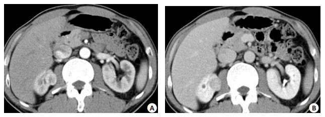
图 1 肿瘤呈分叶状突出肾外
A: CT平扫, 肿瘤呈软组织密度, 分叶状, 周围脂肪密度增高, 肾周筋膜结节状增厚; B:皮髓质期, 增强扫描皮髓质期可见明显不均匀强化; C:实质期中央星状低密度瘢痕区范围缩小; D:排泄早期中央星状低密度瘢痕区进行性范围缩小.

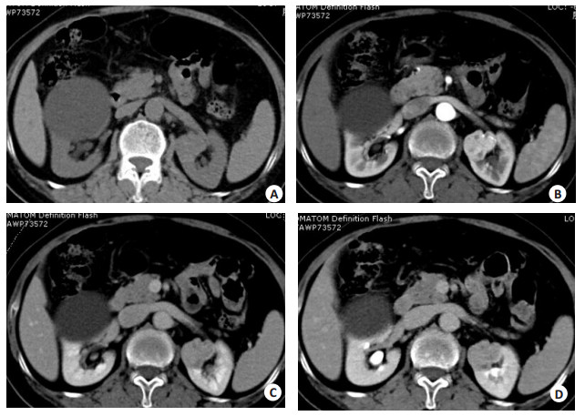
图 3 肿瘤“快进慢出”型表现
A: CT平扫, 肿瘤呈等密度; B:皮髓质期, 明显不均匀强化; C:实质期, 强化程度逐渐缓慢减低; D:排泄期, 强化程度轻度减低, 其内低密度影进行性填充(注右肾前唇囊肿).
-
[1] Rabbani F, Hakimian P, Reuter VE, et al. Renal vein or inferior vena caval extension in patients with renal cortical tumors:impact of tumor histology[J]. J Urol, 2004, 171(3): 1057-61. doi: 10.1097/01.ju.0000112885.66352.e2 [2] 沈剑, 侯建国, 孙颖浩. 14例肾嗜酸性细胞瘤的临床病理分析[J].临床肿瘤学杂志, 2012, 17(1): 55-8. http://mall.cnki.net/magazine/Article/LCZL201201012.htm [3] 方丹波, 任国平, 蔡松良. 17例肾嗜酸性细胞瘤的诊断体会[J].中华外科杂志, 2004, 42(4): 250-1. http://www.cnki.com.cn/Article/CJFDTOTAL-ZHWK200404017.htm [4] 张洁, 马大庆, 贺文, 等.肾嗜酸性细胞腺瘤和肾癌的螺旋CT鉴别诊断[J].中国医学影像技术, 2007, 23(5): 718-20. http://www.cnki.com.cn/Article/CJFDTOTAL-ZYXX200705027.htm [5] 辛越, 毕文杰, 孙庆举, 等.肾脏嗜酸细胞腺瘤的CT诊断[J].临床放射学杂志, 2008, 27(3): 364-7. http://www.cnki.com.cn/Article/CJFDTOTAL-LCFS200803029.htm [6] 强军, 高万勤, 关文华, 等.肾嗜酸细胞腺瘤的CT诊断[J].实用放射学杂志, 2011, 27(1): 90-1, 121. [7] Zippel L. Zur kenntnis Der oncocyten[J]. Virchows Arch, 1942, 2 (3): 360-82. [8] Klein MJ, Qj V. Proximal tubular adenoma of kidney with socalled oncocytic features, A clinicopathologic study of 13 cases of ararely reported neoplasm[J]. Cancer, 1976, 38(2): 906-14. doi: 10.1002/(ISSN)1097-0142 [9] 黎德林, 邓耀良.肾嗜酸细胞腺瘤的诊断进展[J].现代泌尿生殖肿瘤杂志, 2014, 8(2): 125-7. http://www.cnki.com.cn/Article/CJFDTOTAL-PXDM201402020.htm [10] 刘佳, 林建, 韩文科, 等.肾嗜酸性细胞瘤26例临床特征分析[J].中华外科杂志, 2012, 50(7): 642-5. [11] 廖茜, 白人驹, 汪俊萍.肾嗜酸细胞瘤的CT表现与病理对照分析[J].中国医学影像学杂志, 2011, 19(4): 283-6. http://www.cnki.com.cn/Article/CJFDTOTAL-ZYYZ201104016.htm [12] Eiss D, Larousserie F, Mejean A, et al. Renal oncocytoma: CT diagnostic criteria revisited[J]. J Radiol, 2005, 86(12 Pt 1): 1773-82. https://www.researchgate.net/publication/7436885_Renal_oncocytoma_CT_diagnostic_criteria_revisited_in_French [13] Kim JI, Cho JY, Moon KC, et al. Segmental enhancement inversion at biphasic multidetector CT: characteristic finding of small renal oncocytoma[J]. Radiology, 2009, 252(2): 441-8. doi: 10.1148/radiol.2522081180 [14] Mcgahan JP, Lamba R, Fisher J, et al. Is segmental enhancement inversion on enhanced biphasic MDCT a reliable sign for the noninvasive diagnosis of renal oncocytomas [J]. AJR Am J Roentgenol, 2011, 197(4): W674-9. doi: 10.2214/AJR.11.6463 [15] O'malley ME, Tran P, Hanbidge A, et al. Small renal oncocytomas: is segmental enhancement inversion a characteristic finding at biphasic MDCT[J]. AJR Am J Roentgenol, 2012, 199(6): 1312-5. doi: 10.2214/AJR.12.8616 [16] 王芳, 陆菁菁, 秦明伟, 等.肾嗜酸细胞腺瘤的CT表现及病理基础[J].实用放射学杂志, 2008, 24(9): 1230-2. http://www.cnki.com.cn/Article/CJFDTOTAL-SYFS200809031.htm [17] Ambos MA, Bosniak MA, Valensi QJ, et al. Angiographic patterns in renal oncocytomas[J]. Radiology, 1978, 129(3): 615-22. doi: 10.1148/129.3.615 -

 下载:
下载:
 点击查看大图
图(4)
计量
- 文章访问数: 678
- HTML全文浏览量: 234
- PDF下载量: 5
- 被引次数: 0