


Add vessel and merge views of spiral CT volume to reconstruct the value of hemorrhagic cerebral vascular disease
-
摘要:
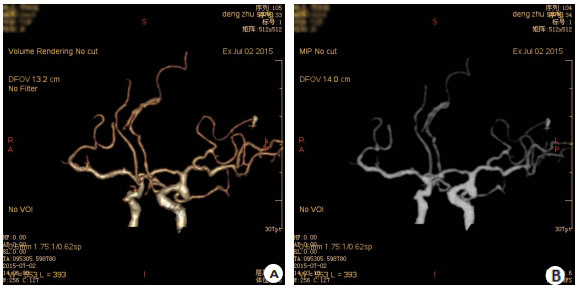
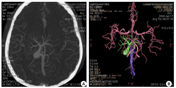
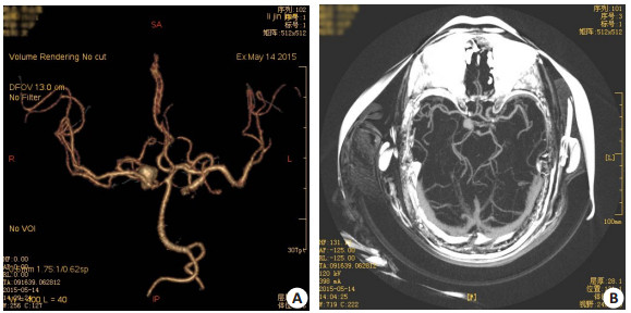
目的探讨采用多排螺旋CT血管生成、图像融合法容积重建出血性脑血管病变的价值。 方法采用16排螺旋CT进行头部容积扫描,利用“血管生长”及“图像融合”法对25例出血性脑血管病变的图像进行容积再现三维重建,分析出血性脑血管病变CTA的影像表现,评价其在出血性脑血管病变诊断和治疗中的优势和不足。 结果25例患者MSCT发现动脉瘤18例,显示瘤体、瘤颈、载瘤动脉和与周围血管及颅骨的关系清晰、确切;动静脉畸型(AVM)3例,显示畸形血管团的部位、大小、供血动脉来源,引流静脉的分支情况,空间立体结构清晰;静脉性血管畸形(CCVM)2例,不仅显示病灶特有的“海蛇头”征,而且完整地显示颅内静脉畸形的组成及引流静脉全程的三维影像;烟雾病1例,MSCT显示颈内动脉远段闭塞及近段Willis环血管渐进性狭窄,脑底部异常血管网形成;不明原因1例。 结论多排螺旋CT采用血管生成、图像融合法进行脑内血管容积重现,能安全、可靠、快速、无创清晰显示脑血管病变,为临床诊断及治疗提供了可靠的信息。正逐渐代替或部分代替全脑血管造影检查,可作为自发性脑出血患者病因诊断的首选检查方法。 Abstract:ObjectiveTo explore the value of add vessel and merge views of spiral CT volume to reproduce hemorrhagic cerebral vascular disease. MethodsUsing 16 -slice spiral CT volume scan of the head, Volume rendering of the 25 cases of hemorrhagic cerebral vascular disease,image of blood vessel growth and image fusion method,Analysis of the performance of the hemorrhagic cerebral vascular disease CTA image, To evaluate the strengths and weaknesses of hemorrhagic cerebral vascular disease diagnosis and treatment. ResultsMSCTA, 18 aneurysms, 3 AVM, and,2 cerebral CVM were found in 25 patients, The aneurysmal body and neck, parent artery and relationship with adjacent vessels and skulls were displayed clearly and exactly by MSCTA, The position, shape, sources of the feeding artery, branch of the draining vein and stereochemical structure of the nidus of AVM were showed clearly. The sensitivity and specificity for MSCTA of detection of SAH with were 95% and 100%,respectively. ConclusionMSCTA, as a simple, fast, non-invasive, safe and reliable technique of Cerebral Angiography, can take place of DSA on some degree, and be regarded as the first choice on etiological diagnose of superacute SAH. -
Key words:
- cerebral vascular disease /
- tomography /
- spiral computed /
- angiography
-
[1] 悦保仕,温平贵.多层螺旋CT三维血管造影容积重建成像在颅内动脉瘤中的诊断价值[J].实用放射学杂志,2005,21(9):904-7. [2] 洪汝建,陈爽,冯晓源.16层CT血管造影在颅内动脉瘤诊断及术后评价中的应用[J].临床放射学杂志,2005,24(4):310-3. [3] 白人驹,马大庆,张雪林,等.医学影像诊断学[M].2版.北京:人民卫生出版社,2006:81. [4] 陈旭,胡海菁,刘成辉,等.三维CT血管成像在颅内动静脉畸形诊治中的价值[J].中国脑血管病杂志,2009,10(4):198-202. [5] 胡海菁,陈旭,李春芳,等.MSCT在自发性蛛网膜下腔出血超级性期诊疗中的应用[J].中国介入影像与治疗学,2008,5(1):19-22. [6] 曹代荣,游瑞雄,杨焱,等.脑静脉血管瘤16层螺旋CT及CTV诊断[J].放射学实践,2007,22(5):455-8. -

 下载:
下载:
 点击查看大图
图(3)
计量
- 文章访问数: 691
- HTML全文浏览量: 226
- PDF下载量: 3
- 被引次数: 0