


Imaging characteristics of pulmonary inflammatory pseudotumor in patients with complications after puncture via CT scan
-
摘要:
目的穿刺易出现并发症的肺部炎性假瘤的影像学特征,为提高CT引导穿刺安全性提供参考 方法分析15例穿刺导致并发症(A组)及32例未出现并发症(B组)患者的一般临床特征及影像学特点,比较比较两组差异。 结果A组以女性为主,而B组以男性为主,两组性别构成有统计学差异(P=0.036);A组以青年为主,B组以中年为主,两组年龄差异有统计学意义(P=0.002);A组病程显著短于B组,假瘤体最大直径显著大于B组(P=0.033),且CT值显著低于B组(P=0.009)。A组以左肺假瘤为主,而B组以右肺稍多,两组好发肺侧差异有统计学差异(P=0.039);A组多数无完整包膜,而B组多数有包膜(P=0.021)。A组有纵膈淋巴结肿大患者稍多,而B组大多数无淋巴结肿大(P=0.029)。A组多数假瘤呈不规则外形,但B组多数呈圆形或接近圆形,外形差异有统计学意义(P=0.027);A组假瘤多数呈不均匀密度,而B组大多数呈均匀密度,差异有统计学意义(P=0.032)。 结论对于发病时间较短的青年女性肺部炎性假瘤患者,尤其是发生于左肺、瘤体直径较大、外形不规则、密度不均匀、CT值偏低、包膜不完整,伴有纵膈淋巴结肿大者,应警惕CT引导穿刺所致并发症的发生。 Abstract:Objective To conclude the imaging characteristics of the patients with pulmonary inflammatory pseudotumor who suffered from complications after puncture via CT scan. Methods analysis of 15 cases of complications puncture (group A) and 32 cases (group B) does not appear complications in patients with the clinical features and imaging characteristics of comparison to compare differences between the two groups. Results in group A is given priority to with women, and group B is given priority to with men, two groups of gender constitute A statistically significant (P=0.036); Is given priority to with the youth group A and group B is given priority to with middle-aged, age differences between the two groups was statistically significant (P=0.002). Course significantly shorter than group B, group A false tumors had the largest diameter was significantly more than group B (P=0.033), and computed tomography (CT) value was significantly lower than that of group B (P=0.009). Give priority to with the left pulmonary pseudotumor, group A and group B with A bit more right lung, good hair lung side similar between the two groups was statistically difference (P=0.039); Most complete capsular, group A and group B most envelope (P=0.021). A group of patients with mediastinal lymph node enlargement is A bit more, and most of group B without lymph node enlargement (P=0.029). Most of the group A pseudotumor showed irregular shape, but group B most assumes the circular or nearly circular, shape difference was statistically significant (P=0.027); Pseudotumor most uneven density, group A and group B most homogeneously density, difference was statistically significant (P=0.032). Conclusion The onset time shorter young female patients with pulmonary inflammatory pseudotumor, especially in the left lung, tumors had larger diameter, irregular shape, uneven density, CT value is low, coated incomplete, associated with mediastinal lymph node enlargement, should be vigilant the complications caused by CT guided puncture. -

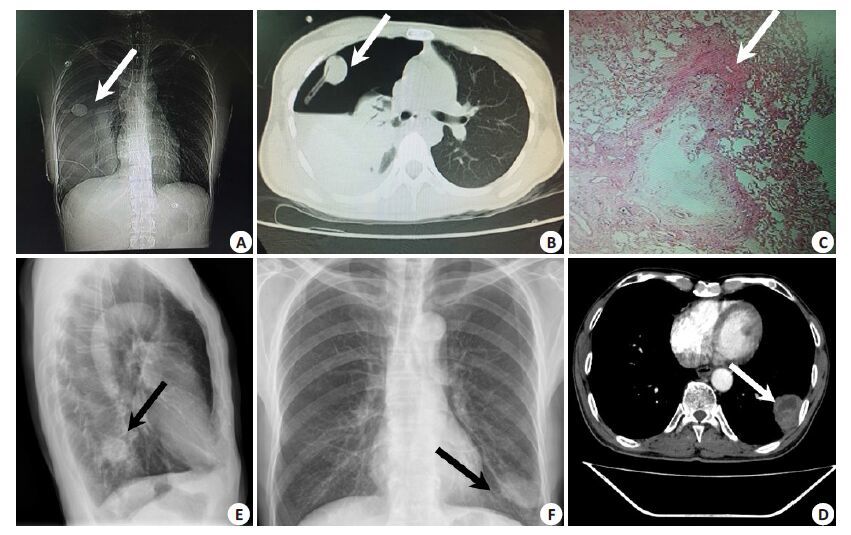
图 1 出现与未出现并发症患者的影像学及病理学检查结果(箭头)
A: 甲患者,女性22岁,右肺上叶中高密度信号; B: 甲患者CT引导下行病灶穿刺术,患者在术中出现血气胸、呼吸困难; C: 甲患者急诊胸腔镜下行肺叶切除并病灶切除术,术后病理切片提示肺泡上皮及纤维组织增生,间质呈炎症性改变并肉芽肿,提示炎性假瘤; D: 乙患者,男性48岁,侧位胸片提示左肺下叶背段类圆形密度增高影; E: 乙患者相片正位提示左肺下叶椭圆形高密度影; F: 增强CT提示左肺下叶背段一密度增高影,部分与胸膜相连。
表 1 两组患者一般情况及定量影像学资料比较(x±s)
分组 n 性别 年龄(岁) 病程(年) 最大直径(cm) 平扫CT值(Hu) 男 女 出现并发症组(A组) 15 4 11 27.29±6.17 1.68±0.73 3.34±1.52 24.81±13.79 无并发症组(B 组) 32 26 6 43.26±13.49 3.53±1.29 2.19±0.58 35.63±18.42 χ2/t χ2=2.397 -6.294 -2.513 2.237 -4.039 P 0.036 0.002 0.029 0.033 0.009  下载: 导出CSV
表 2 两组患者定性影像学资料比较(n)
分组 n 部位 完整包膜 肿大淋巴结 外形 密度 左肺 右肺 有 无 有 无 圆或类圆形 不规则 均匀 出现并发症组(A组) 15 11 4 5 10 9 6 3 12 5 无并发症组(B 组) 32 15 17 23 9 4 28 27 5 26 χ2 1.967 2.783 2.538 2.626 2.291 P 0.039 0.021 0.029 0.027 0.032
下载: 导出CSV
-
[1] 王荣品, 赵振军, 张金娥, 等. MSCT鉴别诊断肺门区炎性块影与中央型肺癌[J]. 中国医学影像技术, 2009, 25(5): 779-82. http://www.cnki.com.cn/Article/CJFDTOTAL-ZYXX200905022.htm [2] 李盛祥, 巩湘浩, 曹火乃. CT引导下经皮肺穿刺活检对肺部小病灶(≤3cm) 的诊断价值[J]. 南华大学学报:医学版, 2008, 36(4): 473-5. http://www.cnki.com.cn/Article/CJFDTOTAL-HYYY200804018.htm [3] 吕淑华, 郭中威. CT引导下经皮肺穿刺活检诊断老年患者肺炎性假瘤的价值[J]. 中国当代医药, 2013, 20(20): 114-5. http://www.cnki.com.cn/Article/CJFDTOTAL-ZGUD201320063.htm [4] 夏国亮, 赵记明, 赵兴康, 等. 肺炎性假瘤的CT诊断[J]. 医学影像学杂志, 2005, 15(11): 948-50. http://www.cnki.com.cn/Article/CJFDTOTAL-XYXZ199401000.htm [5] 杨贤卫, 周伟生, 何蓉. CT引导下经皮肺穿刺切割活检对肺部病变的诊断价值[J]. 中国介入影像与治疗学, 2006, 3(3): 161-4. http://www.cnki.com.cn/Article/CJFDTOTAL-JRYX200603000.htm [6] 王兵, 张德超, 程贵余, 等. 肺部炎性假瘤的诊断及外科治疗(附51例报告) J]. 癌症, 2005, 24(2): 219-21. http://xuewen.cnki.net/CJFD-AIZH200502020.html [7] 邵亚军, 朱红艳, 王西惠, 等. CT引导下经皮肺穿刺活检的应用价值及其并发症[J]. 现代肿瘤医学, 2014, 22(6): 1340-1. http://www.cnki.com.cn/Article/CJFDTOTAL-SXZL201406034.htm [8] 张福康, 马军, 陈境弟, 等. 肺炎性假瘤的CT表现与误诊分析[J]. 影像诊断与介入放射学, 2011, 20(3): 186-9. http://www.cnki.com.cn/Article/CJFDTOTAL-YXZD201103009.htm [9] 丁凯, 周华富, 廖寿合, 等. 肺炎性假瘤的误诊分析与手术治疗:附53例报告[J]. 中华肺部疾病杂志:电子版, 2010, 3(1): 24-7. http://www.cnki.com.cn/Article/CJFDTOTAL-ZFBD201001008.htm [10] 吴志祥, 刘韧. 肺部炎性假瘤的诊断与外科治疗[J]. 医学临床研究,2009, 26(7): 1316-7. -

 点击查看大图
图(1) / 表(2)
计量
- 文章访问数: 562
- HTML全文浏览量: 208
- PDF下载量: 0
- 被引次数: 0